Abstract 19633: Comparison of Angiotensin II Receptor Blocker versus Beta-blocker Therapy in Genetically Classified Children With Marfan Syndrome
Erica E Gonzalez, Lisa C D’Alessandro, Nitya K Viswanathan, Shaine A Morris, Dianna M Milewicz
Circulation. 2015;132:A19633
Abstract
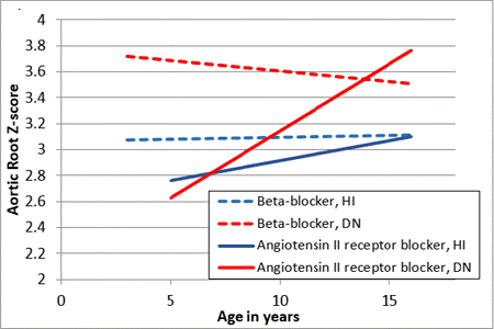
Background: Children with Marfan syndrome (MFS) treated with angiotensin II receptor blocker (ARB) or beta-blocker (BB) therapy show a reduction in the rate of aortic root (AR) growth, yet substantial variability in effect remains. Adult data suggests that patients with haploinsufficient (HI) mutations may be more responsive to ARBs compared with dominant negative (DN) mutations. This study sought to assess the effect of genotype on the rate of AR growth in children treated with either ARB or BB.
Methods: A retrospective review of all individuals with MFS assessed at our institution since 1980 was undertaken; those with ≥2 echocardiograms a minimum of 1 year apart and a documented disease-causing fibrillin-1 (FBN1) mutation were included. One patient with infantile MFS was excluded given extremely AR growth. Genotypes were classified by predicted protein effect as either HI or DN. Multivariable longitudinal linear regression analysis was used to compare average rate of AR growth by genotype and medical therapy using serial echocardiographic data.
Results: Overall 41 children were included, including 18 with HI and 23 with DN mutations. Males comprised 58%. There was no difference in median age at first echo (HI 5.4 years, IQR 3.9-9.3 vs. DN 5.2 years, IQR 3.4-10.6 years, p=0.97), follow-up time (HI 5.8 years, IQR 4.6-8.2 vs. DN 4.9 years, IQR 3.3-8.7, p=0.35), AR dimension (HI 26 mm, IQR 23-29 vs DN 26 mm, IQR 22-37, p=0.81), or AR z-score (HI 3.0, IQR 2.3-4.0 vs DN 3.4, IQR 2.4-4.2, p=0.58). AR growth rate was greater in children with DN mutations (z-score annual change HI -0.04 vs DN +0.07, p<0.0001). When evaluating AR growth only in patients on ARB or BB monotherapy, patients on ARB with DN mutations grew significantly faster than those on BB therapy with DN mutations (p=0.02).
Conclusions: In our cohort, patients with MFS and DN mutations had slower growth on BB therapy compared to those on ARB therapy. Genetic testing may play an important role in targeted medical therapy in MFS.
Article Information
vol. 132 no. Suppl 3 A19633
Published By:
American Heart Association, Inc.
Online ISSN:
History:
- Originally published November 6, 2015.
Copyright & Usage:
© 2015 by American Heart Association, Inc.
Author Information
- 1Cardiology, Texas Children’s Hosp/Baylor College of Medicine, Houston, TX
- 2Cardiology Pediatric, Boston Children’s Hosp/Milford Regional Med Cntr, Milford, MA
- 3Dept of Internal Medicine, Div of Med Genetics, The Univ of Texas Health Science Cntr at Houston Med Sch, Houston, TX
Abstract 19428: Evaluating the Risk of Rheumatic Heart Disease Among Family Members of Children Identified With Latent RHD in School-based Echocardiographic Screening
Twalib Aliku, Andrea Beaton, Amy Scheel, Alison Tompsett, Peter Lwabi, Craig Sable
Circulation. 2015;132:A19428
Abstract
Background: Poorly understood genetic susceptibility factors influence who develops acute rheumatic fever (ARF) and who develops the long-term complication, rheumatic heart disease (RHD). Risk of RHD in family members – and thus the need for screening of first degree relatives – once an index case has been identified, is not known.
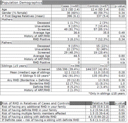
Methods: Sixty RHD positive children (30 borderline 30 definite RHD) and 67 age & gender matched controls were recruited from previous school-based echocardiographic screening. All first-degree relatives ≥ 5 years were invited for echocardiographic screening (2012 World Heart Federation Criteria). Absent family members were recorded with reason for absence including death. Continuous variables were compared using 2-tailed independent samples t-test, categorical variables by Fisher’s Exact Test, and relative risk was used to compare cases and controls.
Results: A total of 454/733 (62%) family members were screened (106 mothers, 48 fathers & 300 siblings). Definite RHD was more common in childhood siblings of cases than controls (p=0.05), and children with RHD were 4.5 times as likely to have a sibling with RHD compared to controls. Mothers had more RHD than fathers without regard to child’s screening status.
Conclusions: Siblings of children identified with latent RHD are at greater risk of having RHD and screening of these children should be considered. While no adult RHD screening data exists from sub-Saharan Africa, this study suggests that the prevalence of occult RHD in adult women is quite high (9.4%). Genotyping studies are needed to fully understand individual and family susceptibility to RHD.
Article Information
vol. 132 no. Suppl 3 A19428
Published By:
American Heart Association, Inc.
Online ISSN:
History:
- Originally published November 6, 2015.
Copyright & Usage:
© 2015 by American Heart Association, Inc.
Author Information
- 1Cardiology, Gulu Univ, Gulu, Uganda
- 2Cardiology, Children’s National Med Cntr, Washington, DC
- 3Undergraduate, Virginia Tech, Blacksburg, VA
- 4Cardiology, The Uganda Heart Institute, kampala, Uganda
Abstract 18815: High Dose Aspirin is Associated With Decrease of Hemoglobin and Do Not Affect the Disease Outcome in Acute Stage of Kawasaki Disease
Ho-Chang Kuo, Kai-Sheng Hsieh
Circulation. 2015;132:A18815
Abstract
Introduction: Kawasaki disease (KD) is a systemic vasculitis of unknown etiology. Although intravenous immunoglobulin (IVIG) has been shown to reduce the incidence of coronary artery lesions (CAL) effectively, the role and appropriate dose of aspirin during acute phase is still unclear. Thus, this study was conducted to evaluate the effect of treatment and the change of serum hepcidin with or without high dose aspirin in the acute stage of KD.
Hypothesis: High dose aspirin is not benefit for acute stage of KD.
Methods: Retrospective analysis of KD patients from two medical centers during 1999-2009 in Taiwan. All patients were initially treated with a single dose of IVIG (2 g/kg) during a 12-h period. High dose aspirin was given (> 50 mg/kg/day) until the fever subsided then low-dose aspirin (3-5 mg/kg/day) was prescribed until all signs of inflammation resolved at group 1. Low-dose aspirin was prescribed without high-dose aspirin at group 2. A total of 851 KD patients (group 1, N= 305, group 2, N= 546) were enrolled in this study. patients.
Results: There were no significant difference between group 1 and group 2 including gender distribution (p=0.51), IVIG resistant rate (31/305 vs. 38/546, p=0.07), CAL formation rate (52/305 vs. 84/546, p=0.67), and total hospital days (6.3 ± 0.2 vs. 6.7 ± 0.2, p=0.13). There were also no significant difference between total white blood counts, hemoglobin levels, platelet counts, CRP and serum hepcidin levels before IVIG treatment between group 1 and group 2 (all p>0.1). After IVIG treatment, there were significant lower hemoglobin and higher CRP as well as lower decrease of CRP levels in group 1 than in group 2 (p=0.006, <0.001 and =0.012, respectively). Furthermore, there were also high serum hepcidin and lower decrease of hepcidin levels in group 1 than in group 2 (p=0.04, and =0.02, respectively).
Conclusions: These results provide evidence supporting high dose aspirin in acute phase of KD may affect the improvement of inflammatory markers. It also caused lower hemoglobin level that was associated with higher hepcidin level after IVIG treatment. It seems unneeded of high dose aspirin in acute phase of KD.
Article Information
vol. 132 no. Suppl 3 A18815
Published By:
American Heart Association, Inc.
Online ISSN:
History:
- Originally published November 6, 2015.
Copyright & Usage:
© 2015 by American Heart Association, Inc.
Author Information
- 1Pediatrics and Kawasaki Disease Cntr, Kaohsiung Chang Gung Memorial Hosp and Chang Gung Univ College of Medicine, Kaohsiung, Taiwan
- 2Pediatrics, Kaohsiung Chang Gung Memorial Hosp, Kaohsiung, Taiwan
Abstract 14661: Early Predictive Markers of Right Ventricular – Pulmonary Vascular Dysfunction in Preterm Infants With Bronchopulmonary Dysplasia
Meghna D Patel, Philip T Levy, Brittney Dioneda, Timothy J Sekarski, Mark R Holland, Aaron Hamvas, Gautam K Singh
Circulation. 2015;132:A14661
Abstract
Introduction: Improved survival of extremely preterm infants has led to an increased recognition of bronchopulmonary dysplasia (BPD) associated with abnormal cardio-pulmonary hemodynamics. Reliable non-invasive quantitative markers for early recognition of BPD in preterm infants are lacking. The aim of this study was to evaluate quantitative echocardiographic measures of right ventricular hemodynamics (RV) as early predictive markers in infants at risk for development of BPD.
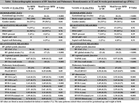
Methods: A prospective study was conducted in 115 preterm infants (< 29 weeks gestational age) enrolled through the Prematurity and Respiratory Outcomes Program (NCT01435187). Echocardiographic measures of RV function and pulmonary hemodynamics were evaluated at 32 and 36 weeks post-menstrual age (PMA). RV systolic function was assessed with fractional area of change (FAC), tricuspid annular plane systolic excursion (TAPSE), and global and free wall longitudinal strain. RV morphometrics were assessed with RV dimensions and areas. Pulmonary hemodynamics were assessed by pulmonary artery acceleration time (PAAT). Commonly used qualitative measures (tricuspid regurgitation velocity and septal flattening) were evaluated for comparison. BPD was classified by the NIH workshop definition.
Results: RV global and free wall strain, FAC, TAPSE, and PAAT were significantly reduced, and RV morphometrics were significantly larger at 32 and 36 weeks PMA in preterm infants who developed BPD (Table 1). Qualitative measures were present in < 20% of patients, and only septal flattening was significant at 32 weeks PMA in the BPD group.
Conclusions: Quantitative echocardiographic measures of RV function and pulmonary hemodynamics are reliable early predictive markers of subsequent development of BPD in preterm infants. These measures are superior to commonly used qualitative measures and can be useful tools for early identification of preterm infants at high risk for BPD.
Article Information
vol. 132 no. Suppl 3 A14661
Published By:
American Heart Association, Inc.
Online ISSN:
History:
- Originally published November 6, 2015.
Copyright & Usage:
© 2015 by American Heart Association, Inc.
Author Information
- Meghna D Patel1;
- Philip T Levy2;
- Brittney Dioneda1;
- Timothy J Sekarski1;
- Mark R Holland3;
- Aaron Hamvas4;
- Gautam K Singh1
- 1Pediatric Cardiology, Washington Univ Sch of Medicine, St. Louis, MO
- 2Neonatology, Goryeb Children’s Hosp, Morristown, NJ
- 3Radiology, Indiana Univ-Purdue Univ Indianapolis, Indianapolis, IN
- 4Neonatology, Northwestern Univ Feinberg Sch of Medicine, Chicago, IL
Abstract 13850: Severity Assessment of Coronary Artery Aneurysm by Z-score of the Internal Diameter in Kawasaki Disease
Masaru Miura, Naoya Fukushima, Tetsuji Kaneko, Tohru Kobayashi, Kenji Suda, Hiroyuki Yamagishi, Taichi Kato, Shinya Shimoyama, Hitoshi Kato, Tsutomu Saji, Kenji Hamaoka, Yuichi Nomura, Kenji Waki, Ryuji Fukazawa, Keiichi Hirono, Shigeto Fuse
Circulation. 2015;132:A13850
Abstract
Introduction: The severity of coronary artery aneurysms (CAA) in patients with Kawasaki disease (KD) has been recently classified according to the z-score. However, it is not known whether this classification can predict coronary events such as stenosis, obstruction, and thrombosis.
Methods: In this multicenter retrospective study, data on height, weight, CAA diameter measured by echocardiography in the acute phase, and the clinical course in KD patients 18 years of age or younger who received a coronary angiography between 1992 and 2011 were collected. Time-dependent occurrence of coronary events was analyzed by Kaplan-Meier method according to small (z-score, < 5.0), medium (≥ 5.0 to < 10.0), and large (≥ 10.0) CAA using a 5 increment scale scheduled to be included in the new American Heart Association criteria. Cox regression analysis was used to identify risk factors for coronary events. The occurrence rate of major cardiac events such as angina pectoris, myocardial infarction, and cardiac death was also analyzed.
Results: Data were analyzed for 1,002 patients from 44 institutions. Both the body surface area and CAA diameters were available in 741 cases for the right coronary artery (RCA) and 609 cases for the left anterior descending artery (LAD). Coronary events occurred in 83 (11.2%) of the RCA group and 57 (9.4%) of the LAD group, while major events occurred in 30 cases (3.0%). The 10-year event-free survival rate for coronary events for small, medium, and large aneurysms was 100, 95.5, and 64.9% in the RCA group, and 100, 94.4, and 63.5% for aneurysms in the LAD group, respectively. The rate of major cardiac events was 98.5, 98.1, and 87.6% for the RCA group, and 100, 97.5, and 86.8% for the LAD group, respectively. Cox regression analyses showed that the z-score of the CAA diameter was an independent risk factor for coronary events for the RCA [large versus medium aneurysm; hazard ratio (HR) 2.8, 95% confidence interval (CI) 1.5 to 5.3, p = 0.002] and the LAD [HR 3.2, 95% CI 1.6 to 6.5, p = 0.015] groups.
Conclusions: The severity assessment of CAA using the 5-increment z-score for coronary arterial diameter can predict the time-dependent occurrence of coronary events in patients with KD.
Article Information
vol. 132 no. Suppl 3 A13850
Published By:
American Heart Association, Inc.
Online ISSN:
History:
- Originally published November 6, 2015.
Copyright & Usage:
© 2015 by American Heart Association, Inc.
Author Information
- Masaru Miura1;
- Naoya Fukushima1;
- Tetsuji Kaneko2;
- Tohru Kobayashi3;
- Kenji Suda4;
- Hiroyuki Yamagishi5;
- Taichi Kato6;
- Shinya Shimoyama7;
- Hitoshi Kato8;
- Tsutomu Saji9;
- Kenji Hamaoka10;
- Yuichi Nomura11;
- Kenji Waki12;
- Ryuji Fukazawa13;
- Keiichi Hirono14;
- Shigeto Fuse15
- 1Div of Cardiology, Tokyo Metropolitan Children’s Med Cntr, Fuchu, Tokyo, Japan
- 2Clinical Rsch Support Cntr, Tokyo Metropolitan Children’s Med Cntr, Fuchu, Tokyo, Japan
- 3Cntr for Clinical Rsch and Development, National Cntr for Child Health and Development, Tokyo, Japan
- 4Dept of Pediatrics, Kurume Univ Sch of Medicine, Kurume, Fukuoka, Japan
- 5Dept of Pediatrics, Keio Univ Sch of Medicine, Tokyo, Japan
- 6Dept of Pediatrics, Nagoya Univ Graduate Sch of Medicine, Nagoya, Aichi, Japan
- 7Div of Cardiology, Gunma Children’s Med Cntr, Shibukawa, Gunma, Japan
- 8Dept of Cardiology, National Cntr for Child Health and Development, Tokyo, Japan
- 9Dept of Pediatrics, Toho Univ Omori Med Cntr, Tokyo, Japan
- 10Dept of Pediatric Cardiology and Nephrology, Kyoto Prefectural Univ of Medicine, Graduate Sch of Med Science, Kyoto, Japan
- 11Dept of Pediatrics, Kagoshima Univ Faculty of Medicine, Kagoshima, Japan
- 12Dept of Pediatrics, Kurashiki Central Hosp, Kurashiki, Japan
- 13Dept of Pediatrics, Nippon Med Sch, Tokyo, Japan
- 14Dept of Pediatrics, Univ of Toyama, Faculty of Medicine, Toyama, Japan
- 15Dept of Pediatrics, NTT East Japan Sapporo Hosp, Sapporo, Hokkaido, Japan
Abstract 13271: Aortic Inflammation Continues in Patients With A History of Kawasaki Disease and Persistent Arterial Aneurysms
Kenji Suda, Nobuhiro Tahara, Akihiro Honda, Tomohisa Nakamura, Hironaga Yoshimoto, Shintaro Kishimoto, Yoshiyuki Kudo, Motofumi Iemura, Yoshihiro Fukumoto
Circulation. 2015;132:A13271
Abstract
Introduction: Kawasaki disease (KD) is well known vasculitis that primarily affects small to middle sized arteries such as coronary arteries and/or peripheral arteries. However, little evidence showed inflammation of large vasculature such as the aorta in patients with KD. Measurements of 18F-fluorodeoxyglucose (FDG) uptake evaluated by positron emission tomography (PET) and X-ray computed tomography (CT) could be useful to identify inflammatory activity of the vessel wall.
Hypothesis: We hypothesized that aortic inflammation continues long after KD.
Methods: FDG-PET/CT was performed in 19 patients with a history of KD. Of 19 patients, 11 patients still had persistent coronary and/or systemic vascular aneurysms (KD-An) and the remaining 8 revealed regression of arterial aneurysms (KD-Reg). Patients suffered from KD at 2.8 ± 3.2 years old and underwent FDG-PET at 22.2 ± 8.0 years old. FDG-PET was also performed in 5 control with age 14.1 ± 2.6 years old. Vascular inflammation was measured by blood-normalized standardized uptake value, known as a target-to-background ratio (TBR). Also demographic and laboratory data were collected.
Results: Aortic FDG uptake was distinctly intense in patients with a history of KD and persistent vascular aneurysms. The Aortic TBR was significantly higher in KD-An (1.50 ± 0.30) than KD-Reg (1.11 ± 0.13, p = 0.004) or controls (1.03 ± 0.25, p=0.003). Although Control was significantly younger and had significantly lower body mass index, there was no significant correlation between these values and TBR. Also there was no significant difference in laboratory data including lipid profiles and glycemic status among 3 groups.
Conclusions: Vascular inflammation of the aortic wall continues long after KD with persistent arterial aneurysms.
Article Information
vol. 132 no. Suppl 3 A13271
Published By:
American Heart Association, Inc.
Online ISSN:
History:
- Originally published November 6, 2015.
Copyright & Usage:
© 2015 by American Heart Association, Inc.
Author Information
- Kenji Suda1;
- Nobuhiro Tahara2;
- Akihiro Honda2;
- Tomohisa Nakamura2;
- Hironaga Yoshimoto1;
- Shintaro Kishimoto1;
- Yoshiyuki Kudo1;
- Motofumi Iemura3;
- Yoshihiro Fukumoto2
- 1DEPT OF PEDIATRICS & CHILD HLTH, Kurume Univ Sch of Medicine, Kurume City, Japan
- 2DEPT OF Cardiovascular Medicine, Kurume Univ Sch of Medicine, Kurume City, Japan
- 3Dept of Pediatric Cardiology, St Mary’s Hosp, Kurume City, Japan
Abstract 10890: Aortic Stiffness Predicts Clinical Outcome and Rate of Aortic Root Growth in Marfan Syndrome
Elif Seda Selamet Tierney, Jami C Levine, Lynn A Sleeper, Mary J Roman, Timothy J Bradley, Steven D Colan, Shan Chen, Michael Jay Campbell, Meryl S Cohen, Julie De Backer, Haleh Heydarian, Arvind Hoskoppal, Wyman W Lai, Aimee Liou, Edward Marcus, Arni Nutting, Aaron K Olson, Gail D Pearson, David Parra, Mary E Pierpont, Beth F Printz, Reed E Pyeritz, William Ravekes, Angela M Sharkey, Shubhika Srivastava, Luciana Young, Ronald V Lacro
Circulation. 2015;132:A10890
Abstract
Background: The NHLBI Pediatric Heart Network randomized trial of atenolol vs. losartan in Marfan Syndrome demonstrated no significant treatment difference in the rate of change in body surface area adjusted maximum aortic root diameter z score (AoRz).
Objectives: To report trial results on aortic stiffness and to determine whether aortic stiffness predicts clinical outcome and change in AoRz.
Methods: 608 patients (6 mo – 25 yr) who met original Ghent criteria and had AoRz > 3 were enrolled. Echocardiograms obtained at 0, 6, 12, 24 & 36 months were centrally interpreted. Aortic dimensions were measured by 2D imaging, and stiffness indices were calculated for aortic root (AoR) and ascending aorta (AA). Where appropriate, stiffness measurements were indexed to 1/sqrt(R-R interval) to adjust for heart rate. Data were analyzed by multivariable mixed effects modeling and Cox regression.
Results: The rate of change over three years in heart rate-adjusted AoR stiffness index differed by treatment (p = 0.016), with a decrease in the atenolol group and no significant change in the losartan group (-0.29 ± 0.14 vs. 0.14 ± 0.14/year). There was no significant treatment effect on the rate of change for AA stiffness index or for elastic modulus (AoR and AA). In the entire cohort, baseline AoR but not the AA stiffness index predicted the rate of change in ARz, with above-average stiffness index (≥ 10) predicting a slower annual decline of AoRz (-0.08 ± 0.02 vs. -0.15 ± 0.01 for below-average stiffness, p < 0.001), even after adjusting for baseline age. Elastic modulus was not a significant predictor of rate of change in AoRz. AoR elastic modulus >122 kPA (75th %ile) independently predicted the composite outcome of AoR surgery, dissection or death (hazard ratio 2.17, 95% CI 1.02 – 4.63, p = 0.04), controlling for baseline age and AoRz. Crude 3-year event rates were 10.4% vs. 3.2% for higher vs. lower elastic modulus groups, respectively.
Conclusions: Atenolol reduced AoR stiffness over 3 years, while losartan did not. In this medically-treated cohort, higher baseline AoR stiffness was associated with a smaller decrease in AoRz, and a greater hazard of AoR dissection/surgery/death. These data suggest that aortic stiffness measures may identify patients at higher risk and guide management.
Article Information
vol. 132 no. Suppl 3 A10890
Published By:
American Heart Association, Inc.
Online ISSN:
History:
- Originally published November 6, 2015.
Copyright & Usage:
© 2015 by American Heart Association, Inc.
Author Information
- Elif Seda Selamet Tierney1;
- Jami C Levine2;
- Lynn A Sleeper3;
- Mary J Roman4;
- Timothy J Bradley5;
- Steven D Colan1;
- Shan Chen6;
- Michael Jay Campbell7;
- Meryl S Cohen8;
- Julie De Backer9;
- Haleh Heydarian10;
- Arvind Hoskoppal11;
- Wyman W Lai12;
- Aimee Liou13;
- Edward Marcus14;
- Arni Nutting15;
- Aaron K Olson16;
- Gail D Pearson17;
- David Parra18;
- Mary E Pierpont19;
- Beth F Printz20;
- Reed E Pyeritz21;
- William Ravekes22;
- Angela M Sharkey23;
- Shubhika Srivastava24;
- Luciana Young25;
- Ronald V Lacro1,
- for the Pediatric Heart Network Investigators
- 1Pediatrics, Boston Children’s Hosp, Harvard Med Sch, Boston, MA
- 2Cardiology, Boston Children’s Hosp, Harvard Med Sch, Boston, MA
- 3Rsch, New England Rsch Institutes, Inc., Watertown, MA
- 4Cardiology, Cornell Univ, New York, NY
- 5Pediatrics, Univ of Toronto, Toronto, Canada
- 6Biostatitics, New England Rsch Institutes, Inc., Watertown, MA
- 7Pediatrics, Duke Univ Med Cntr, Durham, NC
- 8Pediatrics, Children’s Hosp of Philadelphia, Philadelphia, PA
- 9Pediactrics and Med Genetics, Ghent Univ, Ghent, Belgium
- 10Pediatrics, Univ of Cincinnati, Cincinnati, OH
- 11Pediatrics, Univ of Utah, Salt Lake City, UT
- 12Pediatrics, Columbia Univ, New York, NY
- 13Pediatrics, Baylor College of Medicine, Houston, TX
- 14Pediatrics, Boston Children’s Hosp, Boston, MA
- 15Pediatrics, Med Univ of South Carolina, Charleston, SC
- 16Pediatrics, Univ of Washington, Seattle, WA
- 17Adult and Pediatric Cardiac Rsch Program, National Heart, Lung, and Blood Institute, Bethesda, MD
- 18Pediatrics, Vanderbilt Univ, Nashville, TN
- 19Pediatrics, Univ of Minnesota, Minneapolis, MN
- 20Cardiology, Univ of California, San Diego, San Diego, CA
- 21Medicine, Univ of Pennsylvania, Philadelphia, PA
- 22Pediatrics, Johns Hopkins Univ, Baltimore, MD
- 23Pediatrics, Washington Univ, St. Louis, MO
- 24Pediatrics, Mount Sinai Hosp, New York, NY
- 25Pediatrics, Northwestern Univ, Chicago, IL
Abstract 9979: Health-Related Quality of Life in Children With Marfan Syndrome
Jill C Handisides, Danielle Hollenbeck-Pringle, Karen Uzark, Felicia L Trachtenberg, Victoria L Pemberton, Teresa W Atz, Timothy J Bradley, Sylvia De Nobele, Georgeann K Groh, Michelle S Hamstra, Rosalind Korsin, Jami C Levine, Meghan K Mac Neal, Larry W Markham, Tonia Morrison, Kathleen Mussatto, Aaron K Olson, Mary Ella M Pierpont, Mary J Roman, Reed E Pyeritz, Elizabeth A Radojewski, Michelle Robinson, Elizabeth A Sparks, Kathryn Waitzman, Mingfen Xu, Ronald V Lacro
Circulation. 2015;132:A9979
Abstract
Background: Marfan syndrome (MFS) is an autosomal dominant disorder that affects the heart, aorta, eyes, skeleton, lungs, and other organs.
Objective: To assess quality of life (QL) in a large multicenter cohort of children with MFS.
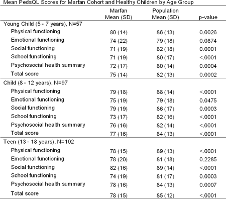
Methods: The Pediatric Quality of Life Inventory (PedsQL) was administered to 256 subjects with MFS ages 5-18 years as an ancillary study to the Pediatric Heart Network’s Marfan Trial, which compared the effects of atenolol vs. losartan on aortic root growth. PedsQL scores were compared to population norms by one-sample t-tests. Scores > 1 SD below the population sample mean represent at-risk status for impaired health-related QL. The impact of treatment arm (atenolol vs. losartan), severity of clinical features, and patient-reported symptoms on QL was assessed by general linear models.
Results: The subjects had a mean age of 11.8±3.9 years and were 62% male, 84% white, and 88% non-Hispanic. Mean PedsQL scores for MFS subjects were significantly lower than population norms.
Overall, scores were in the impaired range for physical QL and psychosocial QL in 34% and 27% of subjects, respectively. QL across multiple domains correlated negatively with frequency of patient-reported symptoms (r=0.32-0.40, p<.0001). Subjects with a reported neurodevelopmental disorder (mainly learning disability, attention deficit disorder, and/or hyperactivity) had lower mean QL scores (5.5-7.4 lower, p<.04). There were no significant differences in QL scores between treatment arms. We found no significant association between QL and aortic root z-score, extent of skeletal involvement, or presence of ectopia lentis.
Conclusions: Children with MFS are at risk for impaired QL. Higher number of patient-reported symptoms had the greatest negative impact on QL, rather than treatment arm or severity of cardiac, skeletal, or ocular findings. Future interventions to address patient symptoms and neurodevelopmental disorders could improve QL for children with MFS.
Article Information
vol. 132 no. Suppl 3 A9979
Published By:
American Heart Association, Inc.
Online ISSN:
History:
- Originally published November 6, 2015.
Copyright & Usage:
© 2015 by American Heart Association, Inc.
Author Information
- Jill C Handisides1;
- Danielle Hollenbeck-Pringle2;
- Karen Uzark3;
- Felicia L Trachtenberg2;
- Victoria L Pemberton4;
- Teresa W Atz5;
- Timothy J Bradley6;
- Sylvia De Nobele7;
- Georgeann K Groh8;
- Michelle S Hamstra9;
- Rosalind Korsin10;
- Jami C Levine1;
- Meghan K Mac Neal11;
- Larry W Markham12;
- Tonia Morrison13;
- Kathleen Mussatto14;
- Aaron K Olson15;
- Mary Ella M Pierpont16;
- Mary J Roman17;
- Reed E Pyeritz18;
- Elizabeth A Radojewski6;
- Michelle Robinson19;
- Elizabeth A Sparks20;
- Kathryn Waitzman21;
- Mingfen Xu22;
- Ronald V Lacro1,
- for the Pediatric Heart Network Investigators
- 1Cardiology, Boston Children’s Hosp, Boston, MA
- 2Pediatric Heart Network, New England Rsch Institutes, Watertown, MA
- 3Cardiology, Univ of Michigan Mott Children’s Hosp, Ann Arbor, MI
- 4Pediatric Heart Network, National Heart, Lung, and Blood Institute, Bethesda, MD
- 5Pediatric Cardiology, Med Univ of South Carolina, Charleston, SC
- 6Cardiology, The Hosp for Sick Children, Toronto, Canada
- 7Cntr for Med Genetics, Ghent Univ Hosp, Ghent, Belgium
- 8Pediatrics, Washington Univ Sch of Medicine, St. Louis, MO
- 9Heart Institute, Cincinnati Children’s Hosp Med Cntr, Cincinnati, OH
- 10Cardiology, Morgan Stanley Children’s Hosp of New York, New York, NY
- 11Molecular Cardiology, Icahn Sch of Medicine at Mount Sinai, New York, NY
- 12Cardiology, Vanderbilt Univ Med Cntr, Nashville, TN
- 13Cardiology, The Children’s Hosp of Philadelphia, Philadelphia, PA
- 14Herma Heart Cntr, Children’s Hosp of Wisconsin, Milwaukee, WI
- 15Cardiology, Seattle Children’s Hosp, Seattle, WA
- 16Cardiovascular Rsch, Children’s Hosp and Clinics of Minnesota, St. Paul, MN
- 17Cardiology, Weill Cornell Med College, New York, NY
- 18Medicine, Perelman Sch of Medicine at the Univ of Pennsylvania, Philadelphia, PA
- 19Pediatric Cardiology, Univ of Utah, Salt Lake City, UT
- 20Genetics, Johns Hopkins Medicine, Baltimore, MD
- 21Cardiology, Lurie Children’s Hosp, Chicago, IL
- 22Pediatric Cardiology, Duke Univ Med Cntr, Durham, NC
Abstract 19270: Heart Failure Incidence is Elevated in Adults With Simple Congenital Heart Defects Compared With the General Population: A Cohort Study
Morten Olsen, Dan E Høfsten, Nicolas L Madsen, Henning B Laursen, Søren P Johnsen, Jørgen Videbæk
Circulation. 2015;132:A19270
Abstract
Heart failure (HF) is a major cause of morbidity and mortality in adults with congenital heart defects (CHD). However, few data exist on the long-term prognosis of simple CHD. We aimed to estimate the incidence of HF and use of HF drugs in individuals with simple CHD compared with the general population.
Methods: We identified a nationwide population-based simple CHD cohort, defined by isolated and uncomplicated secundum atrial septal defect (ASD), persistent ductus arteriosus (PDA), ventricular septal defect (VSD) judged to be either muscular or with normal pulmonary flow resistance, or mild pulmonary stenosis (PS) verified by catheterization. Subjects were born and diagnosed from 1963 through 1973 in Denmark. We included patients with no childhood co-morbidity who were alive at 32 years of age (this age was determined by the establishment of a nationwide prescription register in 1995). For each CHD subject, we identified 10 control individuals from the general population using the Danish Civil Registration System, matched by sex and birth year. A unique personal identifier used in all Danish public registries enabled virtually complete follow-up for migration, death, use of HF drugs, or HF diagnoses as identified in a hospital discharge registry covering all Danish hospitals since 1977. Specific indications for drug use were unknown (could be e.g. hypertension). We computed cumulative incidence of first diagnosis of HF and first filling of a HF drug prescription, for CHD subjects and controls, and the corresponding hazard ratios (HR).
Results: We identified 802 CHD subjects (ASD, n=115; VSD, n=437; PS, n=75; PDA, n=175) and 7,947 controls, 43% male. Cumulative incidence for CHD subjects by age 45 years of HF drug use was 22% (16% for controls), whereas it was 3% (0.4% for controls) for a HF hospital diagnosis. The corresponding HRs comparing CHD subjects with controls were 1.4(95% CI:1.2-1.6) and 6.9(95% CI:4.0-12).
Conclusion: Adults with simple CHDs were at increased risk of HF. The data on HF drug use should be interpreted with caution as other cardiovascular conditions were likely the indications for many of the prescriptions. However; further studies on the effectiveness of medical follow-up programs for patients with simple CHDs appear warranted.
Article Information
vol. 132 no. Suppl 3 A19270
Published By:
American Heart Association, Inc.
Online ISSN:
History:
- Originally published November 6, 2015.
Copyright & Usage:
© 2015 by American Heart Association, Inc.
Author Information
- Morten Olsen1;
- Dan E Høfsten2;
- Nicolas L Madsen3;
- Henning B Laursen1;
- Søren P Johnsen1;
- Jørgen Videbæk1
- 1Dept Clinical Epidemiology, Aarhus Univ Hosp, Århus, Denmark
- 2Dept of Cardiology, Rigshospitalet, Univ of Copenhagen, København Ø, Denmark
- 3Heart Institute, Cincinnati Children’s, Cincinnati, OH
Abstract 19452: Long-term Outcomes in Adults With Congenital Heart Disease Evaluated for Heart Transplantation
Gavin Hickey, Andrea Elliott, Morgan Hindes, Tim Maul, Ravi Ramani, Michael Mathier, Stephen Cook
Circulation. 2015;132:A19452
Abstract
Background: The prevalence of adult congenital heart disease (ACHD) patients with heart failure is increasing. No guidelines exist for evaluation and listing for heart transplant (HT) in ACHD. We sought to review the outcomes of HT committee decisions [accepted and transplanted (T), accepted awaiting transplant (A), deferred (D), ineligible (I)].
Methods: This was a retrospective analysis performed on ACHD (≥ 18 yrs) referred for HT between 2000-2014. Data on demographics, CHD severity, NYHA class, imaging, exercise testing, catheterization and HT committee decision were recorded. Primary outcome was all-cause mortality. Analysis was completed to identify variables that were statistically different amongst groups.
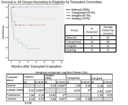
Results: Sixty-two ACHD were included in analysis, A (n= 17) I (n=14) T (n=22) D (n=9), NYHA class III-IV (98%), CHD of great complexity (74%). Median time to HT, after listing, was 3 mo. (mean 5.5). Survival was significantly lower in the group awaiting transplant compared to all other groups (p<0.001): A (18.6%) I (65.3%) T (78.3%) D (100%) (Fig). Among those who had not been transplanted, several variables were significantly different (p<0.05) including: systolic BP, alb, BNP, Hct, right atrial pressure and peak oxygen consumption. For those awaiting transplant with pulmonary hypertension (PH) mortality was 100% but 40% for those without PH (p<0.1).
Conclusion: Waitlist mortality in the ACHD population is high. Given poor outcomes while on the waitlist, earlier listing may improve survival. Specific variables were statically different among patients not transplanted and there was a high mortality in those with PH. These may have contributed to the difference in survival among groups and may impact HT committees in the evaluation and listing for HT.
Article Information
vol. 132 no. Suppl 3 A19452
Published By:
American Heart Association, Inc.
Online ISSN:
History:
- Originally published November 6, 2015.
Copyright & Usage:
© 2015 by American Heart Association, Inc.
Author Information
- Gavin Hickey;
- Andrea Elliott;
- Morgan Hindes;
- Tim Maul;
- Ravi Ramani;
- Michael Mathier;
- Stephen Cook
- Cardiology, Univ of Pittsburgh Med Cntr, Pittsburgh, PA
Abstract 18744: Rising Costs of Adult Congenital Heart Disease Care: Does Gender Matter?
David A Briston, Elisa Bradley, Aarthi Sabanayagam, Ali N Zaidi
Circulation. 2015;132:A18744
Abstract
Introduction: The profile of congenital heart disease (CHD) has shifted, now with more adults than children who have survived. Few studies have provided a global assessment of adult congenital heart disease (ACHD) healthcare cost in the United States.
Methods: Data from the National Inpatient Sample (2002-2012) utilizing diagnostic ICD-9 codes for moderate and complex ACHD were analyzed. Hospital discharges, total billed and reimbursed amounts, gender and age disparities were evaluated.
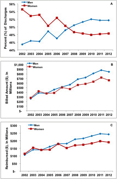
Results: There was an overall increase in ACHD discharges (Moderate CHD: 4,742 vs. 6,545, Severe CHD: 807 vs. 1,115), billed and reimbursed dollar amounts (Billed: $542,703,961 vs. $1,506,945,042, +178%; reimbursed: $221,417,779 vs. $432,797,543, +95%). Women had more discharges in 2002 but not in 2012 (men: women, 2002: 6,503 vs. 7,805; 2012: 7,715 vs. 7,200) [Figure 1A]. Gender-based billed amounts followed a similar trend (2002: $262,918,357 vs. $279,785,604; 2012: $844,923,857 vs. $$662,021,185; p=0.006) as did total reimbursed (2002: $107,766,175 vs. $113,651,604; 2012: $243,183,638 vs. $189,613,905, p=0.008) [Figure 1B,C]. Healthcare expenditure increased across all age groups: this was most prominent in the > 44 vs. 18-44 yr. group (Billed: $617,589,813 vs. $346,652,267, p<0.001; reimbursed: $136,013,528 vs. $75,366,237, p<0.001).
Conclusions: We are the first to report a change in the rate of gender-based ACHD hospitalizations, whereby men now account for more hospitalizations in the U.S. As ACHD discharges, billed and reimbursed amounts continue to rise over the last decade, we are also the first to demonstrate increased expenditure in older (> 44 yrs.) ACHD patients, a pattern that we predict will continue to grow and requires future investigation.
Article Information
vol. 132 no. Suppl 3 A18744
Published By:
American Heart Association, Inc.
Online ISSN:
History:
- Originally published November 6, 2015.
Copyright & Usage:
© 2015 by American Heart Association, Inc.
Author Information
- 1Pediatric Cardiology, Montefiore Med Cntr, Bronx, NY
- 2Div of Adult Congenital Heart Disease, Nationwide Children’s Hosp, Columbus, OH
- 3Montefiore Heart and Vascular Care Institute, Montefiore Med Cntr, Bronx, NY
- 4Montefiore Adult Congenital Heart Disease Program, Montefiore Med Cntr, Bronx, NY
Abstract 18971: Heart Transplantation for Adult Patients With Congenital Heart Disease: Are the Outcomes Still Worse Than in the General Population in 2015?
Diego Moguillansky, Biagio A Pietra, Frederick J Fricker, Mark S Bleiweis
Circulation. 2015;132:A18971
Abstract
Introduction: Short and medium term outcome after heart transplantation has steadily improved over the last few decades. Outcomes for adult patients with congenital heart disease (ACHD) who undergo transplantation are generally considered to be less favorable.
Hypothesis: We hypothesized that the development of heart teams that specialize in ACHD would lead to improved outcomes after transplantation in this population.
Methods: We reviewed the records of all patients undergoing first heart transplant at a university center with a dedicated ACHD team over the last 10 years. Patients undergoing re-transplantation were excluded. We looked at short (30 days) and medium term (1 year) survival after heart transplantation in ACHD patients.
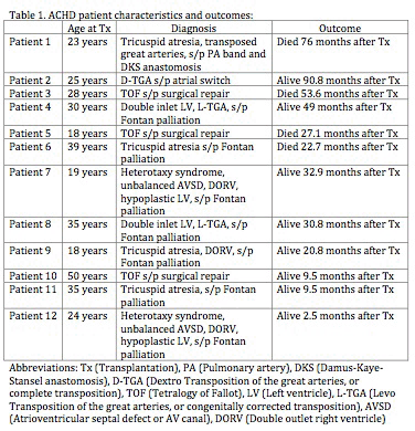
Results: Between 1/1/05 and 6/10/15, 258 patients underwent heart transplantation. Of the 258 patients, 17 were re-transplants and were excluded. Of the remaining 241 patients, 12 were ACHD patients and 229 were transplanted for other diagnosis (general group). In the general group 184 of 212 (86.8%) patients were alive at 1 year (the remaining 17 did not have sufficient follow up to be included in the 1-year survival analysis). In the ACHD group 9 of 9 patients (100%) were alive at 1 year. The remaining 3 ACHD patients with insufficient follow up to be included in the 1-year survival analysis were still alive 2.5-9.5 months after transplant, such that all 12 ACHD patients survived at least 30 days and were discharged home after heart transplant. At the end of the study period 160 of 229 (70%) patients were still alive in the general group, compared to 8 of 12 (66.7%) in the ACHD group.
Conclusions: Short and medium term survival after heart transplantation appears to be no worse for selected ACHD patients compared to the general population. Larger studies with longer follow up are needed to confirm our findings and clarify the intermediate and long-term outcomes of ACHD patients undergoing heart transplantation in the modern era.
Article Information
vol. 132 no. Suppl 3 A18971
Published By:
American Heart Association, Inc.
Online ISSN:
History:
- Originally published November 6, 2015.
Copyright & Usage:
© 2015 by American Heart Association, Inc.
Author Information
- Diego Moguillansky;
- Biagio A Pietra;
- Frederick J Fricker;
- Mark S Bleiweis
- Congenital Heart Cntr, Univ of Florida, Gainesville, Gainesville, FL
Abstract 17639: Predictors of Poor Neurocognitive Performance and Neuropsychological Phenotypes in Adolescents and Young Adults With Congenital Heart Disease
Maria Emilia G Areias, Bruno Peixoto, Ana Carolina Pinheiro, Helena Monteiro, Sara Filipa Araújo, Tânia Carvalho, Stefanie Melo, João Pedro Lopes, Filipa Rodrigues, Ana Catarina Nascimento, Joana Miranda, Filipa Vilacova, Cláudia Moura, Joana Luz Soares, Victor Viana, Jorge Quintas, José Carlos Areias
Circulation. 2015;132:A17639
Abstract
Objectives: to determine the predictors of neurocognitive performance (NP) of patients with Congenital Heart Disease (CHD), analysing its relation to parameters of fetal development, as head circumference (HC), weight (W) and length (L) at birth, neonatal parameters (APGAR 1, 5), quality of life (QOL), psychiatric morbidity (PM), psychosocial adjustment (PSA) and traits of personality (TP); to identify different phenotypes of NP in CHD patients.
Methods: 337 CHD patients, 189 male, ages 12 to 30 years (mean= 16.34 ± 3.12), 117 cyanotic, and 119 healthy controls (56 males, mean=18.41±3.20) participated. Clinical data were collected. Neuropsychological assessment included Wechsler’s Digit Test (direct and reverse) and Symbol Search, Rey’s Complex Figure, BADS’s Key Searching Test, Color-Word Stroop Test, Trail Making Test (A, B) and Logical Memory Task. Participants were interviewed on social support, family educational style, self-image, physical limitations, completed a psychiatric interview (SADS-L) and self-report questionnaires on QOL (WHOQOL-BREF), PSA (YSR and ASR) and TP (NEOPI-R). HC, W and L and APGAR were collected.
Results: CHD patients had a significantly worse NP than healthy controls in all tests, and the cyanotic worse than the acyanotic patients. Several correlations were apparent between fetal parameters (HC, W and L) and neuropsychological abilities in CHD. However, the HC was the main predictor of bad NP later on in CHD patients (R=0.435; R2=0.189; F=14.692; p=0.000; β=0.201; t=2.487; p=0.016). We identified 3 neurocognitive phenotypes in CHD patients, minimally, moderately and globally impaired. The last showed lower HC (p=0.003), W (p=0.022) and L, and a bigger number of retentions in school (p=0.017) than the first. The last reported more aggressive behaviors.than the other 2 (p=0.007; p=0.006) and their caregivers reported more social (p=0.025), thought (p=0.041) and attention problems (p=0.001) in their children than the parents of the other 2.
Conclusion: CHD patients have worse NP than controls; small HC at birth was the main predictor of poor NP; We identified 3 patterns of neurocognitive disability among CHD patients: mild, moderate and severely impaired, all showing significant differences compared to controls.
Article Information
vol. 132 no. Suppl 3 A17639
Published By:
American Heart Association, Inc.
Online ISSN:
History:
- Originally published November 6, 2015.
Copyright & Usage:
© 2015 by American Heart Association, Inc.
Author Information
- Maria Emilia G Areias1;
- Bruno Peixoto2;
- Ana Carolina Pinheiro2;
- Helena Monteiro2;
- Sara Filipa Araújo2;
- Tânia Carvalho2;
- Stefanie Melo2;
- João Pedro Lopes2;
- Filipa Rodrigues2;
- Ana Catarina Nascimento2;
- Joana Miranda3;
- Filipa Vilacova3;
- Cláudia Moura3;
- Joana Luz Soares2;
- Victor Viana4;
- Jorge Quintas5;
- José Carlos Areias6
- 1Social and Behavior Sciences, Instituto Universitário de Ciências da Saúde (CESPU); Cardiovascular Rsch Unit (Faculty of Medicine, Univ of Porto), Gandra-Paredes, Portugal
- 2Social and Behavior Sciences, Instituto Universitário de Ciências da Saúde (CESPU), Gandra-Paredes, Portugal
- 3Pediatric Cardiology, Cntr Hospar S. João, Porto, Portugal
- 4Pediatrics, Cntr Hospar S. João, Porto, Portugal
- 5Criminology, Faculty of Law, Univ of Porto, Porto, Portugal
- 6Pediatric Cardiology, Cntr Hospar S. João; Cardiovascular Rsch Unit (Faculty of Medicine, Univ of Porto), Porto, Portugal
Abstract 14823: Adult Congenital Heart Disease is Associated With a Decreased Prevalence of Morbid Obesity
Joseph B Lerman, John T Doucette, Ira A Parness, Rajesh U Shenoy
Circulation. 2015;132:A14823
Abstract
Introduction: Obesity may be associated with greater morbidity in adults with Congenital Heart Disease (ACHD), than in the general population. While obesity prevalence in pediatric CHD has been well studied, the prevalence of obesity in the American ACHD population is unknown. Our study seeks to determine the prevalence of obesity (BMI > 30) and morbid obesity (BMI > 35) in a cohort of ACHD patients, as compared to matched controls.
Hypothesis: We assessed the hypothesis that ACHD is associated with a higher prevalence of obesity compared to controls.
Methods: Retrospective analysis was performed on all ACHD patients (identified via ICD-9 coding) seen at a tertiary center in 2013. A control group without CHD was randomly created, matching for age, sex, and ethnicity.
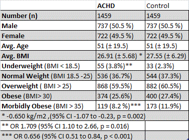
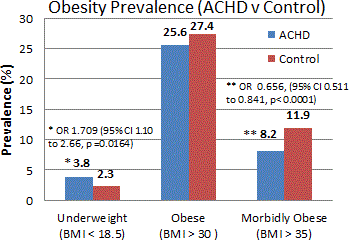
Results: 1459 ACHD patients met inclusion criteria. ACHD patients had a lower mean BMI than controls (-0.650 kg/m2, 95% CI -1.068 to -0.231, p <0.002). Obesity prevalence did not differ significantly between groups: 25.6% in ACHD, 27.4% in controls (OR 0.909, 95% CI 0.766 to 1.079, p = 0.285). ACHD was associated with lower prevalence of morbid obesity (OR =0.656, CI 0.511 to 0.841, p < 0.0001), and higher prevalence of being underweight (BMI < 18.5) (OR 1.709, 95% CI 1.098 to 2.663, p < 0.016). ACHD was also associated with a lower prevalence of obesity amongst 41-65 year olds (OR = 0.722, 95% CI from 0.561 to 0.929, p < 0.011). Severity of CHD (classified as simple vs. complex) was not significantly related to obesity prevalence.
Conclusion: In conclusion, ACHD is associated with a decreased prevalence of morbid obesity, and an increased prevalence of being underweight. ACHD is not significantly associated with obesity prevalence.
Article Information
vol. 132 no. Suppl 3 A14823
Published By:
American Heart Association, Inc.
Online ISSN:
History:
- Originally published November 6, 2015.
Copyright & Usage:
© 2015 by American Heart Association, Inc.
Author Information
- 1Dept of Pediatric Cardiology, Icahn Sch of Medicine at Mount Sinai, New York, NY
- 2Dept of Preventive Medicine, Icahn Sch of Medicine at Mount Sinai, New York, NY
- 3Div of Pediatric Cardiology, Kravis Children’s Hosp at Mount Sinai, Icahn Sch of Medicine at Mount Sinai, New York, NY
Abstract 12897: Long-term Risk of Atrial Fibrillation in Adults Diagnosed With Atrial Septal Defect in Childhood
Zarmiga Karunanithi, Camilla Nyboe, Vibeke E Hjortdal
Circulation. 2015;132:A12897
Abstract
Atrial fibrillation (AF) is very common in patients with atrial septal defects (ASD). The impact of closure on the prevalence and especially the correct timing is controversial. We investigated the risk of AF in patients diagnosed with ASD in childhood, both with and without closure, compared with a general population cohort.
Data on all Danish ASD patients diagnosed before the age of 18 years between 1963 and 1994 was obtained from national registries. Follow-up was continued until outcome, death, emigration or 31st of Dec 2011. Hazard ratios were calculated using Cox proportional hazards regression and cumulative incidences estimated using Fine and Gray’s competing risk regression.
A total of 1,111 patients met the inclusion criteria, and each was matched on age and gender with 10 controls. Mean age at diagnosis was 4.9 years (range 0-17.9 years). Mean follow-up since diagnosis was 26.3 years (range 1-53.3 years). A total of 642 patients had closure performed either surgically or percutaneously with 402 before the age of 10 years.
Patients with an unclosed ASD had an increased risk of AF (HR 16.4, 95% CI: 6.8; 39.8) compared with the general population cohort. For patients with closure the risk was similarly high (HR 18.1, 95% CI: 9.8; 33.5). Closure before (HR 17.9, 95% CI: 7.5; 42.7) and after (HR 18.4, 95% CI: 7.7; 43.8) the age of 10 did not affect the risk of AF later in life. Cumulative incidences after 10 years was 0.6% [0.1; 4.4] and after 50 years 9.8% [6.7; 14.12].
In conclusion, risk of AF is high despite closure in childhood. Closure being insufficient protection against development of AF in adulthood speaks for causality other than the mere presence of an ASD. Furthermore, ASD patients (regardless of closure) call for follow-up investigation past the 5th decade.
Article Information
vol. 132 no. Suppl 3 A12897
Published By:
American Heart Association, Inc.
Online ISSN:
History:
- Originally published November 6, 2015.
Copyright & Usage:
© 2015 by American Heart Association, Inc.
Author Information
- Zarmiga Karunanithi;
- Camilla Nyboe;
- Vibeke E Hjortdal
- Dept. of Cadiothoracic and Vascular Surgery, Aarhus Univ Hosp, Aarhus N, Denmark
Abstract 11940: Aortic Complications Associated With Pregnancy in Marfan Syndrome: The NHLBI GenTAC Registry
Mary J Roman, Ryan E Whitworth, Joseph E Bavaria, Richard B Devereux, Hal Dietz, Kathryn Holmes, Kim A Eagle, Scott A LeMaire, Shaine A Morris, Reed Pyeritz, William J Ravekes, Michael Silberbach
Circulation. 2015;132:A11940
Abstract
Background: Definition of pregnancy risk in women with Marfan syndrome (MFS) has been confounded by ascertainment bias, lack of imaging data, small sample size, and inclusion of women without a known MFS diagnosis before a pregnancy complication. To lessen these limitations, we analyzed MFS women participating in the large GenTAC Registry.
Results: Among 184 women in whom pregnancy information was available, 94 women (51%) had a total of 227 pregnancies (average 2.5, range 1-6). Compared to MFS women who never became pregnant, those with pregnancies were older at time of MFS diagnosis (27 vs. 13 years, p<0.0001) and at time of GenTAC enrollment (47 vs. 37 years, p<0.0001). When comparing ever-pregnant to never-pregnant women with MFS at the time of registry entry, higher historical rates of aortic dissection and prophylactic proximal aortic surgery were observed (24/94 [25.5%] vs. 13/90 [14.4%], p=0.068 and 15/94 [16.0%] vs. 4/90 [4.4%], p=0.014; respectively).
Among the 94 MFS women with pregnancies, 10 (10.6%) experienced a pregnancy-related aortic complication (4 Type A and 3 Type B dissections, one coronary artery dissection, and 2 with significant [≥3 mm] aortic growth, 1 of whom required post-partum prophylactic surgery). Four of the aortic dissections, including all 3 Type B, and the coronary dissection occurred in the post-partum period. Only 3 of 7 women (43%) with an aortic dissection related to pregnancy were aware of their MFS diagnosis before pregnancy. Women with and without pregnancy-associated aortic complications did not differ in phenotypic features. The rate of aortic dissection was higher during pregnancy + post-partum period (3.5 per 100 person-years vs. 0.8 per 100 person-years, odds ratio 4.6 [95% confidence interval 2.1-10.1], p=0.0002).
Conclusions: Pregnancy in MFS is associated with an increased risk of aortic dissection, both Types A and B, particularly in the immediate post-partum period, and more frequent prophylactic surgery. Lack of knowledge of underlying MFS diagnosis before aortic dissection is a major contributing factor. These findings underscore the need for early diagnosis, pre-pregnancy risk counselling, and multi-disciplinary peri-partum management.
Article Information
vol. 132 no. Suppl 3 A11940
Published By:
American Heart Association, Inc.
Online ISSN:
History:
- Originally published November 6, 2015.
Copyright & Usage:
© 2015 by American Heart Association, Inc.
Author Information
- Mary J Roman1;
- Ryan E Whitworth2;
- Joseph E Bavaria3;
- Richard B Devereux1;
- Hal Dietz4;
- Kathryn Holmes5;
- Kim A Eagle6;
- Scott A LeMaire7;
- Shaine A Morris8;
- Reed Pyeritz9;
- William J Ravekes10;
- Michael Silberbach11
- 1Dept of Medicine, Weill Cornell Med College, New York, NY
- 2Biostatistics and Epidemiology Div, RTI International, Rsch Triangle Park, NC
- 3Cardiothoracic Surgery, Hosp of the Univ of Pennsylvania, Philadelphia, PA
- 4Pediatrics, Medicine, and Molecular Biology & Genetics, Johns Hopkins Univ Sch of Medicine, Baltimore, MD
- 5Pediatrics, Oregon Health & Science Univ, Portland, OR
- 6Medicine, Univ of Michigan Health System, Ann Arbor, MI
- 7Cardiovascular Surgery, Texas Heart Institute/Baylor College of Medicine, Houston, TX
- 8Pediatric Cardiology, Texas Children’s Hosp/Baylor College of Medicine, Houston, TX
- 9Genetics, Perelman Sch of Medicine at the Univ of Pennsylvania, Philadelphia, PA
- 10Pediatrics, Johns Hopkins Univ Sch of Medicine, Baltimore, MD
- 11Pediatrics, Doernbecher Children’s Hosp, Portland, OR
Abstract 12047: Quality of Life in Adults With Congenital Heart Disease Worldwide: A Multilevel Study in 15 Countries
Silke Apers, Adrienne H Kovacs, Koen Luyckx, Philip Moons
Circulation. 2015;132:A12047
Abstract
Objectives: Health care should be evaluated not only on the basis of saving lives, but also in terms of improving the quality of life (QOL). However, results regarding QOL in adults with congenital heart disease (CHD) are inconsistent and vary across the world. Methodological differences and limitations hamper the interpretation of these results. Therefore, we aimed (1) to describe QOL in an international sample of adults with CHD using a uniform method; (2) to investigate the association between patient characteristics and variation in QOL; and (3) to explore international variation in QOL and the relationship with country-specific characteristics.
Methods: For this cross-sectional multilevel study, patients were recruited from adult CHD centers or national registries from 15 countries. QOL was assessed by a linear analog scale (0-100). Patient characteristics included sex, age, marital status, educational level, employment status, CHD complexity and patient-reported New York Heart Association (NYHA) class. Country-specific characteristics included national happiness score and six cultural dimensions. Multilevel generalized linear mixed models were conducted and Empirical Bayes estimates for QOL were calculated to account for the sample size in each country.
Results: In total, 4,028 adults with CHD were enrolled (median age=32 y; 53% women; 26% mild, 49% moderate, 26% complex CHD). Median QOL for the total sample was 80. Age, marital status, employment status and NYHA class were significantly associated with variation in QOL (p<.001). Australia had the highest QOL estimate (82) and Japan the lowest (72). Four countries had a QOL estimate of ≥80 (i.e., Australia, Switzerland, USA, Malta). After adjusting for patient characteristics, happiness and cultural dimensions were not significantly associated with variation in QOL.
Conclusions: This is the first international study comprehensively assessing QOL in any subgroup of cardiac patients. We found that QOL in adults with CHD varies across countries and is related to patient characteristics but not to country-specific characteristics. The identification of significant patient characteristics can support the development of interventions targeting the QOL of adults with CHD around the world.
Article Information
vol. 132 no. Suppl 3 A12047
Published By:
American Heart Association, Inc.
Online ISSN:
History:
- Originally published November 6, 2015.
Copyright & Usage:
© 2015 by American Heart Association, Inc.
Author Information
- Silke Apers1;
- Adrienne H Kovacs2;
- Koen Luyckx3;
- Philip Moons1,
- APPROACH-IS consortium and the International Society for Adult Congenital Heart Disease
- 1Cntr for Health Services and Nursing Rsch, KU Leuven – Univ of Leuven, Leuven, Belgium
- 2Toronto Congenital Cardiac Cntr for Adults, Peter Munk Cardiac Cntr, Univ Health Network, Toronto, Canada
- 3Sch Psychology and Child and Adolescent Development, KU Leuven – Univ of Leuven, Leuven, Belgium
Abstract 10903: Extraction Outcomes in Patients With Congenital Heart Disease
Erin A Fender, Ammar M Killu, David O Hodge, Bryan C Cannon, Paul A Friedman, Christopher J Mcleod, Craig S Broberg, Charles A Henrikson, Yong-Mei Cha
Circulation. 2015;132:A10903
Abstract
Introduction: Patients with congenital heart disease (CHD) frequently require implantable cardiac devices. Device infection or malfunction may necessitate lead extraction. Extraction may be challenging due to long lead dwell times, anatomic abnormalities, and prior cardiac surgery. Little is known about extraction outcomes in the CHD population.
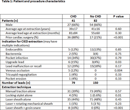
Methods and Results: This retrospective study included 41 CHD patients and 82 age and gender matched controls that underwent lead extractions at two centers between 2001-2014. Only patients with leads older than 12 months were included. There were 79 leads in CHD patients and 150 in controls. Patients with CHD had a mean age of 39±17 years at extraction and on average their leads had been implanted for 81±84 months, this was not significantly different from the control group. Eighty eight percent of CHD patients had 1 or more cardiac surgeries as compared to only 22% of controls (p<0.001). The number of abandoned leads was also significantly different with 16 abandoned leads in the CHD group and 3 in controls (p<0.001). There was no statistically significant difference in extraction techniques between the groups. Complete extraction was achieved in 94% of patients in both groups. There were no CHD group complications. Control group complications included 3 SVC lacerations requiring sternotomy (one was fatal), and 1 ventricular perforations with tamponade requiring sternotomy. None of these patients had a history of cardiac surgery. The average age of leads in patients with a complication was 15.3 years which was older than the mean lead age seen in the CHD and control populations which was 6.75 and 4.6 years respectively.
Conclusions: Lead extraction can be safely performed in patients with CHD. Despite anatomic abnormalities and previous cardiac surgery, the outcome of lead extraction in patients with CHD is comparable to controls. Prior cardiac surgery may lower the risk for extraction complications.
Article Information
vol. 132 no. Suppl 3 A10903
Published By:
American Heart Association, Inc.
Online ISSN:
History:
- Originally published November 6, 2015.
Copyright & Usage:
© 2015 by American Heart Association, Inc.
Author Information
- Erin A Fender1;
- Ammar M Killu1;
- David O Hodge1;
- Bryan C Cannon1;
- Paul A Friedman1;
- Christopher J Mcleod1;
- Craig S Broberg2;
- Charles A Henrikson2;
- Yong-Mei Cha1
- 1Cardiology, Mayo Clinic, Rochester, MN
- 2Cardiology, Oregon Health and Science Universtiy, Portland, OR
Abstract 18774: Increased Epicardial Fat After Fontan Palliation Associated With Decreased Cardiac Output
Adam M Lubert, Jimmy C Lu, Albert P Rocchini, Mark D Norris, Sunkyung Yu, Prachi P Agarwal, Maryam Ghadimi Mahani, Adam L Dorfman
Circulation. 2015;132:A18774
Abstract
Introduction: Epicardial fat produces multiple proinflammatory and proatherogenic cytokines and is associated with adverse cardiovascular events. Inflammation and resultant endothelial dysfunction may play a role in progressive myocardial dysfunction among adults with single ventricle physiology status post Fontan, but the potential impact of epicardial fat volume (EFV) has not been studied. This study aimed to quantify EFV in Fontan patients compared to a control group of repaired tetralogy of Fallot patients (rTOF) and to evaluate its association with ventricular ejection fraction and cardiac index.
Methods: We retrospectively measured EFV on all patients post Fontan, 15 years or older, with cardiac magnetic resonance performed at the University of Michigan from 2007-2014, and 1:1 age, sex, and body mass index (BMI) matched patients with rTOF. EFV was measured manually on cine, steady-state free precession, short-axis images and was indexed to body surface area (BSA). Cardiac index was calculated as the sum of flow in the superior vena cava and inferior vena cava, indexed to BSA. A random subset of studies was re-measured >1 week after initial measurement to assess intraobserver variability.
Results: Fontan patients (n=63, mean age 22.3±6.0, 52% male) had a larger indexed EFV compared to rTOF (mean age 22.2±5.8, 52% male) measuring 75.6±29.2 vs. 60.0±19.9 ml/m2 (p= 0.0003). There was no difference in BMI between the two groups (24.2±5.2 vs 24.2±5.6, p= 0.81). In Fontan patients, indexed EFV was inversely correlated with ventricular ejection fraction (r = -0.26, p = 0.03) and cardiac index (r = -0.34, p = 0.01). Intraobserver variability of the indexed EFV measurements in both groups was excellent (intraclass correlation coefficient of 0.95 and 0.93 in the Fontan group and rTOF group, respectively).
Conclusions: Indexed EFV is higher in individuals post Fontan compared to patients with rTOF and is associated with lower ventricular ejection fraction and cardiac index. Increased epicardial fat could play a role in progressive myocardial dysfunction in this population, but longitudinal studies are necessary to establish any causative role.
Article Information
vol. 132 no. Suppl 3 A18774
Published By:
American Heart Association, Inc.
Online ISSN:
History:
- Originally published November 6, 2015.
Copyright & Usage:
© 2015 by American Heart Association, Inc.
Author Information
- Adam M Lubert1;
- Jimmy C Lu1;
- Albert P Rocchini1;
- Mark D Norris1;
- Sunkyung Yu1;
- Prachi P Agarwal2;
- Maryam Ghadimi Mahani2;
- Adam L Dorfman1
- 1Dept of Pediatrics & Communicable Diseases, Div of Pediatric Cardiology, Univ of Michigan, Ann Arbor, MI
- 2Dept of Radiology, Univ of Michigan, Ann Arbor, MI
Abstract 18682: Increased Oxidative Stress is Associated With Vascular Endothelial Dysfunction and Central and Peripheral Arterial Stiffening in Patients With Fontan Circulation
Clara Kurishima, Seiko Kuwata, Yoichi Iwamoto, Hirotaka Ishido, Satoshi Masutani, Hideaki Senzaki
Circulation. 2015;132:A18682
Abstract
Background: It is well recognized that oxidative stress is responsible for fat accumulation and tissue inflammation, and causes endothelial cell damage that leads to the initiation of atherosclerosis. We and others have previously reported that vascular stiffness is increased with impaired vascular endothelial function in patients after Fontan operation. We hypothesized that increased oxidative stress is associated with vascular endothelial dysfunction and arterial stiffening in patients with Fontan circulation
Methods: Relationship of oxidative stress with endothelial function and arterial stiffness was examined in 16 Fontan patients. Endothelial function and arterial stiffness of forearm artery was measured by flow mediated dilatation. We also measured central arterial stiffness by calculating aortic pulse wave velocity (PWV) during catheter drawback from the ascending aorta to the iliac bifurcation at the time of cardiac catheterization. Oxidative stress was estimated by measuring serum levels of oxidized low-density lipoprotein (LDL) and superoxide dismutase (SOD) activity in the urine as an anti-oxidant.
Results: PWV of the ascending aorta but not of the descending aorta was significantly and positively correlated with oxidized LDL level (P<0.05). Ratio of oxidized LDL level and SOD activity, a balance between oxidative stress and antioxidant activity, was also significantly and positively correlated with the ascending aortic PWV. This was also the case even after controlling for age of the patients by multivariate analysis. Vascular stiffness and endothelial function of the forearm artery were correlated with the balance between oxidative stress and antioxidant activity. Enhanced oxidative stress was related to increased central venous pressure and age.
Conclusion: Increased oxidative stress is associated with vascular endothelial dysfunction and central and peripheral arterial stiffening in patients with Fontan circulation. Antioxidant therapy may improve vascular health and thereby patient’s outcome after Fontan operation.
Article Information
vol. 132 no. Suppl 3 A18682
Published By:
American Heart Association, Inc.
Online ISSN:
History:
- Originally published November 6, 2015.
Copyright & Usage:
© 2015 by American Heart Association, Inc.
Author Information
- Clara Kurishima;
- Seiko Kuwata;
- Yoichi Iwamoto;
- Hirotaka Ishido;
- Satoshi Masutani;
- Hideaki Senzaki
- Pediatric Cardiology, Sitama Med Cntr, Saitama Med Univ, Kawagoe, Japan
Abstract 17227: Late Hepatic Complications Following Fontan Conversion Surgery
Barbara J Deal, Joyce Johnson, Gregory Webster, Sabrina Tsao, Bradley S Marino, John M Costello, Andrew De Freitas, Daniel R Ganger
Circulation. 2015;132:A17227
Abstract
Introduction: Fontan conversion surgery has extended survival for patients with univentricular heart, yet raised concerns regarding durability of hepatic function.
Methods: We reviewed liver outcomes among transplant-free survivors of Fontan conversion with arrhythmia surgery at our institution. Hepatic screening included a combination of annual liver function testing with clinically indicated imaging and/or biopsy.
Results: Between 1994 – 2014, 145 patients underwent Fontan conversion surgery at a median age of 23 (3-47) years, following initial Fontan surgery at median age of 6 (1 -35) years. Initial Fontan surgery types were atriopulmonary (79%), atrioventricular (13%), and lateral tunnel (8%). Early mortality was 2% following Fontan conversion surgery, with 10-year transplant free-survival of 84%. During median follow-up of 9 (1-20) years among 130 transplant-free survivors, significant liver disease developed in 6 patients (4.6%): hepatocellular carcinoma in 5 patients (primary 4, metastatic from appendix 1), and fulminant liver failure in one patient. Median age at diagnosis of hepatocellular carcinoma/liver failure was 35 (28-47) years, a median of 27 (22-33) years following initial Fontan surgery. Carcinoma was detected on annual hepatic screening with ultrasound in 3, and by elevated alpha-fetoprotein level prompting imaging in 2 patients. None of the patients had clinical variceal bleeding. Preoperative status included heterotaxy syndrome in 2, NYHA Classification III-IV in 5, and long standing ascites in 2 patients. Death occurred in 3/6 patients at a mean of 1.4 years following diagnosis of hepatic disease.
Conclusion: Late hepatocellular carcinoma/liver failure was found in 4.6% of non-transplanted Fontan conversion patients, indicating the need for serial monitoring of liver status. Early detection of carcinoma with routine screening using American Association for the Study of Liver Diseases guidelines may allow for improved treatment strategies and prolonged survival.
Article Information
vol. 132 no. Suppl 3 A17227
Published By:
American Heart Association, Inc.
Online ISSN:
History:
- Originally published November 6, 2015.
Copyright & Usage:
© 2015 by American Heart Association, Inc.
Author Information
- Barbara J Deal1;
- Joyce Johnson1;
- Gregory Webster1;
- Sabrina Tsao1;
- Bradley S Marino1;
- John M Costello1;
- Andrew De Freitas1;
- Daniel R Ganger2
- 1Cardiology, Ann & Robert H. Lurie Children’s Hosp of Chicago, Chicago, IL
- 2Hepatology/Gastroenterology, Northwestern Memorial Hosp, Chicago, IL
Abstract 17237: Overweight Associated With Advanced Biventricular Remodeling and Increased Morbidity in Adults With Repaired Tetralogy of Fallot
Jonathan D Kochav, Brian Ghoshhajra, Doreen DeFaria Yeh, Sean Singer, Pedro V Staziaki, Ami B Bhatt
Circulation. 2015;132:A17237
Abstract
Background: Individuals with repaired tetralogy of Fallot (rTOF) often develop progressive pulmonary regurgitation (PR) and right ventricular (RV) dilation after initial repair, placing them at risk of arrhythmia, right heart failure, and sudden cardiac death. MRI research has led to body surface area (BSA)-indexed RV end-diastolic volume cutoffs (RVEDV>160ml/m2) for timing of pulmonary valve replacement (PVR), a strategy potentially limited by rising rates of obesity in this population.
Hypothesis: Indexing RV volumes to actual BSA may underestimate disease progression in overweight patients.
Methods: This retrospective analysis identified adults with rTOF and significant PR who underwent MRI for evaluation of RVEDV prior to consideration of PVR. Charts were reviewed for major adverse clinic events including death, heart failure, and sustained arrhythmia. Chamber volumes were indexed to BSA, as well as ideal BSA (defined by weight set to achieve BMI of 25).
Results: 58 adults with rTOF who met inclusion criteria were identified; 43% were overweight (BMI>25). Compared with normal weight individuals, overweight patients were older at MRI (44±12 vs 30±10 years, p<0.001) and definitive repair (9±8 vs 3±3 years, p=0.002), and more likely to have staged repair (56% vs 24%, p=0.01). Overweight patients had more advanced RV (RVEF 40±11% vs 55±33%, p=0.03) and LV (LVEF 53±12% vs. 59±13%, p=0.02) systolic dysfunction, as well as larger LVEDV (84±26 vs 65±15ml/m2, p=0.001) and RVEDV (152±50 vs 139± 61ml/m2, p=0.37) when indexed to ideal BSA. Overweight patients had higher incidence of arrhythmia (58% vs 20%, p=0.04), and decompensated HF (15% vs 0%, p=0.06) prior to MRI. Indexing RVEDV to ideal BSA in the overweight population led to reclassification of 4 patients meeting criteria for PVR (15 vs 11, p<0.001) meeting established criteria for PVR (RVEDV>160ml/m2), 3/4 of whom had clinical arrhythmia prior to MRI.
Conclusions: Overweight patients with rTOF not only demonstrate advanced biventricular remodeling, but also experience negative clinical outcomes. Indexing RV volume to actual BSA may underestimate chamber size, whereas indexing to ideal BSA may identify high risk patients at risk for cardiac outcomes who could benefit from PVR.
Article Information
vol. 132 no. Suppl 3 A17237
Published By:
American Heart Association, Inc.
Online ISSN:
History:
- Originally published November 6, 2015.
Copyright & Usage:
© 2015 by American Heart Association, Inc.
Author Information
- Jonathan D Kochav1;
- Brian Ghoshhajra2;
- Doreen DeFaria Yeh3;
- Sean Singer2;
- Pedro V Staziaki2;
- Ami B Bhatt3
- 1Internal Medicine, Massachusetts General Hosp, Boston, MA
- 2Cardiovascular Imaging, Dept of Radiology, Massachusetts General Hosp, Boston, MA
- 3Adult Congenital Heart Disease Program, Cardiology, Massachusetts General Hosp, Boston, MA
Abstract 14183: Adult Outcomes in Tetralogy of Fallot Repair With Pulmonary Annular Preserving Technique
Robin A Ducas, Louise Harris, Rachel Wald, Camilla Kayedpour, Govind Krishna Kumar Nair, Edward Hickey, Candice Silversides
Circulation. 2015;132:A14183
Abstract
Background: Tetralogy of Fallot (ToF) is the most common cyanotic congenital lesion. The original ToF repair involved patching the pulmonic annulus (TAP); resulting in chronic severe pulmonic regurgitation. To prevent pulmonary regurgitation and its late sequelae, the pulmonary annulus preserving technique (ToF-AP) was developed. Outcomes in the adult population with ToF-AP have yet to be characterized.
Methods: This was a retrospective study of all adults (birthdate 1982-1996) with childhood repair of ToF followed at our center. We excluded patients with ToF pulmonary atresia, atrioventricular canal defect, absent pulmonary valves, or conduit repair. The primary (composite) cardiac events of interest included death, ventricular arrhythmia, congestive heart failure or cerebrovascular accident. Secondary outcomes included atrial arrhythmia, pulmonary valve replacement (PVR), ventricular function, and exercise capacity. A log rank test was used to determine differences in the primary endpoint between adults with ToF-AP and TAP.
Results: A total of 206 adults were included; 94 with ToF-AP [25± 4years] and 112 with TAP [25±5 years]. The average age at primary repair was 30 months, and approximately 30% of patients in both groups had a previous palliative shunt. There was a trend toward less adverse cardiac events in ToF-AP vs TAP group (1% vs. 6%, p=0.075). There was 1 death in the ToF-AP and 2 in the TAP group. Atrial arrhythmias occurred in 4% ToF-AP vs. 7% of the TAP group (p=0.55). At last follow up, preserved right ventricular systolic function by echocardiography was more common in the ToF-AP vs. TAP group (50% vs 32%, p=0.008). There was no difference in maximal predicted VO2 between groups (69% ToF-AP vs. 65% TAP). Fewer patients with ToF-AP underwent PVR in adulthood compared to TAP. (11% vs. 39%, p<0.001). Similarly, over a lifetime, PVR was less common in patients with ToF-AP vs TAP (17% vs. 64%, p<0.001).
Conclusion: While most young adults with TOF do well, there is a subset of patients with complications. Cardiac complications in adults with ToF-AP are less frequent compared to those with TAP. Adults with ToF-AP are almost 4 times less likely to undergo PVR in childhood or early adulthood compared to patients with TAP.
Article Information
vol. 132 no. Suppl 3 A14183
Published By:
American Heart Association, Inc.
Online ISSN:
History:
- Originally published November 6, 2015.
Copyright & Usage:
© 2015 by American Heart Association, Inc.
Author Information
- Robin A Ducas1;
- Louise Harris1;
- Rachel Wald1;
- Camilla Kayedpour1;
- Govind Krishna Kumar Nair1;
- Edward Hickey2;
- Candice Silversides1
- 1Cardiology, Univ of Toronto, Toronto, Canada
- 2Cardiovascular Surgery, The Hosp for Sick Children, Toronto, Canada
Abstract 14692: Evaluation of Subaortic Right Ventricular Function in ccTGA: How Do Echocardiography Parameters Compare to CMR?
Zakariya Albinmousa, Lan-Chau Kha, Alessia D Carlo, Alton Wong, Andrew Crean, Rachel Wald, William Wilson, Lucy S Roche
Circulation. 2015;132:A14692
Abstract
Background: Subaortic right ventricular (RV) functional assessment in patients with congenitally corrected transposition of the great arteries (ccTGA) is important for both long-term outcome and as an indication for tricuspid valve replacement. Cardiac magnetic resonance (CMR) imaging is considered the reference standard for assessment of subaortic RV systolic function. However, two-dimensional echocardiography (2DE) remains the most frequently used imaging modality in clinical practice.
Objective: To compare 2DE and CMR parameters of RV function in patients with ccTGA.
Methods: We identified adults (≥18) with the diagnosis of ccTGA who underwent consecutive CMR and 2DE imaging within 6 months between 2005 and 2015. Patients with tricuspid valve replacement or pacemaker were excluded. 2DE images were reviewed and the following RV parameters re-measured by a single observer: fractional area shortening (FAC), tricuspid annular plane systolic excursion (TAPSE), tricuspid annular systolic velocity (Ts’), the rate of systolic RV pressure increase (dp/dt) and myocardial acceleration during isovolumic contraction (IVA). RVEF as measured by CMR was recorded.
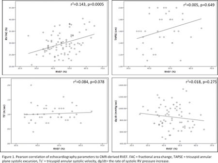
Results: There were 82 matched 2DE and CMR studies in 42 ccTGA patients (50% male). Median age at 2DE was 33.1 years (IQR = 22.7- 48.2 year). Pearson correlation analysis demonstrated a weak correlation between FAC and RVEF (r2=0.143, p=0.0005). Other 2DE parameters failed to show any correlation with RVEF (Figure 1).
Conclusion: In general, 2DE parameters for the assessment of subaortic RV systolic function correlate poorly with CMR measured RVEF in patients with ccTGA. This is important information for selection and interpretation of various modalities in the long-term follow-up of this patient population.
Article Information
vol. 132 no. Suppl 3 A14692
Published By:
American Heart Association, Inc.
Online ISSN:
History:
- Originally published November 6, 2015.
Copyright & Usage:
© 2015 by American Heart Association, Inc.
Author Information
- Zakariya Albinmousa1;
- Lan-Chau Kha2;
- Alessia D Carlo1;
- Alton Wong1;
- Andrew Crean1;
- Rachel Wald1;
- William Wilson1;
- Lucy S Roche1
- 1Div of Cardiology, Toronto Congenital Cardiac Cntr for Adults, Univ of Toronto, Toronto, Canada
- 2Dept of Radiology,, Univ of Toronto, Toronto, Canada
Abstract 13751: Categorizing the Burden of Arrhythmia in Adults With Congenital Heart Disease
David C Peritz, Vincent J Gonzalez, April Edwards, Jennifer E Nelson, Sunita J Ferns
Circulation. 2015;132:A13751
Abstract
Introduction: Advances in surgical and medical therapies have increased the life expectancy for patients with congenital heart disease. Arrhythmias in these patients are common and multi-factorial, often due to a disruption of the hearts’ intrinsic electrical system and degenerative remodeling, secondary to varying pressure and volume loads. The aim of this study is to better characterize the association between type of structural heart disease (SHD) and clinically significant arrhythmias.
Methods: We identified all adults with diagnosed congenital heart disease seen at a large tertiary care referral center between 2000 and 2014. Patients were categorized based on gender, SHD, and type of arrhythmia. We used the American Thoracic Society guidelines to group SHD into discrete categories and grouped arrhythmias as tachyarrhythmias (atrial/ventricular) and bradyarrhythmias.
Results: We identified 1355 individuals with at least one congenital structural heart defect and at least one arrhythmia. Of those, the most common structural abnormality was left-sided outflow tract obstruction and the least common was congenitally corrected transposition of the great arteries. The most common category of arrhythmia identified was atrial arrhythmia and the least common was sinoatrial node dysfunction (Figure). The most common association between SHD and arrhythmia was atrial arrhythmia in patients with left-sided outflow tract obstruction.
Conclusions: This study is one of the largest single-center reports linking specific structural heart defects to type of rhythm disturbance. These data may help providers better anticipate future arrhythmia development based on a patient’s underlying structural defect and offer prognostic information to patients and families. Future studies to more closely evaluate the relationship between arrhythmia patterns and specific risk factors within each category of SHD are needed.
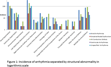
Article Information
vol. 132 no. Suppl 3 A13751
Published By:
American Heart Association, Inc.
Online ISSN:
History:
- Originally published November 6, 2015.
Copyright & Usage:
© 2015 by American Heart Association, Inc.
Author Information
- 1Internal Medicine & Pediatrics, Univ of North Carolina at Chapel Hill, Chapel Hill, NC
- 2Surgery, Div of Cardiothoracic Surgery, Univ of North Carolina at Chapel Hill, Chapel Hill, NC
- 3Pediatrics, Div of Cardiology, Univ of North Carolina at Chapel Hill, Chapel Hill, NC
Abstract 13046: Treat and Repair Strategy in Patients With Atrial Septal Defect and Severe Pulmonary Arterial Hypertension
Teiji Akagi, Yasuhumi Kijima
Circulation. 2015;132:A13046
Abstract
Introduction: An effective therapeutic strategy in patients with atrial septal defect (ASD) and significant pulmonary arterial hypertension (PAH) remains controversial.
Hypothesis: PAH-specific medications contribute to improvement of PAH. Subsequent transcatheter ASD closure may lead to further reduction of PAP.
Methods: Among 646 patients with ASD, 22 who were complicated by definite PAH had successful transcatheter ASD closure. Among them, eight patients received PAH-specific medications prior to the transcatheter procedure (PHM group) and 14 patients did not (non-PHM group).
Results: The mean age at the procedure in the PHM group was 37±15 years. Initially, the PHM group had significantly higher mean pulmonary arterial pressure (PAP) and pulmonary vascular resistance (PVR) compared with the non-PHM group (62±21 vs. 35±8 mmHg, p<0.01; 9.6±3.8 vs. 4.1±1.1 Wood units, p<0.01). PAP and PVR in the PHM group significantly decreased to 41±10 mmHg (p<0.01) and to 4.0±0.8 Wood units (p<0.01), respectively, after treatment with PAH-specific medications. In the PHM group, after ASD closure, further reduction of estimated systolic PAP was observed (72±19 to 41±11 mmHg, p<0.01).
Conclusions:Treat and repair is a valid therapeutic strategy for patients with ASD and significant PAH.
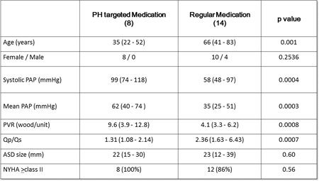
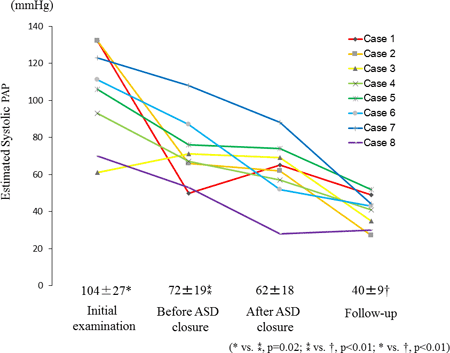
Article Information
vol. 132 no. Suppl 3 A13046
Published By:
American Heart Association, Inc.
Online ISSN:
History:
- Originally published November 6, 2015.
Copyright & Usage:
© 2015 by American Heart Association, Inc.
Author Information
- 1Adult Congenital Heart Disease Cntr, Okayama Univ Hosp, Okayama, Japan
- 2Cardiovascular Medicine, Okayama Univ Hosp, Okayama, Japan
Abstract 12727: The Clinical Impact of Eicosapentaenoic Acid/Arachidonic Acid Ratio in Adult Patients With Congenital Heart Disease
Miki Kanoh, Kei Inai, Eriko Shimada, Tokuko Shinohara, Mikiko Shimizu, Tetsuko Ishii, Hisashi Sugiyama, In-Sam Park
Circulation. 2015;132:A12727
Abstract
Background: Recent studies showed that intake of long-chain n-3 polyunsaturated fatty acids reduces the incidence of cardiovascular events. Furthermore, low levels of eicosapentaenoic acid (EPA) are associated with heart failure (HF), arrhythmia, and cardiac sudden death. However, there have been few studies regarding the clinical implication of the EPA/arachidonic acid (AA) ratio in adult patients with congenital heart disease (CHD).
Objectives and methods: The present study was a prospective study of 64 adult patients with CHD. We measured serum EPA and AA and divided the patients into two groups according to their EPA/AA ratio: <0.25, low group; ≥0.25, high group. Patients were observed for a mean duration of 13 months. We investigated the association between the EPA/AA ratio and clinical data and analyzed the prognostic predictive value of the EPA/AA ratio in these patients.
Results: There were no significant differences between the low (n = 36) and high (n = 28) groups in age, sex, blood pressure, systemic ventricular ejection fraction, medications, or serum levels of hemoglobin A1c, creatinine, low-density lipoprotein-cholesterol, high-density lipoprotein-cholesterol, triglyceride, or B-type natriuretic peptide. A low EPA/AA ratio had significant prognostic value for arrhythmia (hazard ratio [HR] 3.6, 95% confidence interval [CI] 1.1-14.8, p = 0.03) and composite events of arrhythmia and HF hospitalization (HR 5.6, 95% CI 1.53-27.8, p < 0.01). Subgroup analysis revealed that the patients with two-ventricle circulation in the low group (n = 23) were at significantly higher risk of arrhythmia (HR 6.7, p = 0.02), HF hospitalization (HR 16.2, p < 0.01), and composite events of arrhythmia and HF hospitalization (HR 6.7, p = 0.03) than those in the high group (n = 23). In patients with single-ventricle circulation, serum EPA levels were significantly lower compared with patients with two-ventricle circulation.
Conclusions: The EPA/AA ratio had a significant predictive value for cardiac events in adult patients with CHD, especially in patients with two-ventricle circulation. The effect of EPA treatment for patients with CHD should be elucidated in the future.
Article Information
vol. 132 no. Suppl 3 A12727
Published By:
American Heart Association, Inc.
Online ISSN:
History:
- Originally published November 6, 2015.
Copyright & Usage:
© 2015 by American Heart Association, Inc.
Author Information
- Miki Kanoh;
- Kei Inai;
- Eriko Shimada;
- Tokuko Shinohara;
- Mikiko Shimizu;
- Tetsuko Ishii;
- Hisashi Sugiyama;
- In-Sam Park
- pediatric cardiology, Tokyo Women’s Med Univ Hosp, Tokyo, Japan
Abstract 10259: Echocardiographic Predictors of Mortality in Adults With a Fontan Circulation
Rachael Cordina, Katherine von Klemperer, Aleksander Kempny, Roxy Senior, David S Celermajer, Sonya Babu-Narayan, Michael Gatzoulis, Wei Li
Circulation. 2015;132:A10259
Abstract
Introduction: Transthoracic echocardiographic (TTE) parameters that predict mortality have not been well-defined in Fontan-adults. Atrioventricular valve systolic to diastolic duration ratio (AVV S:D) is an echocardiographic indice that reflects global cardiac performance but has not been studied in this setting. AVV S:D may be useful in this heterogeneous group as it does not rely on geographic assumption or segmental analysis.
Methods: Fontan-adults (≥18 years) in sinus or A-paced V-sensed rhythm seen at our institution since 2005 were studied prospectively. Clinical data were recorded from time of index TTE. Subjects were censored at death or most recent review. Nil had cardiac transplant.
Results: In total, 128 patients (64 male) were included, mean age was 27 ± 8 years, 107 had dominant left ventricle (LV), 18 had dominant right ventricle (RV) and 3 were biventricular with large ventricular septal defect. Forty-eight had atriopulmonary connection (APC), 64 had total cavopulmonary connection (TCPC) and 16 had TCPC conversion. Time since first repair was 22 ± 7 years. Mean NYHA Class was 1.4 ± 0.6. During mean follow-up of 4.3 ± 2.8 years, 12 patients died (1 dominant RV, 11 dominant LV). Eight deaths were due to heart failure, 2 were sudden death and 2 were due to liver failure. In univariate analysis the strongest TTE predictors of mortality were AVV S:D, presence of restrictive filling using pulse and tissue Doppler parameters, subjective grade of systolic ventricular function, fractional area change and moderate to severe AVV regurgitation. Results with p ≤ 0.2 are shown in Figure 1. Cox regression analysis suggested NYHA Class and AVV S:D were independent predictors of mortality (p<0.0001 for both).
Conclusions: TTE indices are predictive of mortality in Fontan-adults. Ventricular performance assessed by AVV S:D was the most important parameter and should be incorporated into routine clinical assessment of these patients.
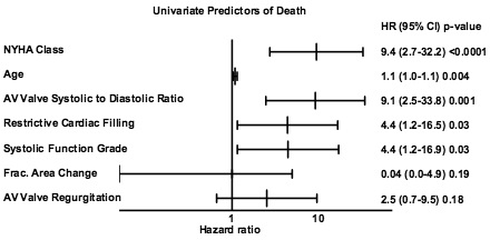
Article Information
vol. 132 no. Suppl 3 A10259
Published By:
American Heart Association, Inc.
Online ISSN:
History:
- Originally published November 6, 2015.
Copyright & Usage:
© 2015 by American Heart Association, Inc.
Author Information
- Rachael Cordina1;
- Katherine von Klemperer2;
- Aleksander Kempny2;
- Roxy Senior3;
- David S Celermajer1;
- Sonya Babu-Narayan2;
- Michael Gatzoulis2;
- Wei Li2
- 1Cardiology, Royal Prince Alfred Hosp, Sydney, Australia
- 2Adult Congenital Heart Disease Unit, Royal Brompton Hosp, London, United Kingdom
- 3Echocardiography, Royal Brompton Hosp, London, United Kingdom
Abstract 19719: Super Flexible Heart Replica With Vacuum Casting Technique Reproduces Real Structure and Texture of the Heart Fascinating Simulation Surgery in Complicted Congenital Heart Disease
Isao Shiraishi, Ken-ichi Kurosaki, Suzu Kanzaki, Katsunori Hatanaka, Hajime Ichikawa
Circulation. 2015;132:A19719
Abstract
Introduction: Precise understanding of 3D anatomy is crucial for successful surgical operation in complicated congenital heart disease (CHD). We have developed a new technology that reproduces accurate and extremely flexible polyurethane biomodels of complicated CHD suitable for simulation surgery and medical education.
Hypothesis: A novel 3D printing technique employing with stereolithography followed by vacuum casting is suitable for simulation surgery in complicated CHD.
Methods: Thirty five replicas of CHD were produced with this technique; diagnosis included tetralogy of Fallot, pulmonary atresia with ventricular septal defect, double outlet right ventricle, persistent truncus arteriosus, hypoplastic left heart syndrome, interrupted aortic arch, congenitally corrected transposition of the great arteries, and so on. Three-dimensional volumetric datasets of MSCT angiography of CHD were converted into standard triangulated language files to make plastic stereolithographic biomodels representing outer and inner surface of the heart. Then, another outer mold was made with urethane using the outer surface biomodels as a cast. Then, rubbery polyurethane was injected between the outer and inner molds under the vacuum condition. After solidification, the molds were carefully removed and the final biomodels were obtained.
Results: Wide variety of accurate and flexible biomodels of complicated CHDs from neonates to adults were reproduced. This technology allowed surgeons to precisely understand the internal chambers of the heart and allowed them to perform simulation surgery by way of cutting and suturing like a real heart tissue.
Conclusions: These super flexible and accurate polyurethane biomodels were instructive for surgeons to understand the complex structures and hemodynamics of the disease and also helpful for planning the individual surgical operation of complicated CHD.
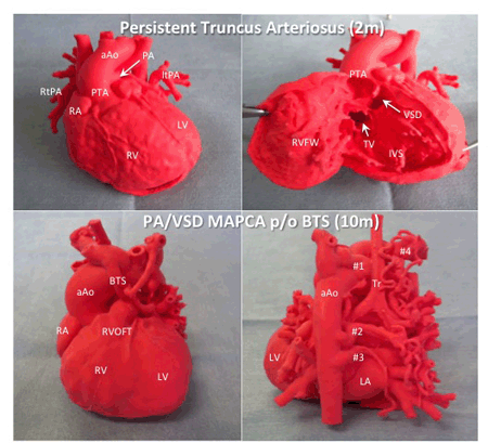
Article Information
vol. 132 no. Suppl 3 A19719
Published By:
American Heart Association, Inc.
Online ISSN:
History:
- Originally published November 6, 2015.
Copyright & Usage:
© 2015 by American Heart Association, Inc.
Author Information
- 1Pediatric Cardiology, National Cerebral and Cardiovascular Cntr, Suita, Japan
- 2Radiology, National Cerebral and Cardiovascular Cntr, Suita, Japan
- 3crossEffect, crossEffect Inc., Kyoto, Japan
- 4Pediatric Cardiac Surgery, National Cerebral and Cardiovascular Cntr, Suita, Japan
Abstract 19011: Initial Field Testing of a Cloud-based Cardiac Auscultation Information Management System in Rural Southwest Kunming Province, Peoples Republic of China
Lee A Pyles, Jihua Pan, Xiaoju Yu, Ke Liu, Jing Wang, Bistra Zheleva, Andreas Tsakistos, Weiguang Shao, Pouya Hemmati, Quan Ni
Circulation. 2015;132:A19011
Abstract
Introduction: Auscultation (Ausc) is used around the world to screen for congenital heart disease (CHD). We describe initial field testing of a cloud-based Ausc information management system in rural China school children with limited access to cardiac care . Our goal is to develop an economical system to facilitate access to cardiac care for underserved children in low- and middle-income countries.
Hypothesis: Web/cloud based distant transmission of auscultation exam can facilitate screening for congenital heart disease.
Methods: A team of cardiology staff from a tertiary hospital conducted field screening for CHD) in rural school children in Yunnan Province, People’s Republic of China. Each child with a murmur with traditional Ausc underwent echo screening and digital Ausc (Thinklabs, USA). Heart sounds were collected from 5 positions (URSB, ULSB, LLSB, apex and mid-LSB), and uploaded to a server using a tablet application (HeartLink System, USA). Blinded Ausc 3-5 months later was compared with echo finding at on-site screening.
Results:8000 children were screened with standard Ausc over 4 days by a team from Kunming Medical University and First University Hospital. Heart sounds were recorded for 149 subjects. The best vibratory quality of an innocent Still murmur was heard at LLSB in 52,at mid-LSB in 51, apex in 10 and URSB/ULSB in 4. Digital Ausc found pathologic murmurs in 11 of 14 subjects with echo-diagnosed CHD. Three missed were ASD called venous hum in 2 of 3; likely related to respiratory artifact at the URSB/ULSB. Accuracy was 136/149 or 91% with 10 false positive. Positive predictive value 52% and negative predictive value 98%. The graphical phonocardiogram facilitates visual analysis of S2 splitting and the duration and timing of the cardiac murmur.
Conclusions: The cloud-based digital Ausc system is able to collect, transmit, organize and display phonocardiograms for remote CHD screening and consultation. Difficulty with respiratory contamination of the signal is noted. VSD and Still murmur were successfully differentiated. The visual phonocardiographic display can aid S2 analysis for splitting. The pilot study demonstrates the potential use of this system to extend pediatric cardiology expertise to underserved rural areas around the world.
Article Information
vol. 132 no. Suppl 3 A19011
Published By:
American Heart Association, Inc.
Online ISSN:
History:
- Originally published November 6, 2015.
Copyright & Usage:
© 2015 by American Heart Association, Inc.
Author Information
- Lee A Pyles1;
- Jihua Pan2;
- Xiaoju Yu3;
- Ke Liu2;
- Jing Wang2;
- Bistra Zheleva4;
- Andreas Tsakistos4;
- Weiguang Shao5;
- Pouya Hemmati6;
- Quan Ni5
- 1Pediatrics, West Virginia Univ, Morgantown, WV
- 2Cardiology, Kunming Med Univ Hosp, Kunming, China
- 3Cardiology, Kunming Med Univ, Kunming, China
- 4International Programs, Children’s HeartLink, Inc, Minneapolis, MN
- 5Volunteer, Children’s HeartLink, Inc, Minneapolis, MN
- 6Med Sch, Univ of Minnesota, Minneapolis, MN
Abstract 18284: Incidence of Epilepsy is Elevated in Children and Young Adults With Congenital Heart Defects Compared With the General Population: A Nationwide Cohort Study
Morten Olsen, Bradley S Marino, Michelle Leisner, Jessica G Woo, Nicolas L Madsen
Circulation. 2015;132:A18284
Abstract
Perioperative seizures related to surgery for congenital heart defects (CHD) are well described; however, few data exist on the long-term risk of epilepsy in patients with CHD. We aimed to estimate the incidence of epilepsy in children and young adults with CHD compared with the general population.
Methods: Utilizing data from the Danish National Registry of Patients (DNRP) we identified all patients diagnosed with CHD before the age of 15 years between 1980 and 2010 who were born during the same period. The DNRP is a nationwide hospital discharge registry covering all Danish hospitals. Previously validated methodology using the DNRP was applied to measure the outcome, epilepsy, as well as presence of extra cardiac defects (ECD) and/or syndromes. We used the Danish Medical Birth Registry to identify preterm birth (gestational age<37 weeks). For each CHD subject, we identified 10 controls from the general population using the Danish Civil Registration System, matched by sex and birth year. A unique personal identifier assigned at birth and used in all Danish public registries enabled virtually complete follow up for migration, death, or epilepsy until January 1, 2013. We computed cumulative incidences and hazard ratios (HR) (split at 5 years of age to obtain proportional hazards) of time from CHD diagnosis (index date for controls) to epilepsy.
Results: We identified 14,665 CHD subjects with a median age at diagnosis of 2 (IQR 19) months. By 15 years of age, the cumulative incidence of epilepsy was 4% among CHD subjects. The HR of epilepsy among CHD subjects compared with the control cohort was 3.7 (95% CI: 3.2-4.3) below 5 years of age, and 2.4 (95% CI: 2.1-2.7) from 5 to 33 years of age. In the older age group, HR for patients with severe CHD was 2.8 (95% CI: 2.3-3.5), and for mild and moderate CHD was 2.2 (95% CI: 1.8-2.6). After exclusion of all subjects with ECDs and/or syndromes and preterm birth, corresponding HRs were 2.2 (95% CI: 1.6-3.0) and 1.7 (95% CI: 1.3-2.2), respectively.
Conclusion: The epilepsy risk was markedly increased in CHD subjects compared with the age and gender matched controls. These findings add evidence to support the importance of developing neuro-protective measures and potentially long-term epilepsy surveillance strategies in the CHD population.
Article Information
vol. 132 no. Suppl 3 A18284
Published By:
American Heart Association, Inc.
Online ISSN:
History:
- Originally published November 6, 2015.
Copyright & Usage:
© 2015 by American Heart Association, Inc.
Author Information
- 1Dept of Clinical Epidemiology, Aarhus Univ Hosp, Århus, Denmark
- 2Divs of Cardiology and Critical Care Medicine, Ann and Robert H. Lurie Children’s Hosp of Chicago, Chicago, IL
- 3Heart Institute, Cincinnati Children’s, Cincinnati, OH
Abstract 17186: Regional Extracellular Volume Fraction Can Identify Cardiac Muscle Changes in Patients With Duchenne Muscular Dystrophy
Laura Olivieri, Peter Kellman, Russell Cross, Kanishka Ratnayaka, Andrew E Arai, Michael S Hansen, Chris Spurney
Circulation. 2015;132:A17186
Abstract
Background: Duchenne muscular dystrophy (DMD) is an X-linked inherited disorder causing dilated cardiomyopathy with variable onset and progression. Our aim was to compare native T1 mapping and extracellular volume fraction (ECV) in boys with DMD to controls to identify markers of preclinical myocardial disease.
Methods: With IRB approval and informed consent/assent, 17 boys with DMD had cardiac magnetic resonance imaging (MRI) with contrast. Ejection fraction (EF), presence of late gadolinium enhancement (LGE), native T1, and ECV mapping were obtained using a modified Look-Locker sequence on a 1.5T MR scanner (Figure 1). Results were compared with age and gender matched controls with no history predisposing to cardiac fibrosis (i.e. bypass, myocarditis, etc).
Results: Seventeen DMD subjects (age 14 ± 5 years; EF 56% ± 8%) were compared with 13 normal controls (age 17 ± 4 years) with normal EF and no LGE. DMD subjects had low-normal EF (average EF 56% ± 8%) and 10 of 17 (59%) had evidence of LGE, all in the lateral location. Comparing native T1 in DMD vs controls, septal native T1 was 1060 ± 71 ms versus 990 ± 34 ms (p < 0.01) and lateral native T1 was 1090 ± 93 versus 978 ± 37 ms, respectively (p < 0.01). Comparing ECV values in DMD vs controls, septal ECV was 29.6 ± 7.8 and 25.9 ± 3.4 (p = 0.03) and lateral ECV was 32.7 ± 8.7 and 24.4 ±3.6 (p < 0.01), respectively. All DMD patients with abnormal EF and/or presence of LGE also had higher ECV values.
Conclusions: ECV is strongly-related to DMD and differs significantly compared to normals, particularly in the lateral wall. ECV mapping may be a useful technique for identifying preclinical myocardial changes in DMD.
Figure 1. A) Precontrast T1 map, B) post contrast T1 map and C) ECV map with scale on the right in the short axis projection of a boy with Duchenne muscular dystrophy. Arrows indicate elevated values laterally.

Article Information
vol. 132 no. Suppl 3 A17186
Published By:
American Heart Association, Inc.
Online ISSN:
History:
- Originally published November 6, 2015.
Copyright & Usage:
© 2015 by American Heart Association, Inc.
Author Information
- Laura Olivieri1;
- Peter Kellman2;
- Russell Cross1;
- Kanishka Ratnayaka1;
- Andrew E Arai2;
- Michael S Hansen2;
- Chris Spurney1
- 1Children’s National Heart Institute, Children’s National Health System, Washington, DC
- 2National Heart, Lung and Blood Institute, National Institutes of Health, Bethesda, MD
Abstract 15653: Optimizing the Screening Algorithm for Critical Congenital Heart Disease: A Data-Driven Approach
Matthew Oster, Caglar Caglayan, Regina Simeone, Pinar Keskinocak, Turgay Ayer
Circulation. 2015;132:A15653
Abstract
Introduction: Screening for critical congenital heart disease (CCHD) using pulse oximetry is performed with various algorithms in the United States, with none of these based on robust data, including the one endorsed by the AHA and other organizations.
Hypothesis: Existing algorithms will have differing results for CCHD screening, and the test characteristics of these algorithms can be improved through modifications.
Methods: After collecting de-identified, patient-level pulse oximetry values from existing national and international screening programs, we compared the commonly used algorithms in the US: the American Academy of Pediatrics (AAP) algorithm (endorsed by the AHA), the New Jersey (NJ) algorithm (slight modification of the AAP algorithm), and the Tennessee (TN) algorithm (screening first on the foot only). We then considered alternate algorithms by modifying the pulse oximetry values that would be considered a pass, fail, or retest result in the existing algorithms. We compared the algorithms according to sensitivity (Se), false positive rate (FP), and area under the curve (AUC).
Results: There were 75,748 screenings, with 57 true positives. Performance characteristics of the 3 common algorithms are summarized in the Table. The NJ algorithm had the greatest Se but also the highest FP, and the TN algorithm had the lowest Se but also the lowest FP. Modifications of these algorithms yielded 2632 potential algorithms with Se ranging from 28% to 91%, FP from 0.05% to 60%, and AUC from 0.64 to 0.82. Of these, 224 algorithms met our optimal algorithm criteria of Se >50%, FP 0.75.
Conclusions: Current algorithms for CCHD screening vary in their test performance characteristics, and there are opportunities to modify these algorithms to improve test performance. Before identifying an optimal algorithm, further work is needed to collect more data (including more robust data on false negatives) and to compare the cost and ease of use of potential algorithms.
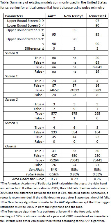
Article Information
vol. 132 no. Suppl 3 A15653
Published By:
American Heart Association, Inc.
Online ISSN:
History:
- Originally published November 6, 2015.
Copyright & Usage:
© 2015 by American Heart Association, Inc.
Author Information
- 1Sibley Heart Cntr Cardiology, Children’s Healthcare of Atlanta, Emory Univ Sch of Medicine, Atlanta, GA
- 2H. Milton Stewart Sch of Industrial and Systems Engineering, Georgia Institute of Technology, Atlanta, GA
- 3National Cntr on Birth Defects and Developmental Disabilities, Cntrs of Disease and Control and Prevention, Atlanta, GA
Abstract 14024: Results of a Phase I / II Multicenter Investigation of Udenafil in Adolescents After Fontan Palliation
David J Goldberg, Victor Zak, Bryan H Goldstein, Shan Chen, Michelle S Hamstra, Elizabeth A Radojewski, Eileen Maunsell, Seema Mital, Shaji C Menon, Kurt R Schumacher, R. M Payne, Mario Stylianou, Jonathan R Kaltman, Stephen M Paridon
Circulation. 2015;132:A14024
Abstract
Introduction: Many pediatric clinical trials fail due to use of sub-therapeutic dosing. This phase I / II clinical trial was designed to evaluate the short-term safety, pharmacokinetics (PK), and preliminary efficacy of udenafil, a phosphodiesterase type 5 inhibitor, in adolescents following Fontan palliation to inform dose selection for a phase III clinical trial.
Methods: This was a dose escalation trial with 5 dosing cohorts of 6 subjects each at 37.5 mg, 87.5 mg, and 125 mg daily, as well as 37.5 mg and 87.5 mg twice daily (bid) for 5 days. Efficacy testing (echocardiography, exercise stress test, endothelial function assessment) was performed at baseline and on day 5. A control cohort of 6 subjects underwent exercise testing without drug administration. Serum samples for PK analysis were collected on days 6-8. Adverse events (AE) were recorded throughout the 8-day study period and for 3 months thereafter. Comparison between baseline and day 5 testing was performed using ANOVA and visual trend lines. PK analysis was performed using a non-linear mixed effects model.
Results: The trial enrolled 36 subjects at a mean age of 15.8 years (58.3% male). Subject characteristics were similar between cohorts. No drug-related serious AEs were reported during the study period, while 24 subjects had non-serious AEs possibly or probably related to study drug. Headache was the most common, occurring in 20 of the 30 subjects. Other possibly or probably medication related side effects occurring in more than one subject included: facial flushing (10 subjects), nasal congestion (7), spontaneous penile erection (6), nausea (3), and abdominal discomfort (2). The 87.5 mg bid cohort achieved the highest steady state serum concentration (172 ng/mL, range 117-202), the highest maximal concentration (506 +/- 188 ng/mL), and the greatest area under the curve for 24 hours (6701 +/- 1591 ng*hr/mL). Efficacy test results did not change significantly in any cohort.
Conclusion: Udenafil was safe and well tolerated at all dosing levels while the 87.5 mg bid cohort achieved the highest serum drug level. With a 5-day treatment period and small sample size no significant clinical changes were observed. These data suggest that the 87.5 mg bid regimen may be most appropriate for a Phase III clinical trial.
Article Information
vol. 132 no. Suppl 3 A14024
Published By:
American Heart Association, Inc.
Online ISSN:
History:
- Originally published November 6, 2015.
Copyright & Usage:
© 2015 by American Heart Association, Inc.
Author Information
- David J Goldberg1;
- Victor Zak2;
- Bryan H Goldstein3;
- Shan Chen2;
- Michelle S Hamstra3;
- Elizabeth A Radojewski4;
- Eileen Maunsell5;
- Seema Mital4;
- Shaji C Menon6;
- Kurt R Schumacher7;
- R. M Payne8;
- Mario Stylianou9;
- Jonathan R Kaltman9;
- Stephen M Paridon1,
- Pediatric Heart Network Investigators
- 1Cardiology, The Children’s Hosp of Philadelphia, Philadelphia, PA
- 2Biostatistics, New England Rsch Institutes, Watertown, MA
- 3Cardiology, Cincinnati Children’s Hosp Med Cntr, Cincinnati, OH
- 4Cardiology, The Hosp for Sick Children, Toronto, Canada
- 5Cardiology, New England Rsch Institutes, Watertown, MA
- 6Cardiology, Primary Children’s Med Cntr, Salt Lake City, UT
- 7Cardiology, C.S. Mott Children’s Hosp, Ann Arbor, MI
- 8Cardiology, Riley Hosp for Children, Indianapolis, IN
- 9National Heart, Lung, and Blood Institute, National Institutes of Health, Bethesda, MD
Abstract 13595: Folic Acid Protects Embryo From Environmental Effects on Lipid Metabolism During Cardiac and Placental Development
Kersti K Linask, Mingda Han
Circulation. 2015;132:A13595
Abstract
Introduction: Congenital heart defects (CHDs) are the most prevalent of human birth defects with males exhibiting more severe types of CHDs. Cardiac cell and placental trophoblast differentiation are two embryonic processes occurring early between E6.5-E8.0 of mouse gestation (16-19 days of human pregnancy) that can be interfered with by environmental factors to induce anomalies.
Objective: To determine the predominant pathway(s) targeted by exposures to lithium (Li+), homocysteine (HCys) , or alcohol (ethanol, EtOH) that result in CHDs and that can be protected by high dose folic acid (FA) supplementation.
Method: On morning of conception, pregnant mice were placed on a protective, high FA diet (10 mg/kg) or on a health maintenance FA diet (3.3 mg/kg) that did not prevent CHDs. At gastrulation (E6.75) the mice received an i.p. injection of Li, HCys, or EtOH. Pregnant control groups received either the protective high FA or the normal FA maintenance diet and an i.p. injection of physiological saline only. Echocardiography on E15.5 determined embryos with abnormal cardiac function. All abnormal hearts and normal control hearts were removed, total RNA extracted. Microarrays were used to evaluate gene changes.
Results: Significant expression changes among 12 treatment groups with Li and HCys exposures indicated that Gene Ontology (GO) categories involving lipid metabolism were chiefly altered. When the GO sets were sorted according to gender, male embryos displayed greater changes in lipid-related categories than the female. Validation studies demonstrated that Acyl CoA dehydrogenase medium length chain (Acadm) and its protein product important in fatty acid oxidation were modulated by the exposures, including by alcohol. Oil Red O (ORO) localization of neutral lipids in the heart and placenta demonstrated that lipids are highly altered possibly related to altered umbilical blood flow seen with embryos showing abnormal cardiac function. High FA restored normal ORO patterns, placental blood flow, and heart function.
Conclusions: Li, HCys and alcohol exposed embryos display altered lipid metabolism. Early gestational supplementation with high-dose FA protects lipid metabolism at control levels, maintains placental physiology, and prevents CHDs.
Article Information
vol. 132 no. Suppl 3 A13595
Published By:
American Heart Association, Inc.
Online ISSN:
History:
- Originally published November 6, 2015.
Copyright & Usage:
© 2015 by American Heart Association, Inc.
Author Information
- Kersti K Linask;
- Mingda Han
- Pediatrics, USF Morsani College of Medicine, St. Petersburg, FL
Abstract 10386: Clinical and Genetic Risk Factors for Post-Operative Atrial Tachycardia After Congenital Heart Surgery in Infants
Amy M O’Connor, Kim Crum, Andrew H Smith, Prince J Kannankeril
Circulation. 2015;132:A10386
Abstract
Introduction: Atrial tachycardia (AT) after infant congenital heart disease (CHD) surgery has been associated with increased mortality. Common genetic variants in PITX2 (rs2200733) and IL6 (rs1800795) have been associated with post-operative AT in adults, but have not been studied in infants after CHD surgery.
Hypothesis: Genetic variants in PITX2 and IL6 are associated with post-operative AT in infants with CHD.
Methods: Children less than 1 year of age undergoing their first CHD surgery at our center between 9/2007 and 3/2014 who consented to the study were included. Subjects provided a DNA sample and had repeated daily assessment of telemetry with documentation of all arrhythmias and clinical variables. Genotyping was performed in the Vanderbilt genomics core lab using the TaqMan PCR Core Reagent Kit. Children with and without AT were compared using chi-square or unpaired t-test as appropriate.
Results: Of 923 enrolled infants, 141 had post-operative AT (15.3%), 79 of who required treatment (8.6%). AT was associated with increased mortality (12.1% mortality with AT; vs. 5.5% no AT, P = 0.004), longer hospital stay (mean of 49 days with AT; vs. 23 days no AT, P < 0.0001), intensive care unit (ICU) stay (mean of 34 days with AT; vs.13 days no AT, P < 0.0001), and mechanical ventilation duration (mean of 22 days with AT; vs. 8 days no AT, P < 0.0001). Age, epinephrine use, milrinone use, bypass time, cross-clamp time, lactate, and number of pump runs were associated with AT and AT requiring treatment. PITX2 and IL6 genotypes were not significantly associated with AT or AT requiring treatment.
Conclusions: AT is associated with increased mortality and longer hospital stay, ICU stay, and mechanical ventilation duration. Numerous operative factors contribute to AT after infant CHD surgery. PITX2 and IL6 genetic variants were not found to be significantly associated with post-operative AT in infants undergoing CHD surgery in our study.
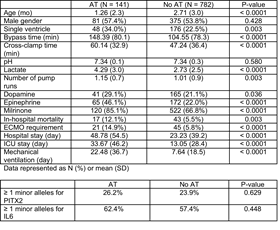
Article Information
vol. 132 no. Suppl 3 A10386
Published By:
American Heart Association, Inc.
Online ISSN:
History:
- Originally published November 6, 2015.
Copyright & Usage:
© 2015 by American Heart Association, Inc.
Author Information
- Amy M O’Connor;
- Kim Crum;
- Andrew H Smith;
- Prince J Kannankeril
- Pediatric Cardiology, Vanderbilt Univ, Nashville, TN
Abstract 10188: Circulating Levels of MIF, GROα And RANTES Chemokines and Interleukin 17E Correlate With Severity of Pulmonary Hypertension in Young Pediatric Patients With Congenital Heart Disease
Leína Zorzanelli, Nair Y Maeda, Mariana M Clavé, Ana M Thomaz, Marlene Rabinovitch, Antonio A Lopes
Circulation. 2015;132:A10188
Abstract
Introduction: Inflammation and immunity are central in the pathogenesis of pulmonary arterial hypertension (PAH), but have not been fully explored in young patients with congenital heart disease and elevated pulmonary artery pressure (PAH-CHD) undergoing surgical repair.
Hypothesis: Cytokines and related proteins may be differentially expressed in PAH-CHD patients with distinct hemodynamic patterns.
Methods: Sixteen patients with PAH-CHD were enrolled (Group 1, age 1.13 (0.76-2.48) years, median and interquartile range). Pulmonary artery pressure was 52 (43-66) mmHg, and pulmonary vascular resistance was 5.2 (4.2-8.9) Wood units•m2. Patients with pulmonary overcirculation, with no need for cardiac catheterization were included for comparison (Group 2, N=31, age 0.71 (0.43-1.02) years). Pulmonary-to-systemic blood flow ratio (echocardiography) in Group 1 and Group 2 was 1.9 (1.3-2.6) and 2.8 (2.3-3.3) respectively (p=0.008). Thirty-six cytokines were analyzed in serum using a chemiluminescence array.
Results: In the whole patient group (N=47), MIF chemokine (macrophage migration inhibitory factor) was significantly increased compared to pediatric controls (respective densities 7510±2755 pixels and 5697±2051 pixels, mean±SD, p=0.027). In patients, GROα chemokine (growth-regulated oncogene alpha) was elevated early in life, but decreased exponentially with the age (R2=0.21, p=0.001), while interleukin 17E (also called IL-25) increased progressively (R2=0.24, p<0.001). MIF was specifically increased in Group 1 compared to Group 2 and controls (respectively, 8494±619 pixels, 6618±477 pixels and 6548±726 pixels, age-adjusted mean±SEM, p=0.037). In contrast, RANTES chemokine (regulated on activation, normal T cell expressed and secreted) was specifically elevated in Group 2 compared to Group 1 and controls (respectively, 74183±3865 pixels, 60130±6455 pixels and 59332±3970 pixels, mean±SEM, p=0.039).
Conclusion: The data indicate a relationship between cytokine levels and severity of the disease (age, groups), with potential pathophysiological implications. Furthermore, involvement of interleukin 17E and MIF emphasize the role of Th2 immune response already described in PAH.
Article Information
vol. 132 no. Suppl 3 A10188
Published By:
American Heart Association, Inc.
Online ISSN:
History:
- Originally published November 6, 2015.
Copyright & Usage:
© 2015 by American Heart Association, Inc.
Author Information
- Leína Zorzanelli1;
- Nair Y Maeda2;
- Mariana M Clavé1;
- Ana M Thomaz1;
- Marlene Rabinovitch3;
- Antonio A Lopes1
- 1Pediatric Cardiology and Adult Congenital Heart Disease, Heart Institute, Univ of São Paulo Sch of Medicine, Sao Paulo, Brazil
- 2Rsch, Pró-Sangue Foundation, Sao Paulo, Brazil
- 3Cardiopulmonary Rsch Laboratory, Stanford Univ Sch of Medicine, Stanford, CA
Abstract 18056: Modeling Duchenne Muscular Dystrophy (DMD) Cardiomyopathy Using Patient-specific Induced Pluripotent Stem Cell-derived Cardiomyocytes
Forum Kamdar, Andre Klaassen Kamdar, Christopher S Chapman, Timothy J Kamp, Joseph C Wu, Naoko Koyano-Nakagawa, Daniel J Garry
Circulation. 2015;132:A18056
Abstract
Introduction: DMD is the most common muscular dystrophy and is characterized by the absence of dystrophin. Cardiomyopathy and associated arrhythmias have emerged as a leading cause of death in DMD.
Hypothesis: We hypothesized that the pathophysiology of DMD cardiomyopathy can be modeled using DMD hiPSC-derived cardiomyocytes (CM), which can be interrogated at the cellular and molecular level.
Methods: Dermal fibroblasts were obtained from patients with genetically confirmed DMD and healthy controls and reprogrammed to hiPSC using human reprogramming transcription factors. DMD and control hiPSC lines were differentiated to CMs using a directed differentiation protocol. Functional analysis including wheat-germ agglutinin co-immunoprecipitation and calcium handling was performed at d60.
Results: DMD hiPSCs demonstrated DYSTROPHIN exon mutations consistent with patient mutations. DMD and control hiPSCs differentiated to beating, electrically coupled CM sheets. Quantitative western blot analysis for dystroglycan complex (DGC) components and novel cardiac DGC proteins Cryab and Cypher demonstrated expression of the majority of DGC and novel cardiac DGC proteins at d60 of differentiation in control hiPSC-derived CM. Dystrophin and DGC proteins including novel cardiac DGC proteins are associated with the DGC in all control hiPSC CM at d60. DMD hiPSC-derived CM have absence of dystrophin and a disrupted DGC. We identified that cypher is absent in DMD hiPSC CM and validated this in human DMD left ventricular tissue. Calcium imaging demonstrated significantly abnormal calcium transients in DMD hiPSC-derived CM correlating to arrhythmias.
Conclusion: Our results suggest that the control hiPSC-derived CMs have a largely nucleated DGC at d60 including novel cardiac DGC associated proteins. DMD hiPSCs have the hallmark absence of dystrophin and a disrupted DGC. The phenotype of increased arrhythmias in DMD patients is also reflected in DMD hiPSC CM. DMD hiPSC-derived CMs can be used as a model for investigation of DMD cardiomyopathy and further evaluation of the molecular and physiologic phenotype of DMD hiPSC cardiomyocytes will serve as a platform for testing current therapies and developing novel therapies for patients with DMD cardiomyopathy.
Article Information
vol. 132 no. Suppl 3 A18056
Published By:
American Heart Association, Inc.
Online ISSN:
History:
- Originally published November 6, 2015.
Copyright & Usage:
© 2015 by American Heart Association, Inc.
Author Information
- Forum Kamdar1;
- Andre Klaassen Kamdar1;
- Christopher S Chapman2;
- Timothy J Kamp3;
- Joseph C Wu4;
- Naoko Koyano-Nakagawa1;
- Daniel J Garry1
- 1Cardiovascular Div, Univ of Minnesota, Minneapolis, MN
- 2Cardiology, Univ of Minnesota, Minneapolis, MN
- 3Cardiovascular Medicine, Univ of Wisconsin-Madison, Madison, WI
- 4Medicine- Cardiovascular Medicine, Stanford Univ, Palo Alto, CA
Abstract 17788: Repaired Coarctation is Associated With Central Hypertension, Increased Vessel Stiffness and Abnormal Wave Reflections – A Novel CMR Hemodynamics Study
Michael A Quail, Rebekah Short, Bejal Pandya, Jennifer A Steeden, Abbas Khushnood, Patrick Segers, Vivek Muthurangu
Circulation. 2015;132:A17788
Abstract
Introduction: The basis of late cardiovascular mortality following coarctation repair is poorly understood. Although hypertension has been implicated, peripheral systolic pressure (pSBP) has not been shown to be definitively higher in this group. This may be because mortality is more related to central systolic pressure (cSBP) than pSBP. We have developed a novel cardiovascular magnetic resonance (CMR) protocol that allows assessment of cSBP and it components: resistance, compliance and wave reflections. The main aims of this study were i) characterize hemodynamic differences between patients and controls ii) define hemodynamic determinants of cSBP in patients and iii) Identify possible biomarkers amongst covariates associated with LV mass (LVM).
Methods: 75 subjects, 50 patients with repaired coarctation, median age 23.5yrs (74% male) and 25 matched controls; 21.0yrs (72% male) were recruited. Ascending aorta area and flow waveforms were obtained using a high temporal resolution (10ms) spiral phase-contrast MR flow sequence. This data was used to derive cSBP and perform wave intensity analysis (WIA) non-invasively using previously validated techniques. The determinants of cSBP and LVM were assessed using multivariable linear regression analysis.
Results: Central SBP was significantly higher in patients compared to controls (115±13 vs 107±9mmHg [mean±sd], p=0.002). However, there was only a trend towards higher pSBP (123±15 vs 117±11mmHg, p=0.052). Patients had reduced arterial compliance, increased characteristic impedance and larger backward compression waves (BCW) than controls; and these parameters were independently associated with cSBP. LVM index was significantly higher in patients than controls (73.1±14.6 vs 58.9±9.7g/m2, p=0.0001). Independent predictors of LVM included cSBP (p=0.001) and BCW (p=0.002), but importantly not pSBP or coarctation index.
Conclusions: Non-invasive assessment of fundamental arterial hemodynamics by CMR is feasible. Using these techniques we have shown elevated cSBP in patients after coarctation repair. Elevated cSBP and BCW are important determinants of increased LVM following coarctation repair. These metrics represent superior biomarkers of afterload than coarctation index and pSBP.
Article Information
vol. 132 no. Suppl 3 A17788
Published By:
American Heart Association, Inc.
Online ISSN:
History:
- Originally published November 6, 2015.
Copyright & Usage:
© 2015 by American Heart Association, Inc.
Author Information
- Michael A Quail1;
- Rebekah Short1;
- Bejal Pandya1;
- Jennifer A Steeden1;
- Abbas Khushnood1;
- Patrick Segers2;
- Vivek Muthurangu1
- 1Cntr for Cardiovascular Imaging, Institute of Cardiovascular Science, Univ College London, London, United Kingdom
- 2IBiTech-bioMMeda, iMinds Med IT, Ghent Univ, Gent, Belgium
Abstract 17405: Phosphodiesterase-based Vasodilation Improves Oxygen Delivery and Clinical Outcomes Following Stage 1 Palliation
Kimberly I Mills, Brian K Walsh, Aditya K Kaza, Hilary Bond, David Wypij, James A DiNardo, Ravi R Thiagarajan, John N Kheir
Circulation. 2015;132:A17405
Abstract
Introduction: The use of systemic vasodilators is known to decrease the incidence of postoperative cardiac arrest in infants with hypoplastic left heart syndrome (HLHS) following stage 1 palliation (S1P) with Blalock-Taussig shunt. Their impact on measured oxygen delivery (DO2) are uncharacterized.
Methods: In 2013, we implemented a treatment algorithm at our institution to standardize care and lower systemic vascular resistance (SVR) in the postoperative period (A). We measured standard hemodynamic variables in consecutive neonates prior to (n=32) and following (n=24) its implementation. 92% of these infants received a Sano modification. We continuously measured oxygen consumption (E-COVX module™, GE Healthcare) and venous oxyhemoglobin saturation (SvO2, PediaSat, Edwards) for Fick-based calculation of DO2 and SVR in a subset (n=21) of patients. Variables were compared pre and post-implementation using linear mixed effects models.
Results: Demographic, anatomic and echocardiographic risk factors were similar between groups, although aortic cross-clamp (ACC) time was significantly shorter following protocol implementation (120.9 vs 91.9 mins, P<0.001). When corrected for ACC time, serum lactate (p<0.001, B) and incidence of postoperative cardiac arrest (P=0.04, C) were lower (B), and SvO2 (P<0.001) was higher following protocol implementation. Systemic cardiac output (β[SE], 0.52 [0.32] L/min/m2; P=0.10) and systemic DO2 (β[SE], 86.7 [55.4] mL/min/m2; P=0.12, D) were higher post-implementation. DO2 was most closely correlated with SVR (r2=0.87), and commonly utilized surrogate variables such as arterial saturation (r2=0.01), SvO2 (r2=0.1), and arterial-venous saturation difference (r2=0.35) correlated poorly.
Conclusions: The implementation of a phosphodiesterase-based treatment algorithm decreased SVR, lactate accumulation and the incidence of cardiac arrest in infants with HLHS following S1P with Sano modification.
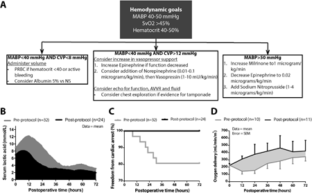
Article Information
vol. 132 no. Suppl 3 A17405
Published By:
American Heart Association, Inc.
Online ISSN:
History:
- Originally published November 6, 2015.
Copyright & Usage:
© 2015 by American Heart Association, Inc.
Author Information
- Kimberly I Mills;
- Brian K Walsh;
- Aditya K Kaza;
- Hilary Bond;
- David Wypij;
- James A DiNardo;
- Ravi R Thiagarajan;
- John N Kheir
- Cardiology, Boston Children’s Hosp, Boston, MA
Abstract 12348: Different Outcomes by First Documented Rhythm After Witnessed Out-of-Hospital Cardiac Arrest in Children
Masahiko Hara, Kenichi Hayashi, Tetsuhisa Kitamura
Circulation. 2015;132:A12348
Abstract
Background: Preventing the tragedy of pediatric out-of-hospital cardiac arrest (OHCA) is an important public health problem, and further accumulation of evidence is needed to improve the outcomes after pediatric OHCA. However, little evidence is available about the prognostic differences by first documented rhythm.
Methods and Results: We enrolled 3968 young (< 18 years old) witnessed OHCA patients between 2005 and 2012 from a prospective nationwide population-based cohort database in Japan. We assessed and compared the neurologically favorable one-month survival defined as Glasgow-Pittsburg cerebral performance category 1 or 2 by first documented rhythm (pulseless ventricular tachycardia/ventricular fibrillation [pVT/VF], pulseless electrical activity [PEA], and asystole). The number of OHCA patients with pVT/VF, PEA, and asystole were 556, 1249, and 2163, respectively. The proportion of overall neurologically favorable one-month survival in patients with pVT/VF, PEA and asystole were 36.5%, 5.0%, and 1.8%, respectively in all study population and 73.8%, 27.7%, and 13.8%, respectively in patients who achieved prehospital ROSC. Figure shows the relationship between time from collapse to first cardiopulmonary resuscitation (CPR) and the endpoint in total population with estimated probabilities of the endpoint (line) and the respective 95% confidence intervals (bands). The proportion of asystole increased as the time from collapse to CPR delayed whereas those of pVT/VF and PEA decreased (Trend p<0.001). Earlier initiation of CPR after pediatric OHCA resulted in higher achievement rate of prehospital ROSC (adjusted odds ratio 0.97, 95% confidence interval 0.95-1.00, p=0.025) which showed much better prognoses than those in total study population.
Conclusions: The pediatric OHCA outcome differed by the type first documented rhythm. Shortening of time to first CPR is crucial for improving outcomes after pediatric OHCA.
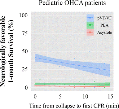
Article Information
vol. 132 no. Suppl 3 A12348
Published By:
American Heart Association, Inc.
Online ISSN:
History:
- Originally published November 6, 2015.
Copyright & Usage:
© 2015 by American Heart Association, Inc.
Author Information
- 1Dept of Cardiovascular Medicine, Osaka Univ Graduate Sch of Medicine, Suita, Japan
- 2Dept of Mathematics, Faculty of Science and Technology, Keio Univ, Yokohama, Japan
- 3Div of Environmental Medicine and Population Sciences, Dept of Social and Environmental M, Osaka Univ Graduate Sch of Medicine, Suita, Japan
Abstract 10384: Validation of a Simple Score to Determine Risk of Hospital Mortality After the Norwood Procedure
Shahryar M Chowdhury, Eric M Graham, Andrew M Atz, Scott M Bradley, Minoo M Kavarana, Ryan J Butts
Circulation. 2015;132:A10384
Abstract
Background: The NIH/NHLBI Pediatric Heart Network Single Ventricle Reconstruction (SVR) trial identified risk factors for hospital mortality after the Norwood procedure. However, the ability to quantify pre-operative risk remains elusive. This study aimed to develop an accurate and clinically feasible score to assess the risk of hospital mortality in neonates undergoing the Norwood procedure.
Methods: All patients (n = 549) in the publically available SVR database were included in the analysis. Patients were randomly divided into a derivation (75%) and validation (25%) cohort. Pre-operative patient, center, and surgeon-related covariates found to be associated with mortality upon univariate analysis (p < 0.2) were included in the initial multivariable logistic regression model. The final model was derived by including only variables independently associated with mortality (p < 0.05). A risk score was then developed using relative magnitudes of the covariates’ odds ratio. The score was then tested in the validation cohort.
Results: A 20-point risk score using 6 variables (see Table) was developed. The derivation and validation cohorts did not differ in age, sex, mortality, and the score covariates. Mean score in derivation and validation cohort were 5.2 ± 3.2 and 5.6 ± 3.5, p = 0.35, respectively. In weighted regression analysis, model predicted risk of mortality correlated closely with actual rates of mortality in the derivation (R2 = 0.87, p < 0.01) and validation cohorts (R2 = 0.82, p 10). Patients were classified as low (score 0-5), medium (6-10), or high risk (>10). Mortality differed significantly between risk groups in the derivation (6% vs. 22% vs. 77%, p < 0.01) and validation (4% vs. 30% vs. 53%, p < 0.01) cohorts.
Conclusion: This mortality score is accurate in determining risk of hospital mortality in neonates undergoing the Norwood operation. The score has the potential to be used in clinical practice to aid in risk assessment prior to surgery.
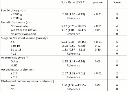
Article Information
vol. 132 no. Suppl 3 A10384
Published By:
American Heart Association, Inc.
Online ISSN:
History:
- Originally published November 6, 2015.
Copyright & Usage:
© 2015 by American Heart Association, Inc.
Author Information
- Shahryar M Chowdhury1;
- Eric M Graham1;
- Andrew M Atz1;
- Scott M Bradley2;
- Minoo M Kavarana2;
- Ryan J Butts1
- 1Pediatrics, Med Univ of South Carolina, Charleston, SC
- 2Surgery, Med Univ of South Carolina, Charleston, SC
Abstract 18446: Treatment With Tetrahydrobiopterin Decreases White Matter Injury in a Mouse Model for In Utero Hypoxia in Congenital Heart Disease
Jennifer Romanowicz, Ludmila Korotcova, Paul Morton, Amrita Cheema, Vittorio Gallo, Richard A Jonas, Nobuyuki Ishibashi
Circulation. 2015;132:A18446
Abstract
Introduction: Reduced oxygen delivery in complex congenital heart disease (CHD) can lead to brain white matter (WM) injury in utero. Currently, no treatment exists. Tetrahydrobiopterin (BH4) is a cofactor for neuronal nitric oxide synthase, and in its absence, toxic peroxynitrite production is favored. Hypoxia activates nitric oxide synthase, which reduces BH4 availability.
Hypothesis: Decreased BH4 levels underlie WM injury in the fetus with CHD, and treatment with BH4 will reduce this injury.
Methods: Mice were divided into three groups: normoxic controls (Nx), hypoxic (Hx), and hypoxic with BH4 treatment (Hx-BH4). Hx and Hx-BH4 mice were kept at 10.5% FiO2 from postnatal day 3 to 11–a period of WM development equivalent to the 3rd trimester in humans. Brain BH4 levels were quantified and compared between Nx (n=11) and Hx (n=12). Densities of cells expressing CNP (marker of oligodendrocytes–cells responsible for myelination) and Caspase3 (apoptosis marker) were quantified in three WM regions and compared between groups (n=3-6 each). Western blot detected myelin basic protein (myelin marker).
Results: Brain BH4 levels were depleted in Hx compared to Nx (-38.4%, p=0.02). CNP+ oligodendrocytes increased after Hx compared to Nx (Fig.1a), consistent with hypoxia-induced proliferation seen previously. BH4 treatment did not limit this proliferation (Fig.1a). Hx had increased WM apoptosis (Fig.1b), which decreased with BH4 treatment (Fig.1b). Remarkably, there was no difference in WM caspase3+ cells between Nx and Hx-BH4 (Fig.1b). There was no difference in effect across WM region. Finally, loss of myelin with hypoxia was mitigated by BH4 treatment (Fig.1c).
Conclusions: Our results show that suboptimal BH4 levels influence hypoxic WM injury. BH4 treatment of phenylketonuria is safe during pregnancy, thus maternal BH4 therapy is feasible. Our data demonstrate that repurposing BH4 for use during fetal brain development has potential to limit WM injury in CHD.
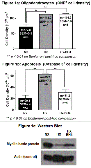
Article Information
vol. 132 no. Suppl 3 A18446
Published By:
American Heart Association, Inc.
Online ISSN:
History:
- Originally published November 6, 2015.
Copyright & Usage:
© 2015 by American Heart Association, Inc.
Author Information
- Jennifer Romanowicz;
- Ludmila Korotcova;
- Paul Morton;
- Amrita Cheema;
- Vittorio Gallo;
- Richard A Jonas;
- Nobuyuki Ishibashi
- Children’s National Heart Institute and Cntr for Neuroscience Rsch, Children’s National Med Cntr, WASHINGTON, DC
Abstract 16838: Circulating miRNAs Can Predict Cardiac Allograft Vasculopathy in Pediatric Heart Transplant Recipients in a Sex-dependent Manner
Scott R Auerbach, Shelley D Miyamoto, Anis Karimpour-Fard, Jane Gralla, Brian L Stauffer, Carmen C Sucharov
Circulation. 2015;132:A16838
Abstract
Introduction: MicroRNAs (miRs) are small noncoding ~22 nucleotide RNAs capable of modulating the expression of many genes. Our hypothesis was that circulating miRs are useful biomarkers of cardiac allograft vasculopathy (CAV) in pediatric heart transplant recipients (PHTR).
Methods: An array for 765 miRs was performed using the Applied Biosystems technology normalized by a global normalization method as determined by the Expression Suite Software. Serum was obtained from 132 PHTR at the time of routine surveillance catheterization with coronary evaluation (intravascular ultrasound [IVUS] performed if ≥ 25 kg). Rejection free CAV+ patients were matched by age at transplant and time since transplant to CAV- controls. Statistical analysis was performed using the Wilcoxon test.
Results: Samples were analyzed from 32 CAV+ PHTR matched to 17 CAV-controls. There were no significant differences between expression of miRs between CAV+ and CAV- PHTR alone, but differences in expression were found when PHTR were separated by sex. miRs significantly upregulated in females (F) included miRs-636 (F with both IVUS and angiographic CAV; p=0.008), -636 and -523 (F with a maximal intimal thickness [MIT] of ≥ 0.5mm; p=0.008 and 0.01, respectively), and -509 and -636 (F with any diagnosis of CAV; p=0.01 and p=0.01, respectively). miRs significantly upregulated in males (M) included miRs-422a (M with both IVUS and angiographic CAV; p=0.007), -422a and let-7e (M with a MIT of ≥ 0.5mm; p=0.003 and p=0.01, respectively), and -422a (M with any diagnosis of CAV; p=0.002). miR-19a was significantly downregulated in in M with any diagnosis of CAV (p=0.04). Boxplots highlighting differential expression of miRs by sex in CAV are shown in Figure 1.
Conclusions: miRs are differentially expressed between PHTR with CAV on the basis of sex. A unique biomarker signature of miRs that are specific to PHTR with CAV would be valuable in the risk stratification of this challenging patient population.
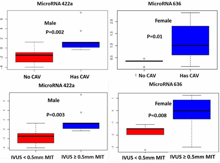
Article Information
vol. 132 no. Suppl 3 A16838
Published By:
American Heart Association, Inc.
Online ISSN:
History:
- Originally published November 6, 2015.
Copyright & Usage:
© 2015 by American Heart Association, Inc.
Author Information
- Scott R Auerbach1;
- Shelley D Miyamoto1;
- Anis Karimpour-Fard2;
- Jane Gralla3;
- Brian L Stauffer4;
- Carmen C Sucharov4
- 1Pediatrics, Div of Cardiology, Univ of Colorado Anschutz Med Campus, Aurora, CO
- 2Pharmacology, Univ of Colorado Anschutz Med Campus, Aurora, CO
- 3Pediatrics, Children’s Hosp Rsch Institute, Univ of Colorado Anschutz Med Campus, Aurora, CO
- 4Medicine, Div of Cardiology, Univ of Colorado Anschutz Med Campus, Aurora, CO
Abstract 14364: Hemodynamic Evaluation of 30 Y-Graft Fontan Completions
Phillip M Trusty, Maria Restrepo, Mark A Fogel, Kirk Kanter, Ajit P Yoganathan, Timothy C Slesnick
Circulation. 2015;132:A14364
Abstract
Background: Fontan completion, resulting in a total cavopulmonary connection (TCPC), is accomplished using either a traditional baffle or bifurcated Y-graft. The use of Y-grafts was originally hypothesized to provide more even hepatic blood flow distribution to the lungs, a factor related to pulmonary arteriovenous malformations. This study evaluates the hemodynamic performance of the largest Y-graft cohort to date, compares those results with a traditional baffle, and highlights Y-graft cases that exhibit both favorable and poor performance.
Methods: Thirty Y-graft and 30 traditional baffle Fontan patients were analyzed. TCPC anatomies and vessel flow waveforms were reconstructed using cardiac magnetic resonance (CMR) images and phase-contrast CMR. Computational fluid dynamic simulations were performed to quantify TCPC power loss, resistance, and hepatic flow distribution (HFD). Comparisons between graft types and subanalysis of favorable versus poor performing Y-grafts were investigated.
Results: The TCPC resistance distribution, HFD distribution, and the effect of LPA stenosis on power loss for Y-grafts and traditional baffles are shown in Fig. 1A-1C. TCPC resistance was significantly lower in traditional baffle patients. HFD was similar overall, with median and range of 40 (1-77) and 51 (1-99) for the traditional baffle and Y-graft, but show a discrepancy at extreme values with more unbalanced flow in the Y-graft cohort. Power loss was more sensitive to LPA stenosis in the Y-graft cohort. Prediction of Y-graft HFD is multi-factorial and six representative cases are shown in Fig. 1D.
Conclusions: Y-grafts do not inherently provide more balanced HFD than traditional baffles. Traditional baffles are more energetically favorable and less sensitive to PA stenosis. Graft type should be considered on an individual basis as hemodynamic performance is based on a combination of factors, including global flow distribution, PA stenosis and SVC positioning.
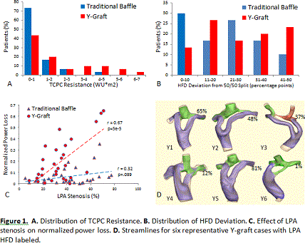
Article Information
vol. 132 no. Suppl 3 A14364
Published By:
American Heart Association, Inc.
Online ISSN:
History:
- Originally published November 6, 2015.
Copyright & Usage:
© 2015 by American Heart Association, Inc.
Author Information
- Phillip M Trusty1;
- Maria Restrepo1;
- Mark A Fogel2;
- Kirk Kanter3;
- Ajit P Yoganathan1;
- Timothy C Slesnick4
- 1Biomedical Engineering, Georgia Institute of Technology, Atlanta, GA
- 2Div of Cardiology, Children’s Hosp of Philadelphia, Philadelphia, PA
- 3Div of Cardiothoracic Surgery, Children’s Healthcare of Atlanta, Atlanta, GA
- 4Dept of Pediatrics, Children’s Healthcare of Atlanta, Atlanta, GA
Abstract 12954: A Novel-Particle Image Velocimetry (PIV) Assay to Measure the Maturity and Functional Cardiomyocytes Contractility
Johnson Rajasingh, Sheeja Rajasingh, Andras Czirok, Saheli Samanta, Dona G Isai, Edina Kosa, Buddhadeb Dawn
Circulation. 2015;132:A12954
Abstract
Introduction: The most important functional property of cardiomyocytes (CMCs) from a therapeutic standpoint is the ability to produce contractile force. Current methods of assessing the same, such as patch clamping and confocal calcium imaging, are labor-intensive, invasive or require fluo-dye in culture medium, which can affect the cells’ contractility. No optimal method is currently available to assess CMC functionality.
Hypothesis: We hypothesized that a novel particle image velocimetry (PIV) method would provide accurate assessment of CMC contractility and maturity.
Methods: We generated induced pluripotent stem cells (iPSCs) from adult human cells using a highly efficient viral-free transfection of DNA and mRNA combination. iPSCs were further differentiated into functional CMCs with defined culture conditions.
Results: To analyze the contractility of iPSC-derived CMCs (iCMCs), we recorded the same areas of culture plate at various time-points using high frame-rate video microscopy (Fig. A, n=11). A novel image analysis technique provided beat patterns time series data of cell displacement, measured relative to a resting reference state (Fig. B, C). As beat patterns of recordings in video microscopic images revealed, the contractility of early CMC nodes was asynchronous in space and irregular in time (Fig. D1-D2, three beating fields are marked as red, green and blue). During subsequent days after the onset of beating, however, the spatially distinct contractile centers became synchronous even as their frequency remained unstable (Fig. D3-D6). The transmission electron microscopic images confirmed a well-developed pattern of sarcomeres in late-stage (day 30) CMCs.
Conclusions: This PIV image analysis is a novel method that enables assessment of CMC contractility multiple times, if necessary, without jeopardizing the biology of cells. This method may also prove beneficial for drug screening and detection of cardiotoxicity in iPSC-derived CMCs.
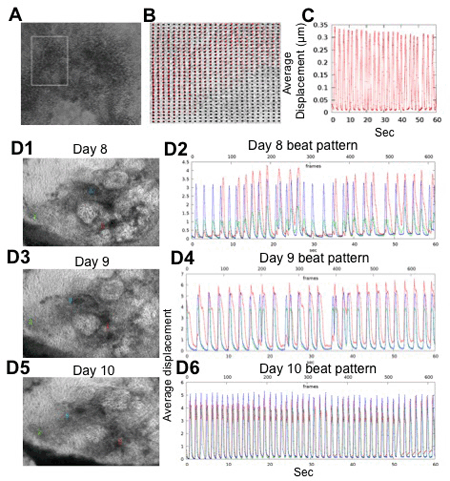
Article Information
vol. 132 no. Suppl 3 A12954
Published By:
American Heart Association, Inc.
Online ISSN:
History:
- Originally published November 6, 2015.
Copyright & Usage:
© 2015 by American Heart Association, Inc.
Author Information
- Johnson Rajasingh1;
- Sheeja Rajasingh1;
- Andras Czirok2;
- Saheli Samanta1;
- Dona G Isai2;
- Edina Kosa2;
- Buddhadeb Dawn1
- 1Medicine, Univ of Kansas Med Cntr, Kansas City, KS
- 2Anatomy and Cell Biology, Univ of Kansas Med Cntr, Kansas City, KS
Abstract 11909: Transcoronary Infusion of Cardiac Progenitor Cells in Hypoplastic Left Heart Syndrome: 3-year Results of the TICAP Trial
Shuta Ishigami, Suguru Tarui, Takuya Goto, Daiki Ousaka, Kenji Baba, Shingo Kasahara, Shinichi Ohtsuki, Shunji Sano, Hidemasa Oh
Circulation. 2015;132:A11909
Abstract
Backgrounds: Hypoplastic left heart syndrome (HLHS) is still one of the severe congenital heart defects. The first results of the TICAP trial (NCT01273857) conducted in our hospital have shown that intracoronary infusion of cardiosphere-derived cells (CDCs) in children with HLHS was feasible and safe; however, the mid-term safety and efficacy of CDCs infusion in these patients remain unsolved. The aim of this study is to evaluate the safety and efficacy of CDCs injection during mid-term period and to determine the factors that may be related to the beneficial effects of cardiac function after cell injection.
Methods and Design: This phase 1 trial is a prospective controlled study. Fourteen patients with HLHS undergoing staged-2 or -3 palliations were enrolled between January 2011, and January 2012. Seven patients assigned to receive intracoronary CDCs infusion 1 month after the cardiac surgery followed by 7 patients allocated to a control with standard care alone. The primary endpoint was to assess the safety and the secondary endpoint was to evaluate the cardiac function and heart failure status.
Results: No complications including tumor formation were reported within 3 years after CDC infusion. Echocardiogram showed a significantly greater improvement in right ventricular ejection fraction (RVEF) in children receiving CDCs than in controls at 3 years (+8.0 ± 4.7% vs. +2.2 ± 4.3%, P=0.03). In cardiac MRI study, improvement of RVEF at 3 years was significantly greater in the CDC-treated group than in controls (+5.7 ± 4.9 vs. -1.0 ± 4.6%, P=0.04). These cardiac function improvements resulted in reduced brain natriuretic peptide levels (P=0.04), lower incidence of unplanned catheter interventions (P=0.04), and higher weight-for-age Z score (WAZ) (P=0.02) at 3 years compared with those of controls. As independent predictors of treatment responsiveness, absolute changes in RVEF at 3 years were negatively correlated with age, WAZ, and RVEF at CDC infusion (age: r=-0.91, P=0.005; WAZ: r=-0.88, P=0.009; EF: r=-0.90, P=0.006).
Conclusions: Intracoronary CDC infusion in patients with HLHS was safe and improved RVEF during 3 years of follow-up. This therapeutic strategy may merit somatic growth enhancement and reduce the incidence of heart failure.
Article Information
vol. 132 no. Suppl 3 A11909
Published By:
American Heart Association, Inc.
Online ISSN:
History:
- Originally published November 6, 2015.
Copyright & Usage:
© 2015 by American Heart Association, Inc.
Author Information
- Shuta Ishigami1;
- Suguru Tarui1;
- Takuya Goto1;
- Daiki Ousaka1;
- Kenji Baba2;
- Shingo Kasahara1;
- Shinichi Ohtsuki2;
- Shunji Sano1;
- Hidemasa Oh3
- 1Dept of Cardiovascular surgery, Okayama Univ Hosp, Okayama, Japan
- 2Dept of Pediatrics, Okayama Univ Hosp, Okayama, Japan
- 3Dept of Regenerative Medicine, Cntr for Innovative Clinical Medicine, Okayama Univ Hosp, Okayama, Japan
Abstract 10894: Cerebral Energy Metabolism and Pools of Neurotransmitters Related to the Use of Deep Hypothermic Circulatory Arrest in Infant Piglets
Masaki Kajimoto, Dolena R Ledee, Aaron K Olson, Nancy G Isern, Christine Des Rosiers, Michael A Portman
Circulation. 2015;132:A10894
Abstract
Introduction: Complex congenital cardiac defect repair in infants often requires deep hypothermic circulatory arrest (DHCA). As immediate or late neurological complications still occur, adjunctive selective cerebral perfusion (SCP) theoretically provides superior neural protection by supplying oxygen and substrate and maintaining energy metabolism.
Hypothesis: SCP preserves glucose utilization, energy metabolism, and glutamate cycling in comparison to DHCA alone.
Methods: Fourteen male Yorkshire piglets (age 34-44 days) were assigned randomly to 2 groups (DHCA or SCP for 60 minutes at 18°C). Cerebral perfusion through 1st branch of aorta was maintained at 40 ml/kg/min during SCP. After the completion of rewarming, 13-Carbon-labeled glucose (13C-glucose) was infused through common carotid artery for 1 hour under continuous cardiopulmonary bypass support and then cerebral tissue was extracted. Gas chromatography-mass spectrometry and nuclear magnetic resonance were used for cerebral metabolic analysis in the frontal cortex of brain.
Results: There were no operative or technical complications in any groups. Cerebral rSO2in simple DHCA dropped substantially to ~ 25% during DHCA, but maintained over 80% in SCP. Apoptosis by TUNEL assay was significantly less in SCP at 2.5 hours after DHCA. ATP levels in the extracted cerebral tissue were similar between 2 groups but glycogen store were higher in SCP. Absolute levels of lactate and citric acid cycle intermediates and their 13C-enrichment were similar in the cerebral tissue between the 2 groups. However, SCP increased absolute and 13C-enrichment for glutamate and compared to DHCA, while gamma aminobutyric acid (GABA) increased in DHCA.
Conclusions: SCP added to DHCA does not provide additional ATP preservation or modify glucose oxidation. However, SCP does alter glucose cycling into alternate pathways, leading to preservation of glycogen, and shifts flux away from GABA, a major inhibitory neurotransmitter, and towards glutamate. These flux changes suggest that SCP decreases synaptic inhibition, which may relate to the reduced neural apoptosis.
Article Information
vol. 132 no. Suppl 3 A10894
Published By:
American Heart Association, Inc.
Online ISSN:
History:
- Originally published November 6, 2015.
Copyright & Usage:
© 2015 by American Heart Association, Inc.
Author Information
- Masaki Kajimoto1;
- Dolena R Ledee1;
- Aaron K Olson2;
- Nancy G Isern3;
- Christine Des Rosiers4;
- Michael A Portman2
- 1Developmental Therapeutics, Seattle Children’s Rsch Institute, Seattle, WA
- 2Pediatrics, Seattle Children’s Rsch Institute, U. Washington, Seattle, WA
- 3Environmental Molecular Sciences Laboratory, Pacific Northwest National Laboratory, Richland, WA
- 4Nutrition, Univ of Montreal and Montreal Heart Institute, Montreal, Canada
Abstract 10987: Minimally Invasive Percutaneous Pericardial ICD Placement in an Infant Piglet Model: Head-to-Head Comparison With an Open Surgical Thoracotomy Approach
Bradley C Clark, Tanya D Davis, Magdy M El-Sayed Ahmed, Nobuyuki Ishibashi, Christopher P Jordan, Timothy D Kane, Peter C Kim, Axel Krieger, Dilip S Nath, Justin D Opfermann, Charles I Berul
Circulation. 2015;132:A10987
Abstract
Introduction: Epicardial ICD placement in infants, small children, and patients with complex cardiac anatomy requires an open thoracotomy and is associated with increased pain, longer length of stay, and higher cost. We compared a surgical epicardial approach with percutaneous pericardial placement of an ICD lead system in an infant piglet model.
Methods: Animals were randomized to undergo either epicardial placement by direct suture fixation through a left mini-thoracotomy or minimally invasive pericardial insertion with thoracoscopic visualization. Pericardial access was achieved through sub-xiphoid insertion of a pericardiocentesis needle followed by 7-French sheath insertion over a guide wire. An ICD lead (Medtronic) was passed through the sheath and fixed to the epicardium. In both groups, the lead was connected to an ICD generator placed within a sub-rectus abdominal pocket. Lead testing and defibrillation threshold testing (DFT) were performed. All piglets underwent a 2 week survival period followed by repeat lead testing and DFT prior to euthanasia.
Results: Minimally invasive pericardial placement was performed in 8 piglets and surgical epicardial placement in 7 piglets (3-4 kg) without procedural morbidity or mortality. Mean initial DFT was 10.5 Joules (range 3-28J) in the pericardial group and 10 Joules (range 5-35J) in the surgical group (p = N.S.). After the survival period, mean DFT was 12 Joules (range 3-20J) in the pericardial group and 12.3 Joules (range 3-35J) in the surgical group (p = N.S.). All lead and shock impedances, R-wave amplitudes, and ventricular pacing thresholds remained stable throughout the survival period and showed no significant difference between the two groups.
Conclusions: Compared to surgically placed epicardial ICD leads, percutaneous pericardial placement shows a nearly identical ability to effectively defibrillate the heart and has demonstrated similar lead stability. This can provide a means of defibrillator implantation without the need for open chest approach and its attendant pain and morbidity. With continued operator experience, the minimally invasive method may provide a viable alternative to epicardial ICD lead placement in infants, children, and adults at risk for sudden cardiac death.
Article Information
vol. 132 no. Suppl 3 A10987
Published By:
American Heart Association, Inc.
Online ISSN:
History:
- Originally published November 6, 2015.
Copyright & Usage:
© 2015 by American Heart Association, Inc.
Author Information
- Bradley C Clark1;
- Tanya D Davis2;
- Magdy M El-Sayed Ahmed3;
- Nobuyuki Ishibashi3;
- Christopher P Jordan4;
- Timothy D Kane5;
- Peter C Kim6;
- Axel Krieger6;
- Dilip S Nath3;
- Justin D Opfermann6;
- Charles I Berul1
- 1Cardiology, Children’s National Health System, Washington, DC
- 2Urology, Children’s National Health System, Washington, DC
- 3Cardiovascular Surgery, Children’s National Health System, Washington, DC
- 4Pediatric Cardiology, Walter Reed National Military Med Cntr, Bethesda, MD
- 5General and Thoracic Surgery, Children’s National Health System, Washington, DC
- 6Sheikh Zayed Institute, Children’s National Health System, Washington, DC
Abstract 19532: Maternal Hyperoxia Increases Cerebral Oxygenation in Fetuses With Complex Congenital Heart Disease: A Functional MRI Study
Wonsang You, Mary Donofrio, David Wessel, Zungho Zun, Josepheen De Asis-Cruz, Gilbert Vezina, Dorothy Bulas, Richard Jonas, Adre du Plessis, Catherine Limperopoulos
Circulation. 2015;132:A19532
Abstract
Introduction: Fetal brain growth is altered in congenital heart disease (CHD). The role of disturbed placental and fetal blood flow/oxygenation remains poorly understood. We compared the effect of maternal hyperoxia on cerebral and placental oxygenation in CHD vs control fetuses using blood oxygenation level dependent (BOLD) functional MRI (fMRI).
Methods: We studied 85 pregnant women (51 healthy /34 CHD fetuses) at 22-39 weeks gestational age (GA), using MRI and Doppler. After 2min baseline recording at normoxia (NO), we made BOLD fMRI measurements in 4 phases, (1) early hyperoxia (HO; 100% O2) for 2min (2) late HO (2min), (3) early return NO (1.75min), (4) late return NO (1.75min). We measured % BOLD signal changes in fetal brain and placenta and correlated these with umbilical artery (UA) pulsatility index (PI) obtained at baseline.
Results: Mean GA of CHD vs control fetuses was 32 4/7 and 32 2/7 (p=0.77). 41.2% had 1-ventricle (1V; all HLHS) vs 2-ventricle (2V) physiology. In controls, brain BOLD signal increased in phase 1 HO and remained elevated through phase 4. In CHD fetuses, the BOLD signal increased in phase 1 and continued to rise through phase 4. Differences in brain BOLD elevation in CHD vs controls (3.1% vs 0.9%, p=0.04) was significant in phase 4 (Fig. 1). Brain BOLD differed between 1V (3.4%, p=0.04) and 2V (0.9%, p=0.15) in phases 3 & 4. Placental BOLD signal increased during HO in control (4.4%, p<0.001) and CHD fetuses (6.4%, p<0.001). UA PI in CHD fetuses was higher than controls (1.11 vs 1.03, p=0.03). UA PI correlated with placental BOLD during HO in healthy and CHD fetuses (p<0.05).
Conclusions: In this first report of placental and fetal brain responses to maternal HO, we show a significantly increased brain BOLD response in CHD fetuses, specifically 1V. These data suggest that maternal HO improves brain oxygenation in CHD fetuses in the short-term. Studies evaluating the prolonged effects of HO are needed to determine the role of HO management in select CHD fetuses.
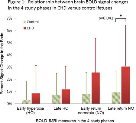
Article Information
vol. 132 no. Suppl 3 A19532
Published By:
American Heart Association, Inc.
Online ISSN:
History:
- Originally published November 6, 2015.
Copyright & Usage:
© 2015 by American Heart Association, Inc.
Author Information
- Wonsang You1;
- Mary Donofrio2;
- David Wessel3;
- Zungho Zun1;
- Josepheen De Asis-Cruz1;
- Gilbert Vezina1;
- Dorothy Bulas1;
- Richard Jonas4;
- Adre du Plessis5;
- Catherine Limperopoulos1
- 1Dept of Diagnostic Imaging & Radiology, Children’s National Med Cntr, Washington, DC
- 2Dept of Cardiology, Children’s National Med Cntr, Washington, DC
- 3Dept of Critical Care Medicine, Children’s National Med Cntr, Washington, DC
- 4Dept of Cardiovascular Surgery, Children’s National Med Cntr, Washington, DC
- 5Dept of Fetal & Transitional Medicine, Children’s National Med Cntr, Washington, DC
Abstract 17826: Long-term Survivors With Transposition of the Great Arteries After Arterial Switch Operation Show No Signs of Adverse Myocardial Remodeling
Heynric B Grotenhuis, Luc Mertens, Barbara Cifra, Eugenie Riessenkampff, Cedric Manlhiot, Lars Grosse-Wortmann
Circulation. 2015;132:A17826
Abstract
Background: The arterial switch operation (ASO) for transposition of the great arteries (TGA) involves coronary translocation. The altered coronary geometry may lead to subclinical myocardial ischemia, which may result in myocardial scarring and diffuse myocardial fibrosis. We hypothesized that myocardial fibrosis is present late after ASO, despite excellent functional outcomes of the ASO in most patients.
Methods: 30 pediatric TGA patients after ASO were prospectively studied by cardiac magnetic resonance imaging (CMR). Late gadolinium enhancement (LGE) was used to detect discrete myocardial scarring. Native T1 relaxation times and extracellular volumes (ECVs) at the mid-ventricular short-axis level of the left ventricle (LV) were used to quantify diffuse myocardial fibrosis. The midventricular short-axis slice was divided into 6 segments. Patients were compared to 21 healthy controls.
Results: Mean ages at ASO and at CMR were 6.3±5.2 days and 15.4±2.9 years, respectively. Mean age of controls at CMR was 14.1±2.6 years. TGA patients showed preserved LV ejection fraction (57±5% vs. 59±4% in controls, p=0.08) and similar indexed LV mass (54±10g/m2 vs. 49±9%, p=0.12). LV end-diastolic and end-systolic volumes were mildly increased (104±20ml/m2 vs. 89±11ml/m2, p=0.01 and 46±13ml/m2 vs. 36±6ml/m2, p<0.01, respectively). None of the TGA patients demonstrated evidence of localized myocardial scarring by LGE. Native T1 times and ECV values for all 6 LV segments were not significantly different to controls. There was no association between global T1 times or global ECV and LV volumes and ejection fraction. Furthermore, native T1 and ECV were not associated with bypass and cross-clamp times at the time of the ASO, or with coronary artery patterns.
Conclusions: Adolescent TGA patients late after ASO have preserved systolic LV function. There is no evidence of myocardial scarring or fibrosis. Our results suggest excellent long-term myocardial and cardiac health after the ASO, paralleling the encouraging clinical outcomes with this procedure.
Article Information
vol. 132 no. Suppl 3 A17826
Published By:
American Heart Association, Inc.
Online ISSN:
History:
- Originally published November 6, 2015.
Copyright & Usage:
© 2015 by American Heart Association, Inc.
Author Information
- Heynric B Grotenhuis;
- Luc Mertens;
- Barbara Cifra;
- Eugenie Riessenkampff;
- Cedric Manlhiot;
- Lars Grosse-Wortmann
- Pediatric Cardiology, The Hosp for Sick Children, Toronto, Canada
Abstract 16408: Acute Changes in Tricuspid Annular Plane Systolic Excursion and Myocardial Deformation in Fetuses With Twin-twin Transfusion Syndrome After Selective Fetoscopic Laser Therapy
Betul Yilmaz, Ryan A Moore, Christopher J Statile, Regina Keller, Foong-Yen Lim, Erik C Michelfelder
Circulation. 2015;132:A16408
Abstract
Introduction: Twin-twin transfusion syndrome (TTTS) is a serious complication affecting 10-15% of monochorionic twin gestations. Right ventricle (RV) function is often abnormal in these fetuses. Tricuspid annular plane systolic excursion (TAPSE) and myocardial strain are used as a marker of RV function in a number of disease processes postnatally.
Hypothesis: Monitoring TAPSE and myocardial deformation may aid in assessing response to selective fetoscopic laser therapy (SFLP) in TTTS. Our objective was to determine whether TAPSE and myocardial deformation are altered after SFLP.
Methods: Fetal echocardiograms of 22 recipient (RT) and donor (DT) twin pairs with TTTS were prospectively evaluated. TAPSE was obtained via M-mode through the lateral tricuspid annulus in the vertical 4-chamber (4C) view. 2D 4C views were acquired utilizing high frame rates (>30 frames/sec) and were interrogated offline using feature-tracking software for strain analysis. Global RV longitudinal strain, TAPSE, and myocardial performance index (MPI) were compared pre and post-SFLP. Reproducibility of both TAPSE and RV strain was assessed.
Results: Initial fetal echo was performed at 20.8 ± 2.3 weeks gestation. Median time between initial study and post-SFLP was 7 days (interquartile range 1 to 11 days). DT showed a slight improvement in strain, but not in TAPSE post-SFLP (Table). There was a significant, acute improvement both in global longitudinal strain and TAPSE among RT after SFLP (Table). Interobserver variability for TAPSE and strain was 4% vs 36%, respectively.
Conclusion: After SFLP, there is an acute improvement in the global RV strain and TAPSE in RT, consistent with improved RV long-axis function or loading conditions. TAPSE is feasible in most fetuses, and is considerably more reproducible than myocardial strain. Further work examining the relationship between RV function and outcome post-SFLP is warranted in TTTS population.
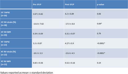
Article Information
vol. 132 no. Suppl 3 A16408
Published By:
American Heart Association, Inc.
Online ISSN:
History:
- Originally published November 6, 2015.
Copyright & Usage:
© 2015 by American Heart Association, Inc.
Author Information
- Betul Yilmaz1;
- Ryan A Moore1;
- Christopher J Statile1;
- Regina Keller1;
- Foong-Yen Lim2;
- Erik C Michelfelder1
- 1Pediatrics, Pediatric Cardiology, Cincinnati Children’s Hosp Med Cntr, Cincinnati, OH
- 2Pediatric Surgery, Cincinnati Children’s Hosp Med Cntr, Cincinnati, OH
Abstract 15413: Comparison of Right Ventricular Functional Indices Using Two-dimensional and Three-dimensional Echocardiography to Predict Outcomes in Pediatric Pulmonary Hypertension
Pei-Ni Jone, SuHong Tong, D. Dunbar Ivy
Circulation. 2015;132:A15413
Abstract
Background: Right ventricular (RV) function is an important determinant of outcomes in pulmonary hypertension (PH) patients. Conventional indices of fractional area change (FAC), tricuspid annular plane systolic excursion (TAPSE), and RV tissue Doppler imaging myocardial performance index (RV TDI MPI) have been used as surrogates of RV function. RV ejection fraction (EF) from real time three-dimensional echocardiography (RT-3DE) has emerged as a quantitative evaluation of global RV function and has correlated well with cardiac magnetic resonance imaging. In this study, 3D RV EF was compared with conventional indices in the serial evaluation of RV function in pediatric PH patients to predict adverse events.
Methods: Forty-eight pediatric PH patients (median age = 10 years (4 months – 27 years)) were evaluated serially (138 visits with median interval visit = 116 days (4 -368 days)) with RT-3DE to follow their ejection fraction (EF) and conventional indices from April, 2014 to May, 2015. Echocardiographic variables include measures of RV function: 3D RV EF, FAC, TAPSE, and RV TDI MPI. Adverse events included: initiation or intensification of intravenous vasodilator therapy, atrial septostomy, Pott’s shunt, or death. Receiver Operating Characteristics (ROC) analyses were performed to identify the best cut-offs in predicting adverse events in serial follow up of pediatric PH patients.
Results: Patients were classified based on their World Health Classification (I = 16, II=16, III=11, IV=3). Two patients were not classified as they were too young. There were 13 adverse events. 3D RV EF was a good predictor of adverse events with highest area under curve (AUC) = 0.79, p<0.001(cut-off value of 38% = sensitivity 69%; specificity of 78%) compared to FAC has an AUC = 0.77, p<0.05 (cut-off value of 33% = sensitivity 63%; specificity of 78%). TAPSE and TV TDI MPI were not statistically significant (AUC = 0.54, p = 0.65; AUC 0.63, p = 0.09 respectively).
Conclusion: 3D RV EF is a good index in predicting adverse events and was better than FAC, TAPSE, and RV TDI MPI in predicting adverse events in serial follow up of pediatric PH patients. 3D RV EF can be used as a noninvasive tool in the serial evaluation of RV function in pediatric PH patients as it is easily obtained clinically.
Article Information
vol. 132 no. Suppl 3 A15413
Published By:
American Heart Association, Inc.
Online ISSN:
History:
- Originally published November 6, 2015.
Copyright & Usage:
© 2015 by American Heart Association, Inc.
Author Information
- 1Pediatric Cardiology, Univ of Colorado Children’s Hosp Colorado, Aurora, CO
- 2Dept of Biostatistics, Univ of Colorado Sch of Public Health, Aurora, CO
- 3Pediatric Cardiology, Children’s Hosp Colorado, Univ of Colorado Sch of Medicine, Aurora, CO
Abstract 16408: Acute Changes in Tricuspid Annular Plane Systolic Excursion and Myocardial Deformation in Fetuses With Twin-twin Transfusion Syndrome After Selective Fetoscopic Laser Therapy
Betul Yilmaz, Ryan A Moore, Christopher J Statile, Regina Keller, Foong-Yen Lim, Erik C Michelfelder
Circulation. 2015;132:A16408
Abstract
Introduction: Twin-twin transfusion syndrome (TTTS) is a serious complication affecting 10-15% of monochorionic twin gestations. Right ventricle (RV) function is often abnormal in these fetuses. Tricuspid annular plane systolic excursion (TAPSE) and myocardial strain are used as a marker of RV function in a number of disease processes postnatally.
Hypothesis: Monitoring TAPSE and myocardial deformation may aid in assessing response to selective fetoscopic laser therapy (SFLP) in TTTS. Our objective was to determine whether TAPSE and myocardial deformation are altered after SFLP.
Methods: Fetal echocardiograms of 22 recipient (RT) and donor (DT) twin pairs with TTTS were prospectively evaluated. TAPSE was obtained via M-mode through the lateral tricuspid annulus in the vertical 4-chamber (4C) view. 2D 4C views were acquired utilizing high frame rates (>30 frames/sec) and were interrogated offline using feature-tracking software for strain analysis. Global RV longitudinal strain, TAPSE, and myocardial performance index (MPI) were compared pre and post-SFLP. Reproducibility of both TAPSE and RV strain was assessed.
Results: Initial fetal echo was performed at 20.8 ± 2.3 weeks gestation. Median time between initial study and post-SFLP was 7 days (interquartile range 1 to 11 days). DT showed a slight improvement in strain, but not in TAPSE post-SFLP (Table). There was a significant, acute improvement both in global longitudinal strain and TAPSE among RT after SFLP (Table). Interobserver variability for TAPSE and strain was 4% vs 36%, respectively.
Conclusion: After SFLP, there is an acute improvement in the global RV strain and TAPSE in RT, consistent with improved RV long-axis function or loading conditions. TAPSE is feasible in most fetuses, and is considerably more reproducible than myocardial strain. Further work examining the relationship between RV function and outcome post-SFLP is warranted in TTTS population.
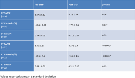
Article Information
vol. 132 no. Suppl 3 A16408
Published By:
American Heart Association, Inc.
Online ISSN:
History:
- Originally published November 6, 2015.
Copyright & Usage:
© 2015 by American Heart Association, Inc.
Author Information
- Betul Yilmaz1;
- Ryan A Moore1;
- Christopher J Statile1;
- Regina Keller1;
- Foong-Yen Lim2;
- Erik C Michelfelder1
- 1Pediatrics, Pediatric Cardiology, Cincinnati Children’s Hosp Med Cntr, Cincinnati, OH
- 2Pediatric Surgery, Cincinnati Children’s Hosp Med Cntr, Cincinnati, OH
Abstract 15413: Comparison of Right Ventricular Functional Indices Using Two-dimensional and Three-dimensional Echocardiography to Predict Outcomes in Pediatric Pulmonary Hypertension
Pei-Ni Jone, SuHong Tong, D. Dunbar Ivy
Circulation. 2015;132:A15413
Abstract
Background: Right ventricular (RV) function is an important determinant of outcomes in pulmonary hypertension (PH) patients. Conventional indices of fractional area change (FAC), tricuspid annular plane systolic excursion (TAPSE), and RV tissue Doppler imaging myocardial performance index (RV TDI MPI) have been used as surrogates of RV function. RV ejection fraction (EF) from real time three-dimensional echocardiography (RT-3DE) has emerged as a quantitative evaluation of global RV function and has correlated well with cardiac magnetic resonance imaging. In this study, 3D RV EF was compared with conventional indices in the serial evaluation of RV function in pediatric PH patients to predict adverse events.
Methods: Forty-eight pediatric PH patients (median age = 10 years (4 months – 27 years)) were evaluated serially (138 visits with median interval visit = 116 days (4 -368 days)) with RT-3DE to follow their ejection fraction (EF) and conventional indices from April, 2014 to May, 2015. Echocardiographic variables include measures of RV function: 3D RV EF, FAC, TAPSE, and RV TDI MPI. Adverse events included: initiation or intensification of intravenous vasodilator therapy, atrial septostomy, Pott’s shunt, or death. Receiver Operating Characteristics (ROC) analyses were performed to identify the best cut-offs in predicting adverse events in serial follow up of pediatric PH patients.
Results: Patients were classified based on their World Health Classification (I = 16, II=16, III=11, IV=3). Two patients were not classified as they were too young. There were 13 adverse events. 3D RV EF was a good predictor of adverse events with highest area under curve (AUC) = 0.79, p<0.001(cut-off value of 38% = sensitivity 69%; specificity of 78%) compared to FAC has an AUC = 0.77, p<0.05 (cut-off value of 33% = sensitivity 63%; specificity of 78%). TAPSE and TV TDI MPI were not statistically significant (AUC = 0.54, p = 0.65; AUC 0.63, p = 0.09 respectively).
Conclusion: 3D RV EF is a good index in predicting adverse events and was better than FAC, TAPSE, and RV TDI MPI in predicting adverse events in serial follow up of pediatric PH patients. 3D RV EF can be used as a noninvasive tool in the serial evaluation of RV function in pediatric PH patients as it is easily obtained clinically.
Article Information
vol. 132 no. Suppl 3 A15413
Published By:
American Heart Association, Inc.
Online ISSN:
History:
- Originally published November 6, 2015.
Copyright & Usage:
© 2015 by American Heart Association, Inc.
Author Information
- 1Pediatric Cardiology, Univ of Colorado Children’s Hosp Colorado, Aurora, CO
- 2Dept of Biostatistics, Univ of Colorado Sch of Public Health, Aurora, CO
- 3Pediatric Cardiology, Children’s Hosp Colorado, Univ of Colorado Sch of Medicine, Aurora, CO
Abstract 14711: Effect of Release of the First Pediatric Appropriate Use Criteria on Outpatient Transthoracic Echocardiogram Ordering Practice
Ritu Sachdeva, Michael S Kelleman, Courtney McCracken, Joseph Allen, Oscar Benavidez, Robert M Campbell, Pamela S Douglas, Benjamin W Eidem, Lara Gold, Leo Lopez, Kenan W Stern, Rory B Weiner, Elizabeth Welch, Wyman W Lai
Circulation. 2015;132:A14711
Abstract
Background: The first pediatric appropriate use criteria (PAUC) were recently published for initial outpatient transthoracic echocardiography (TTE). We sought to determine the effect of release of the PAUC on the appropriateness of TTE ordering patterns of pediatric cardiologists.
Methods: Data were prospectively collected from patients having initial outpatient TTE ordered prior to (Phase I, 6 mo) and 3 months after release (Phase II, 4 mo) of PAUC. Site-investigators determined the indication of the study and assigned appropriateness rating based on the PAUC document [Appropriate (A), May Be Appropriate (M), or Rarely Appropriate (R)] or “Unclassifiable” (U).
Results: A total of 4562 TTEs (2655 Phase I, 1907 Phase II) were ordered by 103 physicians at 6 sites. Overall comparison of appropriateness rating for Phase I and II showed no change in the rate for A or M, but a decline in the rate of R and increase in U. Similar results were noted when comparisons were made for physicians that ordered at least 20 TTEs during each phase (N = 30/103, 29%), Table 1. There was no change in any appropriateness rating at 3 sites, a decline in R at 2, an increase in A at 1, and an increase in U at 2 sites. Overall and site-specific comparisons of rate of R TTEs are shown in Fig 1.
Conclusions: The release of PAUC document had only a small impact on physician ordering behavior for initial TTEs, including a small decrease in R. Overall, these data suggest that more focused interventions are required to fully implement guideline recommendations and improve the appropriate use of pediatric TTE. This information should be helpful in designing educational interventions to reduce the rate of TTEs ordered for R indications.
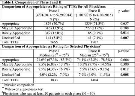
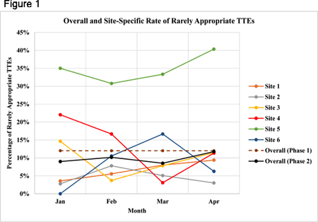
Article Information
vol. 132 no. Suppl 3 A14711
Published By:
American Heart Association, Inc.
Online ISSN:
History:
- Originally published November 6, 2015.
Copyright & Usage:
© 2015 by American Heart Association, Inc.
Author Information
- Ritu Sachdeva1;
- Michael S Kelleman2;
- Courtney McCracken2;
- Joseph Allen3;
- Oscar Benavidez4;
- Robert M Campbell1;
- Pamela S Douglas5;
- Benjamin W Eidem6;
- Lara Gold3;
- Leo Lopez7;
- Kenan W Stern8;
- Rory B Weiner9;
- Elizabeth Welch7;
- Wyman W Lai10
- 1Pediatrics, Emory Univ and Children’s Healthcare of Atlanta, Sibley Heart Cntr Cardiology, Atlanta, GA
- 2Pediatrics, Emory Univ Sch of Medicine, Atlanta, GA
- 3Appropriate Use Criteria Task Force, American College of Cardiology, Washington DC, DC
- 4Pediatrics, Massachusetts General Hosp, Boston, MA
- 5Cardiology, Duke Univ, Durham, NC
- 6Pediatrics, Mayo Clinic, Rochester, MN
- 7Pediatrics, Miami Children’s Hosp, Miami, FL
- 8Pediatrics, Montefiore Med Cntr, Bronx, NY
- 9Cardiology, Massachusetts General Hosp, Boston, MA
- 10Pediatrics, NewYork-Presbyterian, Morgan Stanley Children’s Hosp, New York, NY
Abstract 12052: Myocardial Extracellular Matrix Expansion Detected With Cardiac MRI: A Biomarker of Cardiomyopathic Disease in Duchenne Muscular Dystrophy
Jonathan H Soslow, Stephen M Damon, Kimberly Crum, Mark Lawson, James C Slaughter, Meng Xu, Andrew E Arai, Douglas B Sawyer, David A Parra, Bruce M Damon, Larry W Markham
Circulation. 2015;132:A12052
Abstract
Introduction: Duchenne muscular dystrophy (DMD) cardiomyopathy is a progressive disease for which standard heart failure therapies have limited efficacy. Disease-specific therapies are needed that can be initiated before irreversible myocardial damage ensues. In order to evaluate therapeutic efficacy, surrogate endpoints other than ejection fraction must be found.
Hypothesis: The study hypothesis is that T1 and extracellular volume fraction (ECV) mapping using cardiac magnetic resonance imaging (CMR) can detect diffuse extracellular matrix expansion in DMD patients with normal left ventricular ejection fraction (LVEF) and without late gadolinium enhancement (LGE).
Methods: 28 DMD and 11 healthy control participants were prospectively enrolled. CMR using a modified Look-Locker (MOLLI) sequence was performed before and after contrast administration. T1 and ECV maps of the mid left ventricular myocardium were generated and regions of interest were contoured using the standard 6-segment AHA model. Global and segmental values were compared between DMD and controls using a Wilcoxon rank-sum test.
Results: DMD participants had significantly higher mean native T1 compared with controls (1045ms vs 988ms, p<0.001) (Fig 1A). DMD participants with normal LVEF and without evidence of LGE also demonstrated elevated mean native T1 (1036ms vs 988ms, p<0.001, and 1039ms vs 988ms, p=0.001) (Fig 1C, 1E). Mean post-contrast T1 values were not significantly different between DMD participants and controls. DMD participants had a significantly greater mean ECV than controls (0.31 vs 0.24, p<0.001), even in the settings of normal LVEF (0.29 vs 0.24, p<0.001) and negative LGE (0.30 vs 0.24, p<0.001) (Fig 1B, 1D, 1F).
Conclusions: DMD participants have elevated native T1 and ECV in left ventricular myocardium, even in the setting of normal LVEF and in the absence of LGE. T1 and ECV mapping in DMD have potential to serve as surrogate cardiomyopathy outcome measures for clinical trials.
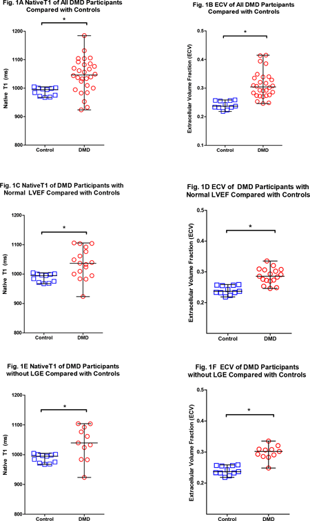
Article Information
vol. 132 no. Suppl 3 A12052
Published By:
American Heart Association, Inc.
Online ISSN:
History:
- Originally published November 6, 2015.
Copyright & Usage:
© 2015 by American Heart Association, Inc.
Author Information
- Jonathan H Soslow1;
- Stephen M Damon2;
- Kimberly Crum1;
- Mark Lawson3;
- James C Slaughter4;
- Meng Xu4;
- Andrew E Arai5;
- Douglas B Sawyer6;
- David A Parra1;
- Bruce M Damon7;
- Larry W Markham1
- 1Pediatrics, Div of Pediatric Cardiology, Vanderbilt Univ Med Cntr, Nashville, TN
- 2Dept of Electrical Engineering and Computer Sciences, Vanderbilt Univ, Nashville, TN
- 3Medicine, Div of Cardiology, Vanderbilt Univ Med Cntr, Nashville, TN
- 4Biostatistics, Vanderbilt Univ Med Cntr, Nashville, TN
- 5National Heart, Lung and Blood Institute (NHLBI), National Institutes of Health (NIH), National Institutes of Health, Bethesda, MD
- 6Dept of Medicine, Div of Cardiovascular Services, Maine Med Cntr, Portland, ME
- 7Depts of Radiology and Radiological Sciences, Molecular Physiology and Biophysics, and Biomedi, Vanderbilt Univ, Nashville, TN
Abstract 9918: Respiration Increases Ventricular Filling at Rest and Exercise via Pulmonary Compliance: A Clinical and Computational Modeling Study
Ethan Kung, Alexander Van De Bruaene, Guido Claessen, Andre La Gerche, Alison Marsden, Pieter De Meester, Sarah Devroe, Jan Bogaert, Piet Claus, Hein Heidbuchel, Werner Budts, Marc Gewillig
Circulation. 2015;132:A9918
Abstract
Introduction: Due to the absence of a sub-pulmonary ventricle, the Fontan circulation is sensitive to respiration-induced changes in intrathoracic pressure. However, the importance of a ‘respiratory pump’ in creating forward flow remains controversial. We aim to investigate the effects and mechanisms of respiration on ventricular filling at rest and exercise using clinical data and computational modeling.
Hypothesis: We assess the hypotheses that (1) changes in intrathoracic pressure due to respiration would aid ventricular filling and output and (2) this effect would be maintained or enhanced during incremental exercise.
Methods: Ten Fontan patients (6 male, 20±4 years) underwent ungated cardiac magnetic resonance imaging at rest and during supine bicycle exercise (3 incremental intensities) to evaluate systemic ventricular volumes. Patient-specific computational simulations using a lumped-parameter network model of Fontan exercise elucidated resting and exercise physiology in details for each patient at each metabolic state tested.
Results: Compared to expiration, inspiration increased EDVi (98±16 to 103±15 mL/m2;P=0.001), SVi (55±9 to 59±9 mL/m2;P=0.001) and cardiac index (3.9±0.7 to 4.2±0.8 L/min/m2;P=0.002), but did not affect ESVi (P=0.096). Respiratory-dependent SVi did not change significantly during incremental exercise (3±2% to 5±3%; P=0.084). Computational modeling showed highest caval vein flow return during end-inspiration, and peak SV during expiration, exposing a phased time-delay mechanism at work. Removal of respiration in simulations corresponded to decreases in cardiac index of 0.39±0.037 L/min/m2.
Conclusions: Inspiration increased ventricular filling at rest and exercise by similar amounts. A phased time-delay between highest caval vein flow and highest SV suggests an indirect mechanism where respiration aids ventricular filling via pulmonary compliance.
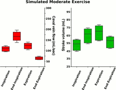
Article Information
vol. 132 no. Suppl 3 A9918
Published By:
American Heart Association, Inc.
Online ISSN:
History:
- Originally published November 6, 2015.
Copyright & Usage:
© 2015 by American Heart Association, Inc.
Author Information
- Ethan Kung1;
- Alexander Van De Bruaene2;
- Guido Claessen3;
- Andre La Gerche4;
- Alison Marsden5;
- Pieter De Meester2;
- Sarah Devroe6;
- Jan Bogaert7;
- Piet Claus8;
- Hein Heidbuchel9;
- Werner Budts2;
- Marc Gewillig10
- 1Mechanical Engineering, Clemson Univ, Clemson, SC
- 2Dept of Adult Congenital Cardiology, Univ Hosps Leuven, Leuven, Belgium
- 3Dept of Cardiovascular medicine, Univ Hosps Leuven, Leuven, Belgium
- 4St Vincent’s Hosp, Dept of Medicine, Univ of Melbourne, Fitzroy, Australia
- 5Mechanical and Aerospace Engineering, Univ of California San Diego, La Jolla, CA
- 6Dept of Anesthesiology, Univ Hosps Leuven, Leuven, Belgium
- 7Dept of Radiology, Univ Hosps Leuven, Leuven, Belgium
- 8Med Imaging Rsch Cntr, Univ Hosps Leuven, Leuven, Belgium
- 9Heart Cntr Hasselt, Heart Cntr Hasselt, Hasselt, Belgium
- 10Pediatric Cardiology, Univ Hosps Leuven, Leuven, Belgium
Abstract 15592: Role of Electroanatomic Voltage Mapping for Risk Stratification in Patients With Repaired Congenital Heart Disease Involving the Right Ventricle
Vincenzo Pazzano, Rosalinda Palmieri, Corrado Di Mambro, Mario S Russo, Massimo S Silvetti, Fabrizio Gimigliano, Salvatore Giannico, Benedetta Leonardi, Fabrizio Drago
Circulation. 2015;132:A15592
Abstract
Introduction: Among adult patients with previous surgical correction for Tetralogy of Fallot or other repaired Congenital Heart Disease (rCHD) involving the RV, ventricular arrhythmias (VA) and sudden cardiac death (SCD) represent a late complication. The imparied haemodynamics that lead to RV dilatation and overload could also alterate its electroanatomic structure.
Hypothesis: 3D electroanatomic mapping (EAM) of the RV could confirm the presence of myocardial electrical abnormalities, allowing to better identify patients at risk for life-threatening arrhythmias.
Methods: 146 patients (age 19.2 ±7.0) with rCHD involving the RV were selected from a population undergoing routine post-surgical follow-up, according to the presence of VA or severe RV dilatation. These patients underwent 3D EAM of the RV. We tested the correlation between size of scar tissue (areas with voltage < 0.5 mV) and several parameters universally accepted by the literature as risk factors for VA in this particular patient population.
Results: In 125 (85.6%) patients, EAM demonstrated areas of low voltage in the antero-lateral RVOT. In 20 of these (16%, 13.7% of the total) the scar extended to the septum. 72 (49.3%) had a peritricuspid scar, and in 20 (13.7%) other areas of the RV were interested. Total low-voltage area, expressed as % of total endocardial area, was significantly higher in patients with history of PVCs [3.2% (±2.6) vs 2.2% (±1.8), p<0.05], complex PVCs at 24h-Holter ECG (Lown class ≥2) [3.4 (±2.5) vs 2.6 (±2.3), p<0.05], exercise-inducible PVCs [3.8 (±2.4) vs 2.6 (±2.2), p=0.01] and history of previous shunt [4.0 (±2.7) vs 2.6 (±2.2), p=0.01]. Scar size was also positively correlated with age (p=0.01), age at correction (p=0.01) and QRS duration on surface ECG (p<0.05).
Conclusions: In patients with rCHD involving the RV it is common to observe endocardial low-voltage areas with variable distribution, not always corresponding to the sites of surgical lesion. Morover, the size of the scar tissue area correlates with some of the parameters which have been already identified as risk factors for life-threatening arrhythmias and SCD in adult patients with CHD. We suggest that EAM should become part of the routine tests for the stratification of arrhythmic risk in this population.
Article Information
vol. 132 no. Suppl 3 A15592
Published By:
American Heart Association, Inc.
Online ISSN:
History:
- Originally published November 6, 2015.
Copyright & Usage:
© 2015 by American Heart Association, Inc.
Author Information
- Vincenzo Pazzano1;
- Rosalinda Palmieri1;
- Corrado Di Mambro1;
- Mario S Russo1;
- Massimo S Silvetti1;
- Fabrizio Gimigliano1;
- Salvatore Giannico2;
- Benedetta Leonardi2;
- Fabrizio Drago1
- 1Dept of Pediatric Cardiology, Bambino Gesù Children’s Hosp, Fiumicino (Roma), Italy
- 2Dept of Pediatric Cardiology, Bambino Gesù Children’s Hosp, Roma, Italy
Abstract 14900: Impact of ICD Programming Changes to Avoid Inappropriate Shocks in the Young
Frank J Zimmerman, Aishwarya Bhan, Anne Freter, Ira Shetty, Lisa Russo, Janis Dennis, Frank T Zimmerman, Adarsh Bhan
Circulation. 2015;132:A14900
Abstract
Introduction: ICD therapy in young pts has been shown to improve mortality but inappropriate ICD shocks (IAS) occur in up to 25% of young pts. Adult studies have shown that changing the VF detection rate (RATE) and/or duration to detection (DUR) results in fewer IAS without increasing mortality. The purpose of this study is to assess the impact of RATE and/or DUR changes to avoid IAS in young pts with ICDs.
Methods: This two-part study initially consisted of a retrospective analysis of IAS in young pts with ICDs at our institution from 2007-2012. A total of 1200 intervals from EGMs recorded from 16 pts with IAS were analyzed and the risk for VF detection was calculated at various RATE and DUR. Analysis of true episodes of VT/VF in 8 pts was also performed. We found that changing the RATE from 200 bpm to 250 bpm resulted in a 49-85% reduction of risk for IAS. Based on this analysis, the RATE was increased to 220-250 bpm in our ICD pts from 2012-2015. The clinical rate of IAS was then compared before and after changing ICD programming to higher RATE.
Results: 64 pts (ages 8y to 57y, mean 21y, 41 male) were included in this study. In the time period from 2007-2012, IAS occurred in 30% of pts (50% due to lead malfunction and 50% due to SVT). ICD programming was changed to VF RATE of 220-250 bpm with DUR of 18/24 in all ICD pts in 2012 and thereafter. There was a significant reduction of IAS with ICD program changes (30% vs 1.5%, p=0.001) with no change in appropriate shock rate (APS) or lead malfunction rate between the two time periods (see table). There were no episodes of syncope or death in those with APS before or after programming changes.
Conclusions: The incidence of IAS in young pts can be reduced by changes in ICD programming. Increasing the RATE has a greater impact on reduction of risk for IAS compared to prolonging DUR and did not result in an increase in syncope or death. These findings may help to standardize programming of ICDs in young pts.

Article Information
vol. 132 no. Suppl 3 A14900
Published By:
American Heart Association, Inc.
Online ISSN:
History:
- Originally published November 6, 2015.
Copyright & Usage:
© 2015 by American Heart Association, Inc.
Author Information
- Frank J Zimmerman;
- Aishwarya Bhan;
- Anne Freter;
- Ira Shetty;
- Lisa Russo;
- Janis Dennis;
- Frank T Zimmerman;
- Adarsh Bhan
- Pediatric Cardiology, Advocate Children’s Heart Institute, Oak Lawn, IL
Abstract 13473: Refinement of a Risk-Standardization Model for Adverse Outcomes Following Congenital Cardiac Catheterization: Insights From from the NCDR® IMPACTTMRegistry
Natalie Jayaram, John A Spertus, Kevin F Kennedy, Robert N Vincent, Gerard R Martin, Jeptha P Curtis, David Nykanen, Phillip Moore, Lisa Bergersen
Circulation. 2015;132:A13473
Abstract
Background: Risk-standardization for adverse events following congenital cardiac catheterization is needed to equitably compare patient outcomes among different hospitals as a foundation for quality improvement. We sought to refine and update the risk-standardization methodology for the NCDR® IMPACTTM (Improving Pediatric and Adult Congenital Treatment) Registry.
Methods: We identified 39,725 consecutive patients within IMPACT undergoing cardiac catheterization between January 2011 and March 2014. New procedure type risk categories were derived by empiric data and expert opinion and markers of hemodynamic vulnerability were empirically explored and identified. A multivariable hierarchical logistic regression model to identify patient and procedural characteristics predictive of a major adverse event (MAE) or death following cardiac catheterization was derived in 70% of the cohort and validated in the remaining 30%.
Results: The rate of MAE or death was 7.1% and 7.2% in the derivation and validation cohorts, respectively. Six independent indicators of hemodynamic vulnerability and six procedure-type risk groups were identified. The final risk adjustment model included procedure type risk group, hemodynamic vulnerability, renal insufficiency, single-ventricle physiology, and coagulation disorder (See Figure). The model had good discrimination with a C-statistic of 0.76 and 0.75 in the derivation and validation cohorts, respectively. Model calibration was excellent with a slope of 0.97 (standard error [SE] 0.04; p-value [for difference from 1]= 0.53) and an intercept of 0.007 (SE 0.12; p-value [for difference from 0]= 0.95).
Conclusion: We updated and refined a risk-standardization model for outcomes following congenital cardiac catheterization using the large, multicenter IMPACT Registry. Our model can be used for quality reporting back to sites participating in IMPACT to guide quality improvement initiatives.
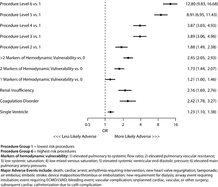
Article Information
vol. 132 no. Suppl 3 A13473
Published By:
American Heart Association, Inc.
Online ISSN:
History:
- Originally published November 6, 2015.
Copyright & Usage:
© 2015 by American Heart Association, Inc.
Author Information
- Natalie Jayaram1;
- John A Spertus2;
- Kevin F Kennedy2;
- Robert N Vincent3;
- Gerard R Martin4;
- Jeptha P Curtis5;
- David Nykanen6;
- Phillip Moore7;
- Lisa Bergersen8
- 1Pediatric Cardiology, Children’s Mercy Hosps and Clinics, Kansas City, MO
- 2Cardiovascular Rsch, Saint Luke’s Mid America Heart Institute, Kansas City, MO
- 3Pediatrics, Children’s Healthcare of Atlanta and Emory Univ Sch of Medicine, Atlanta, GA
- 4Pediatric Cardiology, Children’s National Health System and George Washington Univ Sch of Medicine, Washington, DC
- 5Internal Medicine, Div of Cardiovascular Medicine, Yale Univ Sch of Medicine, New Haven, CT
- 6Pediatric Cardiology, Arnold Palmer Hosp for Children and the Univ of Central Florida, Orlando, FL
- 7Pediatric Cardiology, Univ of California San Francisco, San Francisco, CA
- 8Pediatric Cardiology, Boston Children’s Hosp, Boston, MA
Abstract 12089: Risk Factors for Major Complications in the Implantation of Epicardial Pacemakers in Neonates and Infants
Ahmad S Chaouki
Circulation. 2015;132:A12089
Abstract
Introduction: Complications related to epicardial cardiac rhythm devices in infants have been reported though limited data are available regarding their incidence and associated risk factors.
Hypothesis: Epicardial pacemakers carry a risk for significant complications which is associated with patient and device characteristics.
Methods: Retrospective study of all patients at a single center receiving an epicardial pacemaker at ≤12 months of age at time of implant. Patient characteristics: weight, length, BSA, reason for implantation, prematurity, congenital heart disease and concomitant surgery were obtained. Device characteristics: height, width, length, thickness, mass and volume were also obtained. Wilcoxon Rank-Sum and Fisher’s exact tests were used to compare patient and device characteristics with and without major complications (any re-admission after implant). Using identified characteristics associated with complication, pre-implantation cutoffs for high and low risk for complications were established.
Results: Eighty-six patients were identified, age 73 d (IQR 13-166); 12 (14%) had a major complication, 7 (8%) needed surgical intervention (6 for wound dehiscence and 1 for pericardial effusion) and 3 (3%) required whole system explantation. Younger age (9 vs 89 d, p=0.01), lower weight (2.91 vs 4.44 kg, p=0.004) and lower BSA (0.19 vs 0.24 m2, p=0.03) were associated with major complications. Other patient characteristics were not statistically different. Device height, thickness, mass, volume and dual vs. single chamber system did not differ between groups; however device width differed in patients with complications (p=0.02). Based on these data, a cutoff of >3 kg and ≥ 6 days of age was established as lower risk. Patients with weight ≤ 3 kg and/or <6 days old had an OR of 18.1 (3.6-91.2, p<0.001) for developing a major complication. Using this model resulted in a sensitivity of 83% and positive and negative predictive values of 38% and 97%, respectively.
Conclusions: Young age and low weight at the time of implant are risk factors for major complications while device characteristics appear to play a minor role. Using weight and age cutoffs of 3 kg and 6 days respectively may predict patients at very low risk for developing major complications.
Article Information
vol. 132 no. Suppl 3 A12089
Published By:
American Heart Association, Inc.
Online ISSN:
History:
- Originally published November 6, 2015.
Copyright & Usage:
© 2015 by American Heart Association, Inc.
Author Information
- Ahmad S Chaouki
- Cardiology, Cincinnati Children’s Hosp Med Cntr, Cincinnati, OH
Abstract 11574: Ventricular Arrhythmia Ablation Lesions in Children and Adolescents: Acute Magnetic Resonance Imaging With Outcome Correlation
Elena K Grant, Charles I Berul, Russell R Cross, Jeffrey P Moak, Karin S Hamann, Kendall J O’Brien, Michael Hansen, Peter Kellman, Kanishka Ratnayaka, Laura J Olivieri
Circulation. 2015;132:A11574
Abstract
Introduction: Catheter ablation for arrhythmias is not always 100% effective when using current techniques. Failure of ablation may be secondary to inadequate lesion development. Peri-procedural visualization of ablation lesions may identify inadequate lesions or gaps to guide further ablation and reduce risk of arrhythmia recurrence.
Methods: This pilot feasibility study assessed acute post-procedure ablation lesions by MRI, and correlated these findings with clinical outcomes. Six patients who underwent ventricular tachycardia ablation were transferred immediately post-ablation to a 1.5T MRI scanner for imaging. Late gadolinium enhancement (LGE) imaging was performed using a novel motion-corrected, phase sensitive inversion recovery sequence while free breathing. Immediate and short term arrhythmia recurrences were assessed.
Results: Patient characteristics include median age 14.5 yrs (11 – 17 yrs), median weight 59 kg (43 – 81kg), normal cardiac anatomy (n = 4), anomalous coronary artery post repair (n = 1), and cardiac rhabdomyomas (n = 1). All patients underwent radiofrequency catheter ablation of ventricular arrhythmia with acute procedural success. LGE was identified at the reported ablation site in 5/6 patients, and not seen in 1 patient. The 5 patients with identifiable lesions were arrhythmia- and symptom-free at follow-up, ranging 1-6 months. The 1 patient in whom a lesion could not be identified had recurrence of frequent premature ventricular contractions within 2 hours post-procedure, confirmed on Holter monitoring 1 month later.
Conclusions: Ventricular ablation lesion visibility by MRI in the acute post-procedure setting is feasible, and lesions identifiable with MRI may correlate with clinical outcomes. MRI identification of gaps or inadequate lesions may provide the unique temporal opportunity for additional ablation therapy to decrease arrhythmia recurrence.
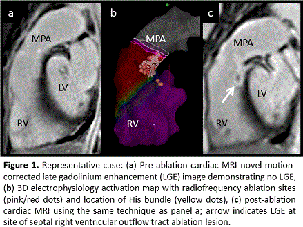
Article Information
vol. 132 no. Suppl 3 A11574
Published By:
American Heart Association, Inc.
Online ISSN:
History:
- Originally published November 6, 2015.
Copyright & Usage:
© 2015 by American Heart Association, Inc.
Author Information
- Elena K Grant1;
- Charles I Berul2;
- Russell R Cross2;
- Jeffrey P Moak2;
- Karin S Hamann2;
- Kendall J O’Brien2;
- Michael Hansen3;
- Peter Kellman3;
- Kanishka Ratnayaka2;
- Laura J Olivieri2
- 1Cardiology, Children’s Healthcare of Atlanta and Emory Univ Sch of Medicine, Atlanta, GA
- 2Cardiology, Children’s National Health System, Washington, DC
- 3National Heart Lung and Blood Institute, National Institutes of Health, Bethesda, MD
Abstract 10404: Incidence and Predictors of Asymptomatic Pericardial Effusion After Transcatheter Closure of Atrial Septal Defect
Jingjin Wang, Mehul Patel, Minghu Xiao, Zhongying Xu, Shiliang Jiang, Hao Wang
Circulation. 2015;132:A10404
Abstract
Introduction: Pericardial effusion (PE) without obvious periprocedural complications (e.g., cardiac perforation, device erosion) may occur after transcatheter closure of secundum atrial septal defects (ASD).It has only been described in case reports, and has not been the subject of systematic study.
Hypothesis: The aim of the study is to investigate the incidence and predictors of PE unrelated to procedural complications, thus providing interventional cardiologists with a novel way to approach new-onset PE after ASD closure.
Methods: We included all patients who had undergone successful percutaneous ASD closure from June 2009 to April 2014 (n=2652) with no preexisting PE or cardiac perforation or erosion. Transthoracic echocardiography (TTE) was performed during the procedure and 1, 3, and 6 months postoperatively.
Results: After device implantation, fifty patients (1.9 %) developed new-onset PE (37 immediately, 13 during follow-up). These patients were asymptomatic, stable hemodynamically, and had no new arrhythmias. PE appeared mild (5.1±1.9 mm) and homogeneously echo-lucent by TTE (Figure). PE diminished spontaneously. Compared with 2602 patients without PE, factors independently predicting asymptomatic PE were the device touching the atrial free wall, device size, patient age, and total defect size (Table). Areas under the receiver operating characteristic curves were 0.78 (p<0.001), 0.66 (p<0.001) and 0.77 (p<0.001) for device size, patient age, and total defect size, respectively.
Conclusion: A new type of PE is firstly reported systematically. Our data provide new insights into new-onset PE after percutaneous ASD closure.
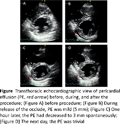
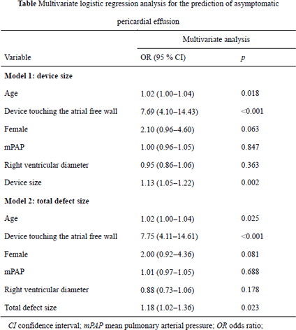
Article Information
vol. 132 no. Suppl 3 A10404
Published By:
American Heart Association, Inc.
Online ISSN:
History:
- Originally published November 6, 2015.
Copyright & Usage:
© 2015 by American Heart Association, Inc.
Author Information
- 1Echocardiography, Fuwai Hosp, National Cntr for Cardiovascular Diseases, Chinese Academy of Med Sciences and Peking Union Med College, Beijing, China
- 2Adult Congenial Heart Disease, Baylor College of Medicine, Houston, TX
- 3Radiology, Fuwai Hosp, National Cntr for Cardiovascular Diseases, Chinese Academy of Med Sciences and Peking Union Med College, Beijing, China
Abstract 18692: Evolution of Diastolic Parameters and Their Relationship to Heart Rate in the 7-15 Week Human Fetus
Claudine M Bohun, Lisa Howley, Jesus Serrano Lomelin, Winnie Savard, Lindsay Mills, Angela McBrien, Dora Gyenes, Tarek Motan, Venu Jain, Lisa K Hornberger
Circulation. 2015;132:A18692
Abstract
Objectives: Fetal echo offers a unique opportunity to investigate cardiac function in humans from the 1st trimester. We sought to explore the evolution left ventricular (LV) diastolic parameters and their relationship to fetal heart rate (HR) from 7-15 weeks gestational age (GA).
Methods: We prospectively performed fetal echo in 137 healthy pregnancies recruited from 7+0 to 15+0 (mean 11.5±2.1) weeks GA. From Doppler studies we measured LV isovolumic relaxation time (IRT), mitral valve (MV) inflow patterns,and duration, diastolic time (IRT+inflow duration), LV ejection time (ET) and systolic time interval (isovolumic contraction time+ET). All variables were compared to GA and HR and were assessed as a proportion of the cardiac cycle length (R-R). Data were analyzed by regression and correlation analyses.
Results: Results: From 7+0-15+0 weeks, HR and GA showed a non-linear relationship (R2=0.55, p<0.001) with low HRs at 7-8 weeks, peaking between 8-9 weeks, then falling thereafter. Diastolic time linearly increased with GA (R2=0.65, p<0.001) and inversely correlated with HR (r=0.77, p<0.001). IRT showed a nonlinear inverse relationship with GA (R2= 0.70, p<0.001), but did not correlate with HR, and IRT/RR increased linearly with GA (R2=0.71, p<0.001). Both inflow duration and inflow duration/R-R linearly increased with GA (R2=0.81,p<0.001). Inflow duration inversely correlated with HR (r=-0.70) and with IRT (r=-0.71) and IRT/R-R (r=-0.83, p<0.001 for all 3). A uniphasic MV atrial systolic inflow signal was present in 20/21 fetuses at 7+1-9+0 weeks, whereas, biphasic MV inflow was seen in 45% at 9+1-10+0 , 76% at 10+1-11+0 , 95% at 11+1-13+0 and 100% after 13+1 weeks GA. Inflow duration was significantly longer (146.9±25.4 vs 85.2±12.6ms, p<0.001) and HR was lower (160±9 vs 170±10 bpm, p<0.001) in fetuses with a biphasic flow pattern. ET demonstrated a weak relationship with GA (R2=0.41) and no relationship with HR, IVRT,or IVRT/R-R, and the systolic time showed a nonlinear trend of decreasing with GA (R2=0.46).
Conclusions: In the 7-15 week fetus, improvements in relaxation suggested by decreasing IRT, decreasing HR, and, to a lesser extent, decreasing systolic time likely contribute to increasing inflow duration and to early ventricular filling.
Article Information
vol. 132 no. Suppl 3 A18692
Published By:
American Heart Association, Inc.
Online ISSN:
History:
- Originally published November 6, 2015.
Copyright & Usage:
© 2015 by American Heart Association, Inc.
Author Information
- Claudine M Bohun1;
- Lisa Howley2;
- Jesus Serrano Lomelin3;
- Winnie Savard4;
- Lindsay Mills4;
- Angela McBrien5;
- Dora Gyenes4;
- Tarek Motan6;
- Venu Jain6;
- Lisa K Hornberger4
- 1Pediatric Cardiology, Univ of Alberta, Stollery Children’s Hosp, Edmonton, Canada
- 2Pediatric Cardiology, Children’s Hosp Colorado, Aurora, CO
- 3Public Health, Univ of Alberta, Edmonton, Canada
- 4Pediatric Cardiology, Univ of Alberta, Stollery Children’s Hosp, Edmonton, Canada
- 5Pediatric Cardiology, The Newcastle upon Tyne Hosps NHS Foundation Trust, Newcastle upon Tyne, United Kingdom
- 6Obstetrics & Gynecology, Univ of Alberta, Edmonton, Canada
Abstract 17047: Pathophysiologic Progression From the Second to Third Trimester in Fetuses With Ebstein’s Anomaly or Tricuspid Valve Dysplasia: Are Early Echocardiographic Features Reliable Indicators of Late Gestation Status?
Elif Seda Selamet Tierney, Doff B McElhinney, Lindsay R Freud, Wayne Tworetzky, Bettina Cueno, Maria C Escobar-Diaz, Catherine Ikemba, Brian T Kalish, Rukmini Komarlu, Stephanie Levasseur, Michael D Puchalski, Gary M Satou, Norman H Silverman, Anita J Moon-Grady
Circulation. 2015;132:A17047
Abstract
Background: Ebstein’s anomaly and tricuspid valve dysplasia (EA/TVD) are associated with high perinatal mortality. Poor hemodynamic status in both early and late gestation is associated with worse outcome. However, it is not known whether EA/TVD fetuses with more favorable physiology earlier in gestation progress to more severe disease in 3rd trimester. The goal of this study was to evaluate whether initial echocardiographic indices in fetuses with EA/TVD presenting at gestational age (GA) <24 wks are reliable indicators of physiologic status later in pregnancy.
Methods: This multi-center, retrospective study included fetuses diagnosed with EA/TVD from 2005-11 at 23 centers. Patients with initial diagnosis at <24 wks gestation and with ≥2 fetal echocardiograms ≥4 wks apart were included. A core laboratory analyzed the echocardiograms. Markers of poor outcome were defined as absence of antegrade flow across the pulmonary valve, pulmonary valve regurgitation (PR), cardiothoracic ratio >0.48, left ventricular (LV) dysfunction or TV annulus Z score >5.6.
Results: The study included 51 fetuses. Median GA at diagnosis was 21 wks [18-24], and the median duration from 2nd to 3rd trimester echocardiograms was 12 wks [4-18]. There were 21 fetuses (41%) with >2 markers of poor outcome at <24 wks, which increased to 32 (63%) in later gestation (p=0.001). Nine of 27 fetuses (33%) with antegrade pulmonary blood flow on first echocardiogram developed anatomic or functional pulmonary atresia during follow-up, and 7 of 39 fetuses (18%) without PR initially developed it later. LV dysfunction was present in 2 patients at <24 wks but present in 14 (37%) later (p<0.01). The only variable associated with worsening physiology, from <2 markers of poor outcome in 2nd trimester to ≥2 markers in 3rd trimester, was larger TV annulus Z-score (4.1±1.0 vs. 2.4±1.9, p=0.008).
Conclusion: These results confirm a wide spectrum of disease presentation and progression in fetuses with EA/TVD. Thus, it is imperative to assess these fetuses serially. In this cohort, there were no early markers of progression other than larger TV annulus Z-score. Great care must be taken in counseling prior to 24 wks, as the absence of factors associated with poor outcome early in pregnancy may be falsely reassuring.
Article Information
vol. 132 no. Suppl 3 A17047
Published By:
American Heart Association, Inc.
Online ISSN:
History:
- Originally published November 6, 2015.
Copyright & Usage:
© 2015 by American Heart Association, Inc.
Author Information
- Elif Seda Selamet Tierney1;
- Doff B McElhinney2;
- Lindsay R Freud3;
- Wayne Tworetzky4;
- Bettina Cueno5;
- Maria C Escobar-Diaz6;
- Catherine Ikemba7;
- Brian T Kalish8;
- Rukmini Komarlu8;
- Stephanie Levasseur9;
- Michael D Puchalski10;
- Gary M Satou11;
- Norman H Silverman1;
- Anita J Moon-Grady12
- 1Pediatrics/Cardiology, Stanford Univ, Palo Alto, CA
- 2Cardiothoracic Surgery, Stanford Univ, Palo Alto, CA
- 3Pediatric Cardiology, Harvard Med Sch, Boston Children’s Hosp, Boston, CA
- 4Pediatrics/Cardiology, Harvard Med Sch, Boston Children’s Hosp, Boston, CA
- 5Pediatrics/Cardiology, Children’s Hosp Colorado, Aurora, CO
- 6Pediatric Cardiology, Universidad Pontificia Bolivariana, Medellin, Colombia, Medellin, Colombia
- 7Pediatrics/Cardiology, UT Southwestern Med Cntr, Palo Alto, CA
- 8Pediatric Cardiology, Harvard Med Sch, Boston Children’s Hosp, Boston, MA
- 9Pediatrics/Cardiology, New York Presbyterian Hosp/ Columbia Univ, New York, CA
- 10Pediatrics/Cardiology, Primary Children’s Hosp/ Univ of Utah Sch of Medicine, Salt Lake City, UT
- 11Pediatrics/Cardiology, UCLA Med Cntr, Los Angeles, CA
- 12Pediatrics/Cardiology, Univ of California, San Francisco, San Francisco, CA
Abstract 17219: Increased Aortic Stiffness Following Intrauterine Exposure to Chronic Hypoxia in Rats
Praveen Kumar, Jude Morton, Sandra T Davidge, Po-Yin Cheung, Lisa K Hornberger
Circulation. 2015;132:A17219
Abstract
Background: We have previously demonstrated in rats that fetal hypoxia is associated with altered left ventricular (LV) diastolic function beginning in early postnatal life (2 weeks). In the present study we hypothesized that intrauterine hypoxia exposure alters aortic stiffness which may contribute to the evolution of myocardial dysfunction after birth.
Methods: Six pregnant rats were exposed to hypoxic conditions (11.5% FiO2) from E15-21and 3 were maintained in normoxic conditions. After delivery, 4 neonatal rats from each group were examined longitudinally at day 1, 3, and week 1, 2, 4, 8 for changes in aortic stiffness. Pups were anesthetized by face mask with 1.5% isoflurane. Aortic stiffness was assessed by measuring the pulse wave velocity (PWV) in the ascending aorta at 3 points: 1. Proximal to the branch of the first brachiocephalic artery 2. At the level of the diaphragm and 3. Just above the aortic bifurcation into the iliac arteries. T1 was the time interval from the peak of the R wave by ECG to onset of flow in the ascending aorta and T2 from peak of the R wave to onset of flow in either the mid aorta or distal aorta. The distance from the suprasternal notch to the subxiphoid process and to the anterior superior iliac spine was measured in each animal. The PWV was calculated using the following equation: [distance/(T2-T1)]. Three consecutive tracings were assessed and the PWV averaged for each study. Statistical analysis was performed using a 2-way ANOVA.
Results: In pups exposed to fetal hypoxia, PWV was significantly increased (p≤0.001) compared to controls at day 1 (4.8±1.5 vs. 2.3±0.2 m/s), day 3 (5.5±0.3 vs. 2.6±0.1 m/s) and week 1 (4.4±0.4 vs. 2.8±0.1 m/s). Interestingly, there was an interaction of time and fetal exposure (p=0.006) whereby PWV values converged from weeks 2 – 8 such that fetal exposure to hypoxia no longer had an effect. No correlation was found between LV diastolic function parameters and aortic PWV in the fetal hypoxia rats at any postnatal age.
Conclusion: Prenatal exposure to hypoxia was associated with increased aortic stiffness during early development. The lack of a relationship between LV diastolic function and aortic stiffness could suggest these represent independent effects of fetal hypoxia exposure.
Article Information
vol. 132 no. Suppl 3 A17219
Published By:
American Heart Association, Inc.
Online ISSN:
History:
- Originally published November 6, 2015.
Copyright & Usage:
© 2015 by American Heart Association, Inc.
Author Information
- 1Dept of Pediatric Cardiology, Univ of Alberta, Edmonton, Canada
- 2Dept of and Obstetrics/ Gynecology, Univ of Alberta, Edmonton, Canada
- 3Dept of Obstetrics & Gynecology, Univ of Alberta, Edmonton, Canada
- 4Depts of Pediatrics, Pharmacology and Surgery, Univ of Alberta, Edmonton, Canada
Abstract 16893: Subclinical Decrease in Myocardial Function in Asymptomatic Infants of Diabetic Mothers: A Tissue Doppler Study
Jenny E Zablah, Dorota Gruber, Denise A Hayes
Circulation. 2015;132:A16893
Abstract
Introduction: Infants of diabetic mothers (IDMs) with cardiac hypertrophy are recognized to have impaired myocardial performance, but less is known about ventricular function in those without hypertrophy. We hypothesized that, in asymptomatic newborns with normal echocardiograms, tissue Doppler imaging (TDI) indices of cardiac function would be decreased in IDMs compared to controls.
Methods: This retrospective case-control study involved IDMs ≥ 36 weeks gestational age, at 0 to 7 days of life. Subjects with cardiorespiratory symptoms, ventricular hypertrophy or dysfunction, or any echocardiographic abnormality (other than a patent ductus arteriosus before 4 days of life, or a patent foramen ovale) were excluded. Each subject was matched with 3 controls (healthy infants of non-diabetic mothers) by age (0-24, 25-48, 49-72, or > 72 hours of life), birth weight (± 0.5 kg), body surface area (± 0.03 m2), and by the ultrasound system utilized. TDI systolic (S’), early diastolic (E’), and late diastolic (A’) velocities were measured at the mitral valve (MV) annulus, basal ventricular septum, and tricuspid valve (TV) annulus, and were averaged from 3 consecutive cardiac cycles. Early Doppler inflow velocity to E’ ratios (E/E’) were calculated.
Results: Seventy cases (39 male) were identified: first 24 hours (h) of life (n=18), 25-48 h (n=22), 49-72 h (n=14), and > 72 h (n=16). Maternal diabetes was gestational in 60 cases, and pre-existing in 10. Median gestational age was 38 6/7 weeks (range 36-41 2/7), median birth weight 3.65 kg (2.56-5.38), and median BSA 0.22 m2 (0.17-0.27). Ultrasound system vendors included Siemens® (n=46), Philips® (n=23), and General Electric® (n=1). Cases were matched with 210 controls.
IDMs had significantly lower S’ (p ≤ 0.05) and E’ (p ≤ 0.01) velocities, and significantly higher E/E’ ratios (p ≤ 0.01) at the MV, basal septum, and TV compared to controls (Wilcoxon rank-sum test). There were no significant differences in A’ values between groups. Intraclass correlation demonstrated 84-99% interobserver and 98-99% intraobserver reliability.
Conclusions: In asymptomatic newborn IDMs without cardiac hypertrophy, TDI suggests a subclinical decrease in systolic and diastolic myocardial function compared to controls.
Article Information
vol. 132 no. Suppl 3 A16893
Published By:
American Heart Association, Inc.
Online ISSN:
History:
- Originally published November 6, 2015.
Copyright & Usage:
© 2015 by American Heart Association, Inc.
Author Information
- 1Pediatric Cardiology, North Shore-LIJ, New Hyde Park, NY
- 2Pediatric Cardiology, North Shore – LIJ, New Hyde Park, NY
- 3Pediatric Cardiology, NorthShore-LIJ, New hyde park, NY
Abstract 16855: Left Ventricular Volumes, Stress, and Strain in Normal Children and Young Adults Measured by 3-Dimensional Echocardiography
Joseph D Kuebler, Meena Nathan, Gerald Marx, Steven Colan, David M Harrild
Circulation. 2015;132:A16855
Abstract
Background: 3D echocardiography (3DE) is increasingly being used clinically to calculate ventricular volumes and function. Normal pediatric values of 3D LV volumes and strain are not well established; moreover, there are no reports of the stress-strain relationship (an index of contractility) based upon 3D technology in this cohort.
Methods: 3D LV datasets were obtained as part of routine echocardiographic examinations in eligible pediatric patients (≤ 21 years of age) between January 2014 and March 2015. Included patients had structurally normal hearts. Exclusion criteria included non-cardiac disorders with a potential impact on ventricular function and family history of cardiomyopathy. Image acquisition was performed using the Philips IE33 with X3/5/7 probes. Strain (3D; circumferential, GCS; and longitudinal, GLS) was analyzed according to a commercial 3D speckle-tracking analysis package (4D LV Analysis 3.1; Tomtec). LV mid-wall global average stress was calculated from the 3D LV volumes and 2D cross-sectional area/long-axis dimensions.
Results: 237 patients were included (age= 0.2 mo-21 y). The correlation between 3D and 2D LV mass and volumes was excellent (mass, R=0.94; end-diastolic volume, R=0.94; end-systolic volume, R=0.90; p<0.001 for all). Mean+-SD strain values (%) were: 3D=-33.9±2.8; GCS=-28.0 ± 3.1; GLS=-20.7 ± 3.0; only GLS varied significantly with age (R=0.26; p<0.001). Overall, 3D strain was inversely linearly related to wall stress (R=0.26; p<0.001); the strongest relationship was present in patients from age 0-5 years (R=0.48; p<0.01). When normalized to stress, absolute LV strain decreased with age (Figure 1: R=0.33; p<0.001).
Conclusions: 3DE may be used to calculate LV stress, strain, and volume parameters. Among strain parameters, age-related changes were seen only in GLS. Examination of the stress-strain relationship using these techniques may yield new insights into maturational changes in myocardial contractility.
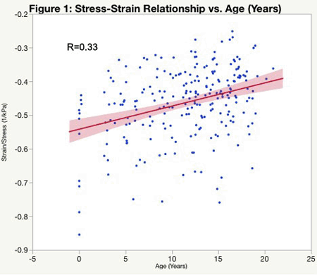
Article Information
vol. 132 no. Suppl 3 A16855
Published By:
American Heart Association, Inc.
Online ISSN:
History:
- Originally published November 6, 2015.
Copyright & Usage:
© 2015 by American Heart Association, Inc.
Author Information
- 1Cardiology, Boston Children’s Hosp, Boston, MA
- 2Cardiac Surgery, Boston Children’s Hosp, Boston, MA
Abstract 16524: Cardiac MRI Evaluation of Myocardial Deformation in Repaired Tetralogy of Fallot Provides Insight Into Right Ventricular Mechanics
Benjamin Goot, Kumaradevan Punithakumar, Edythe Tham, Michelle Noga
Circulation. 2015;132:A16524
Abstract
Introduction: Pulmonary regurgitation or stenosis after repaired tetralogy of Fallot (TOF) impacts the long-term ventricular mechanics. Our objective was to measure RV myocardial deformation using novel CMR software in repaired TOF. We postulate that RV strain will correlate with cardiac MRI (CMR) volumetric data.
Methods: Retrospective study of 55 patients s/p TOF repair compared to 40 normal controls. RV longitudinal strain was measured from the standard 4-chamber view and circumferential strain from the short axis slice apical to the RV outflow tract. Using CMR software developed in-house that utilizes a semi-automatic segmentation program, peak strain was identified. Unpaired t test assessed differences between groups and correlation was performed between strain and volumetric data.
Results: The predominant lesion was regurgitation in 48 (regurgitant fraction (RF) 43±2%), stenosis in 10 (peak gradient 32±2 mmHg), and mixed in 4. Longitudinal strain was reduced in TOF compared to controls while circumferential strain was preserved (Table 1). Correlations found reduced strain with increasing volumes and decreasing right ventricular ejection fraction (RVEF) (Table 2). Longitudinal:circumferential ratio (L/C) increased with increasing volumes and decreasing RVEF. No relationship was found between RV strain and RF.
Conclusions: The relationships between longitudinal and circumferential strain, and the L/C ratio with RV volumes and RVEF suggest that preservation of circumferential strain is important in maintaining RV systolic function. Measurement of RV deformation may provide insight into the RV’s response to volume or pressure overload in TOF, suggesting we should focus on RV mechanical changes rather than RF when managing these patients.
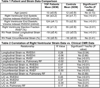
Article Information
vol. 132 no. Suppl 3 A16524
Published By:
American Heart Association, Inc.
Online ISSN:
History:
- Originally published November 6, 2015.
Copyright & Usage:
© 2015 by American Heart Association, Inc.
Author Information
- 1Pediatric Cardiology, Univ of Alberta, Edmonton, Canada
- 2Diagnostic Imaging, Univ of Alberta, Edmonton, Canada
Abstract 14760: Right Ventricular Function in Preterm Neonates: Reference Values for Longitudinal Strain by Two-dimensional Speckle Tracking Echocardiography From Birth to One Year of Life
Philip T Levy, Meghna D Patel, Mark R Holland, Timothy J Sekarski, Amit Mathur, Caroline Lee, Aaron Hamvas, Gautam K Singh
Circulation. 2015;132:A14760
Abstract
Introduction: Right ventricle (RV) systolic function is an important determinant of cardiopulmonary outcomes in premature infants. Two-dimensional speckle tracking echocardiography (2DSTE) derived myocardial strain is a reliable measure of RV systolic function in premature infants, but lacks reference values for clinical application in premature infants. We aimed to determine the maturational (age- and weight- related) changes in RV strain to establish reference values in preterm infants from birth to one year corrected age (CA).
Methods: RV peak global longitudinal strain (pGLS) and RV free wall longitudinal strain (FWLS) were measured in a prospective longitudinal study in 115 preterm infants (< 29 weeks at birth) at 24 and 72 hours of age (HOA), 32 and 36 weeks postmenstrual age (PMA), and one year (CA) by 2DTSE (GE EchoPac) from a RV-focus apical 4-chamber view using a validated protocol. Premature infants that developed chronic lung disease or had a hemodynamically significant PDA were excluded (n=65) from analysis for the reference values.
Results: RV pGLS ranged from -16% at birth to -26% by one year CA and RV FWLS ranged from -18% at birth to -27% to one year CA in healthy preterm infants. RV pGLS and FWLS strain correlated with increasing weight (r=0.87, p < 0.001), PMA in weeks (r=0.85, p < 0.001; r=0.83, p < 0.001), but were independent of gestational age at birth (r=0.4, p=0.38; r=0.3, p=0.5). RV strain was significantly lower in preterm infants with bronchopulmonary dysplasia (p=0.004) at 32 and 36 weeks PMA, and one year CA (Figure). RV strain was independent of gender or need for mechanical ventilation.
Conclusions: This study establishes reference values of RV global and free wall longitudinal strain and tracks their postnatal maturational changes in preterm infants. These measures increase from birth to one year CA and are linearly associated with increasing weight reflecting the postnatal cardiac growth as a contributor to the maturation of RV function.
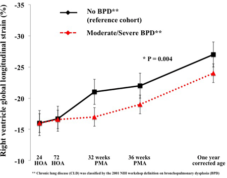
Article Information
vol. 132 no. Suppl 3 A14760
Published By:
American Heart Association, Inc.
Online ISSN:
History:
- Originally published November 6, 2015.
Copyright & Usage:
© 2015 by American Heart Association, Inc.
Author Information
- Philip T Levy1;
- Meghna D Patel2;
- Mark R Holland3;
- Timothy J Sekarski2;
- Amit Mathur1;
- Caroline Lee2;
- Aaron Hamvas4;
- Gautam K Singh2
- 1Neonatology, Washington Univ Sch of Medicine, St. Louis, MO
- 2Pediatric Cardiology, Washington Univ Sch of Medicine, St. Louis, MO
- 3Radiology, Indiana Univ Sch of Medicine, Indianapolis, IN
- 4Neonatology, Northwestern Univ Feinberg Sch of Medicine, Chicago, IL
Abstract 12735: Potential of the Single Beat Estimation for End Systolic Pressure-Volume Relation in Patients With Single Ventricular Heart
Hirofumi Saiki, Seiko Kuwata, Clara Kurishima, Satoshi Masutani, Hideaki Senzaki
Circulation. 2015;132:A12735
Abstract
Background: Estimation of ventricular elastance at the aortic valve opening is fundamental for assessment of non-invasive end-systolic elastance (Ees). Despite conditional variability of time varying elastance (Et) property, the Et during isovolumic contraction (IVC) is less variable and highly adjustable, thus Et has been used to estimate Ees in a number of clinical studies. However, uncertain characteristics of Et due to morphological diversity hindered its application in single ventricular hearts (SV). We tested our hypothesis that Et in early systole is independent of ventricular morphology in SV patients.
Methods: During cardiac catheterization, a total of 78 pressure volume relationships were constructed from 26 children including 18 Fontan patients (right dominant: SRV n=8, left dominant: SLV n=10) and controls (n=8). Et curve, normalized both for Ees and time to Ees, was compared among groups at baseline, dobutamine infusion (DOB, 5 mcg/kg/min) and rapid supraventricular pacing.
Results: Shapes of Et in SRV and SLV at both baseline and DOB were similar, particularly during IVC and ejection phase (Figure). While Et was stable during pacing with additional 20-30 bpm to the baseline heart rate (HR), relaxation delay caused disturbance of Et in some patients at HR greater than 170 bpm. The slope ratio (α) of Et during ejection phase to that during IVC, which determines subtle variations of Et, was significantly correlated with ejection fraction (EF, R=.65, p<.01) and independent of ventricular type, preload and afterload.
Conclusion: Et properties in Fontan patients, both with SRV and SLV, highly resemble those in controls at baseline, with DOB, and physiological pacing during systole. Similar to structurally normal hearts, α can be predicted by EF. While impact of diastolic property on early systole should be accounted for in high HR patients, our results rationalized the utility of normalized Et that provides a fulcrum point to estimate Ees in Fontan circulation.
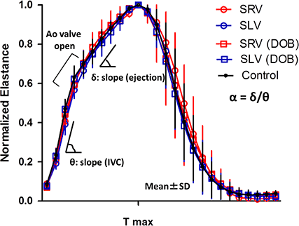
Article Information
vol. 132 no. Suppl 3 A12735
Published By:
American Heart Association, Inc.
Online ISSN:
History:
- Originally published November 6, 2015.
Copyright & Usage:
© 2015 by American Heart Association, Inc.
Author Information
- Hirofumi Saiki;
- Seiko Kuwata;
- Clara Kurishima;
- Satoshi Masutani;
- Hideaki Senzaki
- Pediatric Cardiology, Saitama Med Cntr, Saitama Med Univ, Kawagoe, Saitama, Japan
Abstract 10882: Scaling to Lean Body Mass Accurately Classifies Abnormal LV Mass in Obese Children
Shahryar M Chowdhury, Melissa H Henshaw, Janet Carter, Tyler N Thomas, Andrew M Atz
Circulation. 2015;132:A10882
Abstract
Introduction: Scaling LV mass to BSA or height leads to inaccuracies in the diagnosis of LV hypertrophy in the obese pediatric population. Lean body mass (LBM) is free of the scaling biases seen with BSA and height. Our objectives were to 1) validate predictive equations for LBM, 2) determine the ideal scaling variable for LV mass, and 3) compare the diagnostic accuracy of predicted LBM (LBMp), BSA, and height in detecting abnormal LV mass in obese children.
Methods: Obese subjects ages 4 to 21 were recruited prospectively. Height, weight, sex, race and body mass index z-score were used to calculate LBMp. LV mass was measured by echocardiography. Dual-energy X-ray absorptiometry was used to measure LBM (LBMm). Bland-Altman plots were used to assess agreement between LBMp and LBMm. Linear regression was performed to assess the ideal scaling variable for LV mass. Centile curves of LV mass vs. LBMm as the reference-standard were compared with curves derived from LBMp, BSA, and height2.7 to assess their accuracy in detecting elevated LV mass.
Results: 309 subjects were enrolled for predictive equation validation, 195 had echocardiography performed. Mean age was 11.8 ± 3.4 years. The mean difference between LBMp and LBMm was 1.5% (95% limits of agreement: -16.5%, 19.4%). LBMm had the strongest relationship with LV mass (R2 = 0.83, p < 0.01) compared to LBMp (R2 = 0.81, p < 0.01), BSA (R2 = 0.77, p < 0.01), and height2.7 (R2 = 0.67, p < 0.01). Of the clinically derived variables, LBMp was the only measure to retain a relationship with LV mass upon multivariable regression (partial r = 0.89). LBMp was the most accurate scaling variable in detecting LV hypertrophy (PPV = 85.7%, NPV = 99.3%) compared to BSA and height2.7.
Conclusions: LBM predictive equations are accurate in obese children. LBM is the strongest predictor of LV mass in obese children. LBMp is the most accurate anthropometric scaling variable for LV mass in LV hypertrophy detection. LBMp should be considered for widespread clinical use as the body size correcting variable for LV mass in obese children.
Article Information
vol. 132 no. Suppl 3 A10882
Published By:
American Heart Association, Inc.
Online ISSN:
History:
- Originally published November 6, 2015.
Copyright & Usage:
© 2015 by American Heart Association, Inc.
Author Information
- Shahryar M Chowdhury;
- Melissa H Henshaw;
- Janet Carter;
- Tyler N Thomas;
- Andrew M Atz
- Pediatrics, Med Univ of South Carolina, Charleston, SC
Abstract 16428: Characteristics and Impact of Bystander Cardiopulmonary Resuscitation Following Pediatric Out of Hospital Cardiac Arrest in the United States: A Study From the Cardiac Arrest Registry to Enhance Survival (CARES)
Maryam Y Naim, Rita V Burke, Bryan F McNally, Robert A Berg, Kimberly Vellano, David Markenson, Richard N Bradley, Joseph W Rossano
Circulation. 2015;132:A16428
Abstract
Introduction: Bystander cardiopulmonary resuscitation (BCPR) is associated with improved outcome in adult out-of-hospital cardiac arrest (OHCA). There are few data on the prevalence and impact of BCPR on children.
Hypothesis: We aimed to characterize BCPR in pediatric OHCA and test the hypothesis that BCPR would occur infrequently and would be associated with neurologically favorable survival at hospital discharge from a large cardiac arrest registry in the United States.
Methods: We conducted an analysis of the Cardiac Arrest Registry to Enhance Survival database. Inclusion criteria were age ≤ 18 years of age and non-traumatic OHCA from January 1, 2013 through December 31, 2014. Neurologically favorable survival was defined as a Cerebral Performance Category Scale of 1 or 2.
Results: A total of 2,176 cardiac arrests were evaluated. Most patients were infants (62%) or adolescents (19%). Most arrests occurred at home (86%), were unwitnessed (75%), and had a non-shockable rhythm (93%). BCPR was provided in 49%, most commonly by a family member (71%). BCPR was more common for white (60%) compared to black (42%) and Hispanic children (44%) (p<0.001). Overall, BCPR was associated with a higher rate of neurologically favorable survival (11% vs. 7%, odds ratio [OR]1.6 95% confidence interval [CI] 1.2-2.3). In sub-group analyses, BCPR was associated with a higher rate of neurologically favorable survival for out of home arrests (34% vs. 15%, OR 2.9, 95% CI 1.6-5.3), and arrests presenting in a shockable rhythm (48% vs. 32%, OR 2.0 95% CI 1.0-4.0). For infants BCPR was not associated with survival (6.4% vs. 6.0%, OR 1.1 95% CI 0.7-1.7) or neurologically favorable survival (5.2% vs. 5.0%, OR 1.1 95% CI 0.6-1.8).
Conclusion: BCPR was provided in just under 50% of pediatric OHCAs and was more common for white compared to black and Hispanic children. BCPR was associated with improved survival that was most notable in out of home arrests, with over twice as many patients having neurologically favorable survival. Though infants comprised the largest age group, no effect of BCPR outcome was observed. This impact of BCPR suggests the need for a public health strategy to improve the provision of BCPR, and the need for an alternative strategy for some groups including infants.
Article Information
vol. 132 no. Suppl 3 A16428
Published By:
American Heart Association, Inc.
Online ISSN:
History:
- Originally published November 6, 2015.
Copyright & Usage:
© 2015 by American Heart Association, Inc.
Author Information
- Maryam Y Naim1;
- Rita V Burke2;
- Bryan F McNally3;
- Robert A Berg1;
- Kimberly Vellano3;
- David Markenson4;
- Richard N Bradley5;
- Joseph W Rossano6
- 1Anesthesiology, Critical Care Medicine and Pediatrics, Children’s Hosp of Philadelphia and The Univ of Pennsylvania, Philadelphia, PA
- 2Pediatric Surgery, Children’s Hosp of Los Angeles/ Keck Sch of Medicine, Los Angeles, CA
- 3Emergency Medicine, Emory Univ Hosp, Altanta, GA
- 4Emergency Medicine, Sky Ridge Med Cntr, Lone Tree, CO
- 5Anesthesiology, Critical Care Medicine and Pediatrics, The Univ of Texas Health Science Cntr in Houston, Houston, TX
- 6Pediatrics, Children’s Hosp of Philadelphia and The Univ of Pennsylvania, Philadelphia, PA
Abstract 14827: Nitric Oxide During Cardiopulmonary Bypass Improves Clinical Outcome: A Blinded, Randomized Controlled Trial
Christopher S James, Stephen Horton, Christian Brizard, Charlotte Molesworth, Johnny Millar, Warwick Butt
Circulation. 2015;132:A14827
Abstract
Background: Children requiring cardiac surgery with cardiopulmonary bypass (CPB) frequently develop Low Cardiac Output Syndrome (LCOS), particularly when very young, A pilot study of 16 children by Checchia et al. (2013) showed that the delivery of nitric oxide (NO) to the CPB circuit shortened duration of mechanical ventilation and ICU stay. We hypothesized that administering NO to oxygenator gas flow during CPB would decrease the incidence of LCOS and effect subsequent clinical outcomes.
Methods: We conducted a prospective, blinded, randomized controlled trial in children with congenital heart disease having surgery with CPB. Randomization was stratified by age and ‘blocked’ at six in each group. Children received oxygen alone or 20 ppm gaseous NO and oxygen to the CPB gas administration line. Only the study perfusionist was aware of the allocation and all equipment and devices were otherwise identical in each group; in particular the cardiac surgeon and anesthetist remained blinded to patient allocation.
Results: 198 children were enrolled following written consent. There were no differences in patient characteristics, diagnoses or surgeries between groups. 101 children received NO and had a significant reduction in LCOS (14% vs 31%, p=0.004), use of ECMO (1% vs 8%, p=0.014) and a non-significant reduction in ICU length of stay (48hrs vs 72hrs, p=0.111), compared to the 97 children who did not receive NO. The reduction in LCOS was most pronounced in children less than 6 weeks of age (20% vs 52%, p=0.012) and in those aged 6 weeks to 2 years (6% vs 24%, p=0.026), who also had significantly reduced ICU length of stay (43hrs vs 84hrs, p=0.031). LCOS occurred equally between groups in children greater than 2 years of age (17% vs 21%, p=0.678). There was no difference in the amount of post-operative bleeding in any age group. Children greater than 2 years of age who received NO required fewer blood transfusions (8.3% vs 24.1%, p=0.096).
Conclusions: Delivery of NO to the CPB circuit for children undergoing cardiac surgery significantly reduces the incidence of LCOS, use of ECMO and ICU length of stay by varying degrees, according to age group.
Article Information
vol. 132 no. Suppl 3 A14827
Published By:
American Heart Association, Inc.
Online ISSN:
History:
- Originally published November 6, 2015.
Copyright & Usage:
© 2015 by American Heart Association, Inc.
Author Information
- Christopher S James1;
- Stephen Horton2;
- Christian Brizard3;
- Charlotte Molesworth4;
- Johnny Millar1;
- Warwick Butt1
- 1Intensive Care, Royal Children’s Hosp, Melbourne, Australia
- 2Perfusion, Royal Children’s Hosp, Melbourne, Australia
- 3Cardiac Surgery, Royal Children’s Hosp, Melbourne, Australia
- 4MCRI, Royal Children’s Hosp, Melbourne, Australia
Abstract 14326: Inefficient Ventriculoarterial Coupling in Fontan Patients Measured Noninvasively Using Cardiac Magnetic Resonance
Max E Godfrey, Rahul H Rathod, Ellen M Keenan, Tal Geva, Kimberlee Gauvreau, Andrew J Powell, Ashwin Prakash
Circulation. 2015;132:A14326
Abstract
Introduction: The ventriculoarterial coupling (VAC) ratio, defined as the ratio of arterial elastance (Ea) to ventricular end-systolic elastance (Ees), reflects the mechanoenergetic efficiency of the cardiovascular system. Extreme high or low values of VAC ratio imply inefficient energy transfer. Despite increasing rates of morbidity and mortality in patients with Fontan circulation, little is known about VAC ratio in this population. We used a previously described cardiac magnetic resonance (CMR) method to assess VAC in a large Fontan cohort and examined its relation to outcomes.
Methods and Results: We retrospectively measured VAC from CMR and noninvasive blood pressure data on 195 Fontan patients (age 19.6±10.7 years) and 42 controls (age 15.2±2.2 years). VAC was calculated as Ea/Ees (Ea= mean arterial blood pressure (MBP)/ventricular stroke volume; Ees= MBP/end-systolic volume). Compared with controls, Fontan patients had lower body surface area (BSA) adjusted median Ees (1.54 vs. 2.4, p<0.001) and Ea (1.35 vs. 1.48, p=0.01) and a higher median VAC ratio (0.88 vs 0.62, p<0.001) (Figure). After a median follow-up of 4 years (range 1-10), 20 patients reached a composite endpoint of death or heart transplant listing. As seen in the Figure, extreme values of the VAC ratio were associated with higher risk of reaching the endpoint. In a multivariable Cox regression model controlling for EDV/BSA, which has been shown to be a risk factor, being in the highest tertile of the VAC was no longer a risk factor, but patients in the lowest tertile of VAC ratio showed a trend towards being more likely to reach the endpoint (Hazard Ratio 4.06 vs middle tertile, p=0.08).
Conclusion: In this cohort, Fontan patients demonstrated inefficient VAC as compared with controls. A lower VAC ratio tended to be associated with death or transplantation listing even when controlling for ventricular size. Further investigation of CMR-derived VAC in the Fontan circulation is warranted.
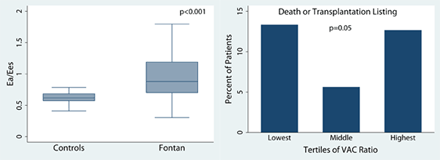
Article Information
vol. 132 no. Suppl 3 A14326
Published By:
American Heart Association, Inc.
Online ISSN:
History:
- Originally published November 6, 2015.
Copyright & Usage:
© 2015 by American Heart Association, Inc.
Author Information
- Max E Godfrey;
- Rahul H Rathod;
- Ellen M Keenan;
- Tal Geva;
- Kimberlee Gauvreau;
- Andrew J Powell;
- Ashwin Prakash
- Cardiology, Boston Children’s Hosp, Boston, MA
Abstract 19315: Geometry, Reintervention, and Growth Patterns in the Reconstructed Aortic Arch With Interdigitating Technique in Patients With Hypoplastic Left Heart Syndrome and Variants
Christoph Haller, Devin Chetan, Matthew Doyle, Arezou Saedi, Rachel Parker, Glen Van Arsdell, Osami Honjo
Circulation. 2015;132:A19315
Abstract
Objectives: The interdigitating technique in aortic arch reconstruction in hypoplastic left heart syndrome and variants (HLHS) is very effective to minimize the recoarctation rate. Little is known on the aortic arch’s growth characteristics and the resulting clinical impact.
Methods: 139 patients with HLHS underwent staged palliation between 2007 and 2014. 72 patients who underwent Norwood arch reconstruction with the interdigitating technique were included. Dimensions of the ascending aorta (AA), transverse arch (TA), isthmus (IA) and descending aorta (DA) in pre-stage II (P1, n=50) and pre-Fontan (P2, n=21) angiograms were measured and geometry and growth characteristics of the aortic arches were analyzed. Correlations between the aortic dimensions and clinical outcomes were assessed.
Results: There were significant increases in diameters in all segments between P1 and P2 (p < .0005). The z-scores in AA, TA and IA were unchanged between P1 and P2 (p = .931/.425/.121), but increased significantly in DA at P2 (p = .039). The percent increase in diameters were comparable among 4 segments (mean, 146% in IA, 144 in DA, p=.648). There were correlations in dimensions and z-scores between P1 and P2 in AA (p = .029/.013) and TA (p = .001/ < .0005), but no correlations were found in IA (p = .140/.747) and DA (p = .075/.432). The most significant tapering in the arch dimension occurred between TA and IA in both time points (P1, 67.3% vs. P2, 61.1%, p=.303). The reverse coarctation index (TA/IA ratio) at P1 (r = .381, p = .042), but not coarctation index (CoAI, IA/DA ratio) at P1 (p = .774) had a significant correlation with post-stage II ventricular function. Balloon dilatation for recoarctation was needed in 2 (2.7%) patients prior to stage II palliation. CoAI at P1 was a predictor for ventricular dysfunction at latest follow-up (p=.017).
Conclusions: Aortic arch growth after interdigitating reconstruction in HLHS is substantial and relatively constant. The isthmus growth is proportional to other segments. Overall reintervention rate for recoarctation is exceptionally low. CoAI prior to stage II palliation may be associated with long-term ventricular function.
Article Information
vol. 132 no. Suppl 3 A19315
Published By:
American Heart Association, Inc.
Online ISSN:
History:
- Originally published November 6, 2015.
Copyright & Usage:
© 2015 by American Heart Association, Inc.
Author Information
- Christoph Haller1;
- Devin Chetan1;
- Matthew Doyle2;
- Arezou Saedi1;
- Rachel Parker1;
- Glen Van Arsdell1;
- Osami Honjo1
- 1Cardiovascular Surgery, The Hosp for Sick Children, Toronto, Canada
- 2Mechanical & Industrial Engineering, Toronto General Hosp, Toronto, Canada
Abstract 18055: Induced Growth of Hypoplastic Cardiac Structures to Facilitate Repair of Congenital Defects
Daniel F Labuz, Lee A Pyles, James A Berry, John E Foker
Circulation. 2015;132:A18055
Abstract
Introduction: Congenital heart defects (CHD) may include hypoplastic valves, ventricles and vessels which complicate repairs. Our hypotheses were that hypoplastic structures are developmental rather than primarily genetic in origin and the correct biomechanical signal would induce catch up growth. Our corollary hypothesis was that the growth signal is provided by flow, not pressure. First stage operations, therefore, were designed to increase flow and induce growth. Once growth was sufficient, a second operation would often complete the repair. We report our results using flow induced growth of hypoplastic structures to facilitate repair in 3 types of CHD.
Methods: Three groups of CHD patients with associated hypoplastic structures: unbalanced atrioventricular canal defects (UAVC); pulmonary atresia with intact ventricular septum (PAIVS); and coarctation of the aorta (CoA) were reviewed. Operations increased flow through the hypoplastic structures, such as creating restrictive septal defects to increase flow through the hypoplastic AV valves and ventricles (UAVC, PAIVS) or relieving obstruction (CoA) to enhance aortic valve and arch flow.
Results: On follow up, virtually all (85/90) hypoplastic structures reached normal size and the rest had nearly comparable growth (Table).
Conclusions: 1) Operations designed to increase flow through hypoplastic structures reliably induced catch up growth of UAVC, PAIVS and CoA lesions.
2) The growth response supported underdevelopment as the cause of hypoplasia and flow as the growth signal to reverse it.
3) Growth induction allowed two ventricle repairs in UAVC and PAIVS patients and avoided extended arch repairs in CoA; all with decreased operative risk.
4) Following catch up growth of the hypoplastic lesions, the outlook improved to that of patients with balanced AVCs, normal sized RVs in pulmonary atresia and well repaired CoA.
5) Once normal size was reached growth continued, producing a durable result.
Article Information
vol. 132 no. Suppl 3 A18055
Published By:
American Heart Association, Inc.
Online ISSN:
History:
- Originally published November 6, 2015.
Copyright & Usage:
© 2015 by American Heart Association, Inc.
Author Information
- 1Surgery, Univ of Minnesota Med Sch – Twin Cities, Minneapolis, MN
- 2Pediatrics, West Virginia Univ Sch of Medicine, Morgantown, WV
- 3Pediatrics, Univ of Minnesota Med Sch – Twin Cities, Minneapolis, MN
Abstract 16949: Failing Fontan: Genesis of a Subpulmonary Neo-ventricle From Engineered Heart Tissue
Daniel Biermann, Alexandra Eder, Hatim Seoudy, Florian Arndt, Tillmann Schuler, Kersten Peldschus, Michael G. Kaul, Hermann Reichenspurner, Thomas Mir, Arlindo Riso, Rainer Kozlik-Feldmann, Arne Hansen, Thomas Eschenhagen, Joerg S. Sachweh
Circulation. 2015;132:A16949
Abstract
Introduction: Fontan palliation is the treatment of choice for patients with morphological or functional univentricular hearts. The unphysiologic and non-pulsatile pulmonary blood flow results in multiorgan complications and a poor long-term outcome. We evaluated graft survival and histomorphology of a pulsatile Fontan conduit generated from Engineered Heart Tissue (EHT) after implantation in a rat model.
Hypothesis: We hypothesized that EHT matures and remains contractile in the setting of venous (Fontan-like) preload.
Methods: EHT was generated from ventricular cardiomyocytes of neonatal Wistar rats, fibrinogen, thrombin and DMEM. After culture for 14 days constructs were implanted around the right superior vena cava of Wistar rats (n=12, 300-350 g). Immunosupression was achieved by daily subcutaneous injection of Cyclosporin A (25 mg/kg BW) and Methylprednisolone (2 mg/kg BW). MRI (Bruker) was used to assess condensation of EHTs in vivo. Animals were euthanized after 7, 14, 28 and 56 days postoperatively for histomorphological analysis. Transmission electron microscopy was used to evaluate sarcomeric integrity of cardiomyocytes within the construct.
Results: In culture, EHTs started beating around day 8 and remained contractile in vivo throughout the experiment (d7=3/3, d14=2/3, d28=3/3, d56=2/3). All animals survived circumferential implantation of EHTs around the right SVC via a right thoracotomy. MRI (d14, n=3) revealed no constriction or stenosis of the SVC by the constructs. Hematoxylin and Eosin staining showed densely packed bundles of cardiomyocytes within the EHT conduit and intense vascularisation. Immunolabeling of actinin and connexin 43 indicated adequate maturation of cardiomyocytes after grafting around the right SVC in rats.
Conclusions: EHTs placed around the superior caval vein of Wistar rats survive and contract for a considerable time after implantation. Histomorphology revealed a matured phenotype of grafted cardiomyocytes and an adequate vascularisation. The functional benefit of a contractile neo-ventricle to propel pulmonary blood flow in Fontan patients remains to be evaluated.
Article Information
vol. 132 no. Suppl 3 A16949
Published By:
American Heart Association, Inc.
Online ISSN:
History:
- Originally published November 6, 2015.
Copyright & Usage:
© 2015 by American Heart Association, Inc.
Author Information
- Daniel Biermann1;
- Alexandra Eder2;
- Hatim Seoudy1;
- Florian Arndt3;
- Tillmann Schuler4;
- Kersten Peldschus5;
- Michael G. Kaul5;
- Hermann Reichenspurner6;
- Thomas Mir3;
- Arlindo Riso1;
- Rainer Kozlik-Feldmann3;
- Arne Hansen2;
- Thomas Eschenhagen2;
- Joerg S. Sachweh1
- 1Paediatric Cardiac Surgery, Univ Heart Cntr Hamburg, Hamburg, Germany
- 2Institute of Experimental and Clinical Pharmacology and Toxicology, Univ Med Cntr Hamburg-Eppendorf, Hamburg, Germany
- 3Paediatric Cardiology, Univ Heart Cntr Hamburg, Hamburg, Germany
- 4Dept of Diagnostic and Interventional Radiology, Univ Heart Cntr Hamburg, Hamburg, Germany
- 5Dept of Diagnostic and Interventional Radiology, Univ Med Cntr Hamburg-Eppendorf, Hamburg, Germany
- 6Cardiovascular Surgery, Univ Heart Cntr Hamburg, Hamburg, Germany
Abstract 14020: Practice Trends in the Care of Infants With Hypoplastic Left Heart Syndrome: A Report From the National Pediatric Cardiology Quality Improvement Collaborative (NPC-QIC)
Waldemar F Carlo, James F Cnota, Jeffrey B Anderson
Circulation. 2015;132:A14020
Abstract
Introduction: The NPC-QIC was established in 2008 to improve outcomes of hypoplastic left heart syndrome (HLHS) patients during the interstage period using quality improvement to reduce unnecessary practice variation. Our objective was to evaluate early and recent interstage practice variation within the collaborative.
Methods: Data from the first 100 patients (6/2008 – 1/2010) representing 18 centers were compared with the most recent 100 patients (1/2014 – 11/2014) from these same centers.
Results: Prenatal diagnosis increased from 72% to 82% (p = 0.09). There were no differences in gestational age or weight at Norwood. A composite of any preoperative risk factor (mechanical ventilation, acidosis, renal insufficiency, arrhythmia, NEC, sepsis) occurred more frequently in the early era (58% vs 34%, p <0.01). While mean age at Norwood was similar (8.3 vs 6.6 days, p = 0.2), the standard deviation for this variable was significantly lower in the recent era (10.5 to 6.4 days, p = 0.03). Use of RV-PA conduit increased (69% to 84%, p = 0.02). Rates of complete discharge communication with both the primary care physician (31 to 97%, p < 0.01) and primary cardiologist (44 to 97%, p < 0.01) increased substantially. There was no change in interstage feeding strategies between eras. Use of interstage outpatient surveillance program increased (76% to 99%, p < 0.01) with all participants in the late era monitoring both oxygen saturation and weight.
Conclusions: Among NPC-QIC centers contributing patients to both early and recent cohorts, there were significant changes in preoperative risk factors, Norwood surgical strategy, discharge communication, and interstage surveillance. Practice variation in timing of Norwood has been reduced. Further study is required to determine an association between these practice trends and decreased mortality.
Article Information
vol. 132 no. Suppl 3 A14020
Published By:
American Heart Association, Inc.
Online ISSN:
History:
- Originally published November 6, 2015.
Copyright & Usage:
© 2015 by American Heart Association, Inc.
Author Information
- 1Pediatrics, UAB, Birmingham, AL
- 2Pediatrics, Cincinnati Children’s Hosp Med Cntr, Cincinnati, OH
Abstract 12803: Long-term Outcomes After Surgical Pulmonary Arterioplasty: Analysis of Re-intervention Rates and Patient-specific Risk Factors
Nicole M Cresalia, Aimee K Armstrong, Jennifer C Hirsch-Romano, Mark Norris, Sunkyung Yu, Albert P Rocchini, Jeffrey D Zampi
Circulation. 2015;132:A12803
Abstract
Introduction: Branch pulmonary artery stenosis (PAS) is common in congenital heart disease. Surgical patch arterioplasty is a conventional therapy, however, limited data exist on long-term outcomes.
Hypothesis: We examined the incidence of and risk factors for re-intervention after surgical arterioplasty for PAS. We assessed the hypothesis that certain characteristics are associated with increased need for re-intervention.
Methods: This retrospective cohort study included patients with two-ventricle physiology who underwent patch arterioplasty for PAS at a single high-volume center from 2004-2013. Freedom from surgical or percutaneous re-intervention for recurrent PAS was estimated. Univariate and multivariable Cox regression were performed to determine risk factors for re-intervention.
Results: Among 116 patients undergoing patch arterioplasty at a median age of 1.2 years, hospital discharge survival was 95%. Of the survivors, 97 (88%) had a median follow-up of 3.6 years (range 15 days to 11 years). PAS re-intervention occurred in 31/97 (32%) at a median time to re-intervention of 1.6 years (range 7 days to 9.3 years). Freedom from re-intervention is shown in Figure 1. In univariate analysis, age less than 30 days at time of arterioplasty, congenital PAS (versus acquired), and bilateral PAS were significantly associated with re-intervention. In multivariate analysis, neonatal age (adjusted hazard ratio [AHR 4.4], p=0.004) and bilateral PAS (AHR 3.3, p=0.003) remained independently associated with re-intervention.
Conclusions: We present the largest cohort study with the longest follow-up time evaluating outcomes after surgical pulmonary arterioplasty for PAS to date. Re-intervention for recurrent PAS following patch arterioplasty is common. In conclusion, patients at highest risk for re-intervention, such as neonates and patients with bilateral PAS, may benefit from frequent monitoring or novel approaches to repair.
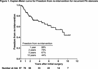
Article Information
vol. 132 no. Suppl 3 A12803
Published By:
American Heart Association, Inc.
Online ISSN:
History:
- Originally published November 6, 2015.
Copyright & Usage:
© 2015 by American Heart Association, Inc.
Author Information
- Nicole M Cresalia;
- Aimee K Armstrong;
- Jennifer C Hirsch-Romano;
- Mark Norris;
- Sunkyung Yu;
- Albert P Rocchini;
- Jeffrey D Zampi
- Pediatric Cardiology, Univ of Michigan, Ann Arbor, MI
Abstract 10018: High on Aspirin Platelet Reactivity in Children Undergoing the Fontan Procedure
Jason T Patregnani, Yaser Diab, Darren Klugman, Michael Spaeder, Robert Freishtat, Pranav Sinha, John Berger
Circulation. 2015;132:A10018
Abstract
Introduction: Despite routine thromboprophylaxis with aspirin, thromboembolism (TE) continues to be a significant complication after the Fontan procedure. Data suggest that suboptimal ex-vivo aspirin-induced platelet inhibition-known as High on-Aspirin Platelet Reactivity (HAPR) is an independent risk factor for breakthrough TE. The occurrence of HAPR may potentially increase the risk of aspirin prophylaxis failure in children who underwent Fontan procedure. However, HAPR has not been adequately studied in this patient population.
Hypothesis: There is a high incidence of HAPR in children after the Fontan procedure.
Methods: We conducted a single-center, prospective, observational pilot study of children requiring Fontan procedure. Thromboelastography with platelet mapping (TEG-PM), platelet CD62P expression by flow-cytometry and urinary 11-dehydro-thromboxane B2 (11-dhTxB2) were measured at baseline prior to starting aspirin at 5 mg/kg/day and after at least 5 days of continuous therapy. HAPR was defined as less than 30% Arachidonic Acid (AA)-induced platelet aggregation inhibition as measured by TEG-PM.
Results: 17 patients (11 males and 6 females, median age 3 years) were enrolled. HAPR was identified in 9/17 (53%) with 3/9 (33%) patients exhibiting no detectable AA-induced platelet aggregation inhibition. There was no significant effect of aspirin on percentage of activated platelets (i.e.: platelets expressing CD62P marker) in children with or without HAPR. In contrast to children with HAPR, aspirin therapy in children without HAPR led to significant reduction in urinary 11-dhTxB2 levels (Table 1).
Conclusions: A significant proportion of children with single ventricle physiology exhibited HAPR in the immediate post-operative period after Fontan procedure. Further studies are needed to examine association of HAPR with increased risk of TE in this patient population.

Article Information
vol. 132 no. Suppl 3 A10018
Published By:
American Heart Association, Inc.
Online ISSN:
History:
- Originally published November 6, 2015.
Copyright & Usage:
© 2015 by American Heart Association, Inc.
Author Information
- Jason T Patregnani1;
- Yaser Diab2;
- Darren Klugman3;
- Michael Spaeder4;
- Robert Freishtat5;
- Pranav Sinha6;
- John Berger1
- 1Dept of Critical Care/Cardiology, Children’s National Med Cntr, Washingon, DC
- 2Dept of Hematology, Children’s National Med Cntr, Washingon, DC
- 3Dept of Critical Care/Cardiology, Children’s National Med Cntr, Washington, DC
- 4Dept of Critical Care, Children’s National Med Cntr, Washingon, DC
- 5Dept of Emergency Medicine, Children’s National Med Cntr, Washingon, DC
- 6Dept of Cardiovascular Surgery, Children’s National Med Cntr, Washingon, DC
Abstract 20169: A Novel Early Warning System to Predict Critical Events in Children With Congenital Heart Disease
Andrew Y Shin, Rui Liu, Vamsi Yarlagadda, Doff McElhinney, Christopher Almond, Xuefeng Ling
Circulation. 2015;132:A20169
Abstract
Background: The outcome of hospitalized patients suffering clinical decline is contingent upon timely recognition. Current algorithms utilize select clinical variables for predictive modeling but have generally excluded congenital heart disease due to heterogeneity of patient and disease. The purpose of this study is to develop a model to predict decline in congenital heart disease patients.
Methods: We analyzed all patients admitted to the Heart Center at Stanford Children’s Health between 9/1/2009 and 12/31/2012. We profiled the phenotypic changes in the EMR and measured entropy defined as the degree of fluctuation in all EMR values at 3-hour intervals. Predictive modeling was performed to identify temporal and directional relationships between entropy and the occurrence of a critical event, compared to the entropy of matched control patients. The entropy index was derived retrospectively in the first two-thirds of patients, and applied prospectively in the final third to determine predictability.
Results: A total of 2,658 encounters occurred. A total of 71 (2.6%) critical events were observed as defined by calls for rapid response team, code blue, and ECMO. Peak entropy within the EMR was observed at mean of 15 (±5.4) hours prior to critical events (Figure). In the prospective arm, the area under the ROC curve for predicting critical events was 0.80 (95% confidence interval [CI]: 0.75-0.83). The sensitivity and specificity derived from the ROC curve was 68.4% and 74.6%, respectively. Only 2% of EMR variables were found in all cases and 14% of all critical events shared 3 or more common variables.
Conclusion: Common EMR variables that cluster uniquely in relation to critical events among children hospitalized for cardiac disease are rare, which poses a challenge to early warning systems that are based on specific variables. Leveraging the EMR to detect increased entropy as a novel predictor of crisis event in this population merits further investigation.
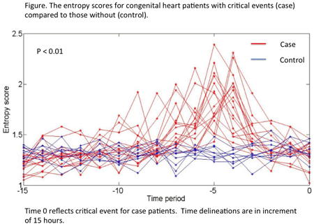
Article Information
vol. 132 no. Suppl 3 A20169
Published By:
American Heart Association, Inc.
Online ISSN:
History:
- Originally published November 6, 2015.
Copyright & Usage:
© 2015 by American Heart Association, Inc.
Author Information
- 1Pediatrics, Stanford Univ, Palo Alto, CA
- 2Surgery, Stanford Univ, Palo Alto, CA
- 3Cardiovascular Surgery, Stanford Univ, Palo Alto, CA
Abstract 18614: Rheumatic Heart Disease Screening in Schools Through Portable Echocardiography: Data From the PROVAR Study
Bruno R Nascimento, Andrea Z Beaton, Maria do Carmo P Nunes, Catherine L Webb, Adriana C Diamantino, Kaciane K Bruno, Cassio O Miri, Gabriel T Pereira, Eduardo L Lopes, Camila G Ferreira, Luciana C Lafeta, Antonio Luiz P Ribeiro, Craig Sable
Circulation. 2015;132:A18614
Abstract
Introduction: The accuracy of clinical examination is limited for early diagnosis of Rheumatic Heart Disease (RHD). The 2012 World Heart Federation (WHF) Criteria provides standardized echocardiographic (echo) guidelines for diagnosis of RHD. There is sparse data on echo prevalence of RHD in Brazil.
Objective: Assess the prevalence of RHD in students (6-18 years) from public schools in low-income areas in Belo Horizonte, Brazil.
Methods: Over 4-days, experts used standard portable echo machines (GE, Vivid-Q) and the 2012 WHF Criteria to determine RHD prevalence. All consented and present children at the visited schools were eligible for participation. Studies identified as abnormal were blindly read by a second cardiologist, with non-agreement adjudicated by a third. RHD prevalence by gender and age (5-11 & 12-18) were compared using Fisher’s Exact Test.
Results: A total of 1381 students were screened across four schools in urban Belo Horizonte, Brazil. The mean age was 13.6±2.8 years, range 5-18 years; 59.3% were female. The prevalence of RHD was 3.8% (53/1381): 3.4% borderline RHD (n=47) and 0.4% definite RHD (n=6). Of these cases, 46 (86.8%) had pathologic mitral regurgitation (≥2cm, seen in 2-views, pan-systolic, and velocity ≥3m/sec) and 7 had pathologic aortic regurgitation (≥1cm, seen in 2-views, pan-diastolic, and velocity ≥3m/sec). One child had mixed mitral and aortic valve disease. There were no cases of mitral or aortic stenosis. There was no difference in RHD prevalence between males and females (p=0.57), but older children had a higher prevalence rate (5-11 years 1.3% vs. 12-18 years 4.5%, p=0.01). Minor congenital heart disease was detected in 8 additional children (5.8%), including one large ASD that was referred for closure.
Conclusions: WHF Criteria make it possible to obtain and accurately compare echo prevalence of RHD among different regions of the world. The PROVAR study shows that, among children living in low-socioeconomic conditions in urban Brazil, RHD remains a significant health burden, comparable to that in other low-resource communities. Screening older children has higher yield. Contemporary data such as these provide justification for investing in comprehensive RHD control programs both in Brazil, and around the globe.
Article Information
vol. 132 no. Suppl 3 A18614
Published By:
American Heart Association, Inc.
Online ISSN:
History:
- Originally published November 6, 2015.
Copyright & Usage:
© 2015 by American Heart Association, Inc.
Author Information
- Bruno R Nascimento1;
- Andrea Z Beaton2;
- Maria do Carmo P Nunes1;
- Catherine L Webb3;
- Adriana C Diamantino4;
- Kaciane K Bruno4;
- Cassio O Miri4;
- Gabriel T Pereira5;
- Eduardo L Lopes5;
- Camila G Ferreira4;
- Luciana C Lafeta4;
- Antonio Luiz P Ribeiro1;
- Craig Sable2
- 1Clínica Médica, Universidade Federal de Minas Gerais, Belo Horizonte, Brazil
- 2Pediatric Cardiology, Children’s National Health System, Washington, DC
- 3Pediatric Cardiology, Univ of Michigan Health System, Traverse City, MI
- 4Cntr de Telessaúde, Hosp das Clínicas da Universidade Federal de Minas Gerais, Belo Horizonte, Brazil
- 5Faculdade de Medicina, Universidade Federal de Minas Gerais, Belo Horizonte, Brazil
Abstract 16644: Interstage Clinical Outcomes and Resource Utilization Among Babies With Single Ventricle: Results of A Randomized Cross-over Study Comparing Standard of Care (Notebook) to Tablet PC-Based Monitoring
Girish Shirali, Lori Erickson, Kathy Goggin, Jonathan Apperson, Michael Bingler, Dawn Tucker, David Williams, Andrea Bradley-Ewing, Vincent Staggs, Amy Lay, Leslie Rabbitt, Richard Stroup
Circulation. 2015;132:A16644
Abstract
Interstage outcomes for babies with single ventricle remain suboptimal. We have developed a tablet PC-based platform (CHAMP) for remote monitoring. This provides immediate access to data and instant alerts to the team. This study evaluates the caregiver experience with CHAMP, and its impact on interstage mortality, morbidity and resource utilization.
Methods: All neonates with single ventricle who were discharged from 5/2014 to 5/2015 were prospectively enrolled. For 1 month after discharge, they were all monitored using a notebook. They were randomized to receive CHAMP at either 1 or 2 months post discharge. A month after randomization, caregivers had to choose either the notebook or CHAMP for the remainder of the inter-stage period. Charts were reviewed for mortality, unplanned readmissions and hospital charges. Caregiver experience was assessed through an exit survey.
Results: We enrolled 24 babies (Norwood, n=11; BT shunt, n=9; no stage I at discharge (balanced circulation), n=3; hybrid, n=1). They were interstage for 3143 days. There was no interstage mortality in either group. While using CHAMP, families transmitted data on 77% of days. Resource utilization is summarized in the Table. CHAMP instant alerts and scheduled daily alerts led to 10 readmissions for issues that were not recognized by caregivers (low saturations (n=6) and poor feeding / weight gain (n=4). When given the option after randomization, 23 of 24 families chose CHAMP. At the end of monitoring, 23 completed an exit survey; when asked what form of monitoring they would choose if they had to do this over, 19 (82%) stated they would choose CHAMP, 3 would choose either, and 1 would choose the notebook.
Conclusions: CHAMP monitoring was associated with significant decreases in unplanned readmission days, ICU days and hospital charges. CHAMP was well-accepted by caregivers, and would appear to facilitate outpatient care for the fragile population of interstage babies with single ventricle.
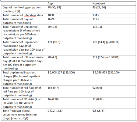
Article Information
vol. 132 no. Suppl 3 A16644
Published By:
American Heart Association, Inc.
Online ISSN:
History:
- Originally published November 6, 2015.
Copyright & Usage:
© 2015 by American Heart Association, Inc.
Author Information
- Girish Shirali1;
- Lori Erickson1;
- Kathy Goggin2;
- Jonathan Apperson1;
- Michael Bingler1;
- Dawn Tucker1;
- David Williams2;
- Andrea Bradley-Ewing2;
- Vincent Staggs3;
- Amy Lay1;
- Leslie Rabbitt1;
- Richard Stroup1
- 1Ward Family Heart Cntr, Children’s Mercy Hosp, Kansas City, MO
- 2Health Outcomes and Health Services Rsch, Children’s Mercy Hosp, Kansas City, MO
- 3Health Services and Outcomes Rsch, Children’s Mercy Hosp, Kansas City, MO
Abstract 15999: Beyond Morbidity and Mortality: Academic Outcomes in Children With Congenital Heart Disease
Matthew Oster, Stephanie Watkins, Kevin Hill, Robert Meyer
Circulation. 2015;132:A15999
Abstract
Introduction: While children with congenital heart disease (CHD) are known to have neurodevelopmental challenges, how these challenges translate to school performance is unknown. The purpose of our study was to compare the academic achievement of children with CHD to that of children without known birth defects.
Hypothesis: Children with CHD would have lower standardized test performance scores and greater need for grade retention than their peers without birth defects.
Methods: We performed a retrospective cohort study comparing educational outcomes for children born 1998-2003 with CHD, identified from the North Carolina (NC) Birth Defects Registry, vs. those without a known birth defect, randomly sampled from NC birth certificates. All children were linked to 3rd grade public school records from the NC Department of Public Instruction through 2012. We performed logistic regression to compare our outcomes of interest: a) meeting standards on the reading and math portions of 3rd grade End of Grade Testing, and b) 3rd grade retention. Models were adjusted for maternal education, public pre-K enrollment, and race/ethnicity.
Results: Of the 5624 subjects with CHD and 10,832 with no birth defect, 51% and 60% were linked, respectively, to 3rd grade standard End of Grade testing records. Compared to children without a birth defect, those with CHD were more likely not to meet proficiency standards in reading (OR 1.4, 95% CI 1.3-1.6), math (OR 1.2, 95% CI 1.1-1.4), or both (OR 1.5, 95% CI 1.3-1.7). (Figure) Similarly, children with CHD were more likely to be retained in 3rd grade (2.8% vs. 1.9%, OR 1.4, 95% CI 1.1-1.9).
Conclusions: Children with CHD have poorer educational achievement as compared to their peers without birth defects. History of CHD should be considered an important factor in determining a student’s need for specialized education services.
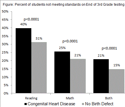
Article Information
vol. 132 no. Suppl 3 A15999
Published By:
American Heart Association, Inc.
Online ISSN:
History:
- Originally published November 6, 2015.
Copyright & Usage:
© 2015 by American Heart Association, Inc.
Author Information
- 1Sibley Heart Cntr Cardiology, Children’s Healthcare of Atlanta, Atlanta, GA
- 2Cntr for Health Promotion and Disease Prevention, Univ of North Carolina – Chapel Hill, Chapel Hill, NC
- 3Pediatrics, Duke Univ Med Cntr, Durham, NC
- 4State Cntr for Health Statistics, North Carolina Div of Public Health, Raleigh, NC
Abstract 15524: Can a Home-based Cardiac Rehabilitation Program Improve the Physical Function Quality of Life in Children With Fontan Circulation?
Roni M Jacobsen, Michael Danduran, Kathleen Mussatto, Jennifer Neubauer, Salil Ginde
Circulation. 2015;132:A15524
Abstract
Introduction: Patients after Fontan operation for complex congenital heart disease (CHD) have reduced exercise capacity and health-related quality of life (HRQOL). Studies suggest hospital-based cardiac rehabilitation programs can improve exercise capacity and HRQOL in patients with CHD; however, these programs have variable adherence rates. The impact of a home-based cardiac rehabilitation program in Fontan survivors is unclear. This pilot study evaluated the safety, feasibility, and benefits of an innovative home-based physical activity program on HRQOL in Fontan patients.
Methods: A total of 14 children, 8-12 yr, with Fontan circulation enrolled in a 12-week moderate/high intensity home-based cardiac physical activity program, which included a home exercise routine and 3 formal in-person assessments at 0, 6, and 12 weeks. Subjects and parents completed validated questionnaires to assess HRQOL. The Shuttle Test Run was used to measure exercise capacity. A Fitbit® Flex Activity Monitor was used to assess adherence to the home activity program.
Results: Of the 14 subjects, 57% were male and 36% had a dominant left ventricle. Overall, 93% completed the program. There were no adverse events. Parents reported improvement in their child’s overall HRQOL (p<0.01), physical function (p<0.01), school function (p=0.01), and psychosocial function (p<0.01). Exercise capacity, measured by Shuttle Run time, improved progressively from baseline to the 6 and 12 week follow up sessions (Figure). Monthly Fitbit® data suggested good adherence to the program.
Conclusion: This 12-week home-based cardiac physical activity program is safe and feasible in Fontan patients. Parent proxy-reported HRQOL and objective measures of exercise capacity significantly improved. A 6 month follow up session is scheduled to assess sustainability. A larger study is anticipated to determine applicability and reproducibility of these findings in other age groups and forms of complex CHD.
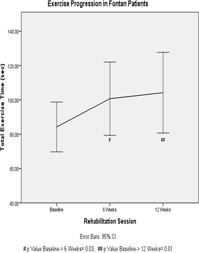
Article Information
vol. 132 no. Suppl 3 A15524
Published By:
American Heart Association, Inc.
Online ISSN:
History:
- Originally published November 6, 2015.
Copyright & Usage:
© 2015 by American Heart Association, Inc.
Author Information
- 1Pediatric Cardiology, Children’s Hosp of Wisconsin/Med College of Wisconsin, Milwaukee, WI
- 2Pediatric Cardiology, Children’s Hosp of Wisconsin, Marquette Univ, Milwaukee, WI
- 3Pediatric Cardiology, Children’s Hosp of Wisconsin, Milwaukee, WI
Abstract 15309: Resource Reduction by Use of Pediatric Chest Pain Standardized Clinical Assessment and Management Plan
Susan F Saleeb, Sarah R McLaughlin, Dionne A Graham, Kevin G Friedman, David R Fulton
Circulation. 2015;132:A15309
Abstract
Using a Standardized Clinical Assessment and Management Plan (SCAMP) for pediatric patients presenting to clinic with chest pain, we evaluated the cost impact associated with implementation of the care algorithm. Previously, we analyzed charges for 406 patients (7-21 yrs) with chest pain seen in 2009, prior to introduction of the SCAMP, and predicted 21% reduction of overall charges had the SCAMP methodology been used. The SCAMP recommended an echocardiogram for history, exam, or ECG findings suggestive of a cardiac etiology for chest pain. Resource utilization for 1520 patients evaluated by the SCAMP from 12/11-4/14 was reviewed in this study. Compared to the 2009 cohort, patients evaluated in the SCAMP presented with similar rates of exertional chest pain (33%, SCAMP vs. 37%, 2009) and significant past medical history (1% vs. 1%). The SCAMP cohort had fewer abnormal physical exam findings (1% vs. 4%) and ECG abnormalities (3% vs. 6%), and more pertinent family history (4% vs. 1%). Echocardiograms were additionally recommended by the SCAMP for exertional syncope (1%), pain worse by supine position, or radiation to the back, jaw, or left arm (5%). Ancillary testing for concurrent symptoms was reduced compared to the 2009 cohort (Figure 1) and compared to predicted: Holter (4% vs. 6%), event monitors (3% vs. 8%), MRI (<1% vs. 1%). Stress testing was not recommended though 4% underwent evaluation. Adherence to SCAMP guidelines for recommended testing approached 80%; slightly fewer echocardiograms were actually performed than recommended. Total testing charges were reduced by an estimated 30% ($740,000) despite a small increase in echocardiogram utilization, and overall charges were reduced by 17% by use of the Chest Pain SCAMP. Given the low incidence of cardiac disease historically as well as detected by the SCAMP algorithm (<1%), further modifications to the algorithm should refine indications for echocardiography, particularly related to exertional symptoms.
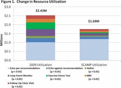
Article Information
vol. 132 no. Suppl 3 A15309
Published By:
American Heart Association, Inc.
Online ISSN:
History:
- Originally published November 6, 2015.
Copyright & Usage:
© 2015 by American Heart Association, Inc.
Author Information
- Susan F Saleeb;
- Sarah R McLaughlin;
- Dionne A Graham;
- Kevin G Friedman;
- David R Fulton
- Cardiology, Boston Children’s Hosp, Boston, MA
Abstract 14733: Influence of High Risk Adrenergic Receptor Genotypes on Outcome in Infants With Single Ventricle
Ronand Ramroop, Dorin Manase, George Manase, Cory Simmons, Jane W Newburger, J William Gaynor, Mark W Russell, Seema Mital
Circulation. 2015;132:A14733
Abstract
Introduction: Variants in adrenergic receptor (ADR) genes are associated with adverse clinical outcomes in patients with heart failure. We evaluated the association of variants in ADR receptor genes with outcomes in infants with single ventricle lesions.
Methods: Infants with single ventricle lesions participating in the Pediatric Heart Network Single Ventricle Reconstruction Trial (randomized to Norwood with modified Blalock-Taussig shunt versus right ventricle-pulmonary artery shunt) underwent genotyping for 4 single nucleotide polymorphisms (SNPs) in 3 ADR genes: ADRB1-A/G (rs1801252), ADRB1-C/G (rs1801253), ADRB2-C/G (rs1042714) and ADRA2A-C/T (rs553668). Linear and logistic regression, deviance tests, t-tests and F-tests were used to analyze association of genotype with clinical outcomes including hospital length of stay (LOS) at Norwood, occurrence of serious adverse events (SAE), and transplant-free survival during 14 months follow-up using a dominant model.
Results: The study included 347 eligible patients (62% male; 83% white). The mean age at Norwood procedure was 6±3.6 days and median Norwood LOS was 8 days. During 14 months follow-up, 147 patients had SAEs, 94 patients died and 14 were transplanted. ADRB1 AA (rs1801252) genotype was associated with longer Norwood LOS. The difference in LOS between AA vs AG/GG was 7.99 days (confidence intervals, 0.27, 15.71; p=0.043). ADRA2A CC (rs553668) genotype was associated with higher odds of SAEs i.e. 103/216 (47.6%) in CC compared to 36/106 (34%) in CT/TT [Odds ratio 1.77 (confidence intervals, 1.09, 2.87), p=0.018]. Transplant-free survival was not different between genotype groups. Combined analysis of risk genotypes did not confer an additive risk of adverse outcomes.
Conclusions: ADR genotypes known to cause adrenergic upregulation were associated with prolonged Norwood hospitalization and/or serious adverse events in infant single ventricles. This may be secondary to adverse effects of adrenergic overexpression on cardiac function and systemic hemodynamics. Analysis is ongoing to replicate these findings for utility as predictive markers for outcomes in infant single ventricles.
Article Information
vol. 132 no. Suppl 3 A14733
Published By:
American Heart Association, Inc.
Online ISSN:
History:
- Originally published November 6, 2015.
Copyright & Usage:
© 2015 by American Heart Association, Inc.
Author Information
- Ronand Ramroop1;
- Dorin Manase1;
- George Manase1;
- Cory Simmons1;
- Jane W Newburger2;
- J William Gaynor3;
- Mark W Russell4;
- Seema Mital1
- 1PEDIATRICS, Hosp for Sick Children, Toronto, Canada
- 2PEDIATRICS, Children’s Hosp Boston, Boston, MA
- 3Surgery, Children’s Hosp of Philadelphia, Philadelphia, PA
- 4PEDIATRICS, CS Mott Children’s Hosp, Ann Arbor, MI
Abstract 12261: Improving Discharge Timeliness on a Pediatric Inpatient Cardiology Unit Through the Implementation of Medical Discharge Criteria Decreases Length of Stay and Reduces Hospital Charges
Andrew Porter, Rhonda Cable, Amanda Kolb, Angela Statile, Christine White, Angela Lorts, Samuel Hanke, Nicolas L Madsen
Circulation. 2015;132:A12261
Abstract
Background: Prior improvement work integrating standardized medical discharge criteria in general pediatric inpatients led to improved discharge timeliness and reduced length of stay (LOS). Our aim was to develop standardized medical discharge criteria on a quaternary pediatric cardiology unit in an effort to improve the timeliness of discharge. We hypothesized that timely discharge would lead to cost savings and a reduced LOS.
Methods and Results: Over 18 months (Jan. 2014 – June 2015), a multi-disciplinary team worked toward the quality improvement aim of increasing the percentage of pediatric cardiology inpatients discharged within 2 hours of achieving medical discharge criteria. We established preset ‘medically ready’ discharge criteria in our hospital EMR, initiated upon admission for all inpatients and modified daily as needed. We organized criteria into 7 categories (congenital heart disease (CHD) medical, CHD post-operative, heart failure/transplant medical, transplant post-operative, electrophysiology (EP) medical, EP post-procedure, and catheterization post-procedure). The median percentage of patients discharged within 2 hours of meeting criteria increased steadily from 35% to 78% over the project period. Concurrently, the median number of patients discharged before noon increased from 18% to 31%. Physician and nursing compliance with necessary process measures was 87% and 89%, respectively. LOS decreased by 8% (3.6 to 3.3 days) from the first 6 months of work to the last 6 months. All-cause readmission remained stable at 15% over the improvement time frame. Review of inpatient charges between Oct. 2014 and June 2015 demonstrated 169 patients (190 discharges) who experienced a delayed discharge based on the set criteria. The sum of charges from care provided after meeting criteria was $231,201 (average of $1,217 per delayed discharge).
Conclusions: Discharge timeliness in pediatric cardiology patients can be improved by standardizing medical discharge criteria. We demonstrate that this can be achieved across all subcategories of pediatric cardiology without increasing the rate of readmission. Improved discharge timeliness not only reduces LOS; it provides an opportunity to substantially decrease medical charges.
Article Information
vol. 132 no. Suppl 3 A12261
Published By:
American Heart Association, Inc.
Online ISSN:
History:
- Originally published November 6, 2015.
Copyright & Usage:
© 2015 by American Heart Association, Inc.
Author Information
- Andrew Porter;
- Rhonda Cable;
- Amanda Kolb;
- Angela Statile;
- Christine White;
- Angela Lorts;
- Samuel Hanke;
- Nicolas L Madsen
- Pediatrics, Cincinnati Children’s Hosp Med Cntr, Cincinnati, OH
Abstract 10945: Impact of Natural or Postoperative Survival Patterns in Long-term Outcome of Adult Survivors With Congenital Heart Disease
Pastora Gallego, Jose M Oliver, Ana E Gonzalez Garcia, Diego Garcia Hamilton, Raquel Yotti Alvarez, Ignacio Ferreira Gonzalez, Francisco Fernandez Aviles
Circulation. 2015;132:A10945
Abstract
Background: Life expectancy of adult survivors with congenital heart disease (CHD) is related to CHD complexity but impact of natural or postoperative survival patterns on long-term outcome have not been reported.
Methods: In a prospective cohort of 3,334 adults (50.6% males) with CHD, followed up to 24 years in a single tertiary centre, we analyzed all-cause mortality, annual death rate, median age of survival (MAS) and standardized mortality ratio (SMR). Data provided by the Spanish national death index were used for mortality analysis. Lesions were classified into 3 groups according to CHD complexity: simple (1,647); moderate (1,283); and severe (404). Patients were then classified into 4 subgroups according to survival pattern: repaired in childhood (1,377); repaired in adulthood (759); CHD that did not require operation (1,035); and inoperable CHD (163).
Results: Median age at first examination was 22 years (IQ range 18-39) and median follow-up time 10.6 years (1-18). At the end of the study 336 patients had died (prevalence 10%; annual death rate 0.89%). In inoperable patients the estimated MAS was significantly lower (48.9 yrs; 95% CI 44-53), but there were not significant differences in MAS between patients repaired in childhood (75.9 yrs), repaired in adulthood (76.0 yrs) or that did not require repair (77.5 yrs). SMR in this latter group was 1.70 (95% CI 1.3-2.1)] but patients repaired in adulthood, repaired in childhood or inoperable had an increased SMR of 2.06 (95% CI 1.7-2.5), 5.87 (95% CI 4.5-7.7) and 24.1 (95% CI 18-33) respectively. The table shows a classification of long-term outcome depending on CHD complexity and survival pattern.
Conclusion: Adult survivors that do not require intervention and patients with repaired simple CHD have excellent life-long prognostic indexes. Excess mortality was demonstrated in repaired moderately complex CHD . Patients with CHD of great complexity have the worst prognosis, especially those that are inoperable.
Article Information
vol. 132 no. Suppl 3 A10945
Published By:
American Heart Association, Inc.
Online ISSN:
History:
- Originally published November 6, 2015.
Copyright & Usage:
© 2015 by American Heart Association, Inc.
Author Information
- Pastora Gallego1;
- Jose M Oliver2;
- Ana E Gonzalez Garcia3;
- Diego Garcia Hamilton3;
- Raquel Yotti Alvarez4;
- Ignacio Ferreira Gonzalez5;
- Francisco Fernandez Aviles4
- 1Intercentre Adult Congenital Heart Disease Unit, Virgen Macarena Univ Hosp, Heart Area, Seville, Spain
- 2Cardiology, Instituto Investigacion Gregorio Marañon, Madrid, Spain
- 3Cardiology, La Paz Univ Hosp, Madrid, Spain
- 4Cardiology, Gregorio Marañon Univ Hosp, Madrid, Spain
- 5Cardiology, Vall d’Hebron Univ Hosp, Barcelona, Spain
Abstract 11011: Long-Term Outcomes of Definitive Surgical Repair for Congenitally Corrected Transposition of the Great Arteries
Yuki Nakamoto, In-Sam Park, Yukihiro Takahashi
Circulation. 2015;132:A11011
Abstract
Background: Congenitally corrected transposition of the great arteries (ccTGA) is characterized by atrioventricular and ventriculoarterial discordance. The indications, timing, and type of repair vary according to the morphology of the heart, the clinical status of the patient, and the patient’s age. Optimal surgical management of patients with ccTGA remains controversial.
Methods: The medical records of all patients with ccTGA who underwent definitive repairs between 1977 and 2013 were retrospectively reviewed. The surgical procedures comprised a physiologic repair in 45 patients, anatomic repair (double switch operation) in 14 patients, and a Fontan-type repair in 12 patients.
Results: The Kaplan-Meier survival at 20 years was similar (75.7% in physiologic repair vs. 83.3% in anatomic repair). However, patients with significant TR showed a worse survival rate than those of the other groups (20 year survival without TVR 96.3%, but only 58.6% with TVR). The freedom from reoperation at 10 years was 78.3% in physiologic repair vs. 66.7% in anatomic repair. The freedom from pacemaker implantation at 10 years was 93.0% in physiologic repair vs. 64.3% in anatomic repair. 14 patients underwent a Fontan-type repair without mortality or heart block.
Conclusions: No differences were observed between long-term survival rates of patients who underwent physiologic versus anatomic repair. The poorest outcome was seen in patients who required tricuspid valve replacement. Performing an anatomic repair should be considered for patients with significant TR. The best outcome was seen in the patients undergoing the Fontan procedure. However, various complications late after Fontan procedure remain a concern.
Article Information
vol. 132 no. Suppl 3 A11011
Published By:
American Heart Association, Inc.
Online ISSN:
History:
- Originally published November 6, 2015.
Copyright & Usage:
© 2015 by American Heart Association, Inc.
Author Information
- 1Pediatric cardiology, Sakakibara Heart Institute, Tokyo, Japan
- 2Pediatric cardiovascular surgery, Sakakibara Heart Institute, Tokyo, Japan
Abstract 15863: Prevalence of Platelet Non-resposiveness to Aspirin by Thromboelastography (teg) in Children With Congenital Heart Disease: An Institutional Experience
Fernando Molina, Stephanie Ghaleb, Cesar Gonzalez de Alba, Sergio Bartakian, John Brownlee
Circulation. 2015;132:A15863
Abstract
Introduction: Aspirin is used to prevent thromboembolism in children with heart defects. Aspirin resistance has been reported in up to 26% of pediatric patients with cardiovascular defects undergoing surgical procedures with significant inpatient variability noted. Because of a patient who had embolic stroke while on therapeutic aspirin doses but in whom aspirin resistance was present on his thromboelastogram (TEG), we chose to obtain a TEG on cardiac patients on aspirin to assess their risk. This study evaluates aspirin resistance noted in these patients.
Hypothesis: Aspirin resistance is high in pediatric patients with heart conditions
Methods: This is a retrospective observational study with IRB approval of 21 patients taking aspirin. Seven Females and 14 males were enrolled, ages 5 months to 27 years. Fourteen had a Fontan surgery. Three had a cavo-pulomanary anastomosis, 1 had HLHS hybrid procedure, 1 had a coronary bypass due to ALCAPA and another had a coronary aneurysm. Aspirin doses were at 5 mg/kg/day but not exceeding 81 mg and had been given at least for one month. Compliance was assured with the patient/family at the time of the clinic visit. Platelet mapping with a TEG was performed on each patient. Aspirin resistance was defined as platelet inhibition below 50%. Variables evaluated were: Level of platelet inhibition(AA plt%), age in months, Weight in Kg, gender and diagnosis. Statistical
Analysis: Chi Square and Pearson Correlation.
Results: Aspirin resistance was present in 77% of these patients (50% in post Fontan and 66% in the remaining patients). There was no significant variation between genders (P 0.576) Correlation between platelet inhibition level for weight were R: -0.3233 (P 0.153) and age were R -0.0923 (P 0.69) both not significant.
Conclusions: The percentage of patients in our group on aspirin with inadequate platelet inhibition is unacceptably high and represents pharmacologic failure based on the TEG. Our findings suggest our cardiac patients may have significantly increased aspirin resistance. A larger cohort is needed to assess if traditional dosing of aspirin for antiplatelet effect is not correct for children.
Article Information
vol. 132 no. Suppl 3 A15863
Published By:
American Heart Association, Inc.
Online ISSN:
History:
- Originally published November 6, 2015.
Copyright & Usage:
© 2015 by American Heart Association, Inc.
Author Information
- Fernando Molina;
- Stephanie Ghaleb;
- Cesar Gonzalez de Alba;
- Sergio Bartakian;
- John Brownlee
- Pediatric Cardiology, Driscoll Children’s Hosp, Corpus Christi, TX
Abstract 15067: Incidence and Outcomes of Neonates With Congenital Heart Disease Complicated by Necrotizing Enterocolitis
Pirouz Shamszad, Shaine A Morris, Deipanjan Nandi, Andrew T Costarino, Bradley S Marino, Joseph W Rossano
Circulation. 2015;132:A15067
Abstract
Introduction: The management of neonates with congenital heart disease (CHD) may be complicated by necrotizing enterocolitis (NEC), however, there is limited multicenter data describing the incidence and outcomes of NEC in the CHD population.
Objective: We aimed to assess the incidence and risk factors for the development of NEC in neonates with major CHD and the impact on survival.
Methods: A retrospective cohort study of neonates with CHD was performed for all index hospitalizations of neonates (<28 days) with major CHD between 2004 and 2014 using the Pediatric Health Information System database. The diagnosis of NEC was determined by the presence of ICD-9 code 777.5x. The incidence of NEC was determined as were risk factors for the development of NEC. Mortality was the primary outcome measure.
Results: Of 38770 neonates with major CHD, 1448 (3.6%) were diagnosed with NEC. The rate of NEC varied between 0-8% by hospital and was not associated with hospital volume (p=0.4). Among neonates with a single, major CHD diagnosis, the rate of NEC was 6% in hypoplastic left heart syndrome (HLHS), 6% in truncus arteriosus (TA) , 4% in tetralogy of Fallot (TOF), 3% in aortic arch obstruction (AO), and 2% in transposition of the great arteries (TGA); these diagnoses accounted for 47% of all NEC. Prematurity and chromosomal anomalies were independently associated with the diagnosis of NEC (p≤0.01 for both). Unadjusted mortality among neonates with NEC was 24% compared to 12% in neonates without NEC (OR 2.4, 95%CI 2.1-2.7). When evaluating changes in adjusted mortality associated with NEC by CHD diagnosis, TOF mortality increased from 8% to 16% (p<.01), TGA increased from 5% to 21% (p<0.01), AO increased from 6% to 20% (p<0.01), HLHS increased from 22% to 28% (p=.07), and TA decreased from 13% to 12% (p=0.7). Median LOS was higher in neonates with NEC than without NEC (54d [IQR 31-93] vs. 18d [IQR 9-34], p<0.01) as was median hospital charge ($600k [IQR 310k-1.1m] vs. $220k [IQR 100k-430k], p<0.01).
Conclusions: The incidence of NEC among neonates with major CHD is highest in HLHS and TA. NEC is associated with significantly higher hospital mortality, LOS, and charges. Determining modifiable factors associated with NEC may allow for interventions to reduce morbidity in this population.
Article Information
vol. 132 no. Suppl 3 A15067
Published By:
American Heart Association, Inc.
Online ISSN:
History:
- Originally published November 6, 2015.
Copyright & Usage:
© 2015 by American Heart Association, Inc.
Author Information
- Pirouz Shamszad1;
- Shaine A Morris2;
- Deipanjan Nandi1;
- Andrew T Costarino3;
- Bradley S Marino4;
- Joseph W Rossano1
- 1Div of Cardiology, Children’s Hosp of Philadelphia, Philadelphia, PA
- 2Pediatric Medicine, Cardiology, Texas Children’s Hosp, Houston, TX
- 3Div of Critical Care, Children’s Hosp of Philadelphia, Philadelphia, PA
- 4Div of Cardiology, Ann & Robert H. Lurie Children’s Hosp of Chicago, Chicago, IL
Abstract 13794: Serial Brain Metabolism in Term Neonates With Congenital Heart Disease: Association With Clinical Factors and Neurodevelopmental Outcomes
Anna L Harbison, Jodie K Votava-Smith, Sylvia Del Castillo, S Ram Kumar, Vincent K Lee, Hollie A Lai, Sharon H O’Neil, Stefan Bluml, Lisa Paquette, Ashok Panigraphy
Circulation. 2015;132:A13794
Abstract
Objectives: Term congenital heart disease (CHD) neonates demonstrate pre-operative (op) abnormal brain metabolism (reduced N-acetylaspartate (NAA), elevated lactate) on long echo MR spectroscopy (MRS). We sought to delineate associations between serial brain metabolism and patient and perioperative clinical factors in term neonates with CHD using short echo MRS. We measured NAA and lactate as well as other metabolites important for brain connectivity such as neurotransmitters glutamate/glutamine and GABA.
Methods: Subjects were prospectively enrolled to undergo pre and post-op 3T short echo single voxel MRS of parietal white matter with absolute quantitation of 15 metabolites using LCModel. Neurodevelopment (ND) was assessed via 18 month Battelle Developmental Inventory. Linear and logistic regression with false discovery rate correction was used for statistical analysis.
Results: Eighty subjects were enrolled 2009-2015 and 21 term CHD infants underwent both pre and post-op MRS. Eight infants had at least one MRS and ND. NAA and glutamate were significantly decreased post-op compared to pre-op (p<0.0001), with no significant difference in other metabolites. Pre-op factors including lower Apgar score, birth weight, head circumference and PaO2 and higher arterial pH and serum lactate were associated with lower NAA (p<0.002). Single ventricle anatomy was associated with low NAA, high myo-inositol and low glutamine/glutamate compared to two ventricles (p<0.01). Longer cardipulmonary bypass time, but not deep hypothermic circulatory arrest, was associated with reduced NAA (p<0.001). Post-op, global alteration in multiple serial brain metabolites (NAA, lactate, glutamate/glutamine, GABA, myo-inostol) were associated with longer ICU and hospital stay (p<0.03). In those with ND testing, high GABA correlated with low cognitive domain score, while high glutamine correlated with low motor score (p<0.03).
Conclusion: In term CHD neonates, serial brain metabolism by MRS demonstrates alterations beyond NAA, including neurotransmitters GABA and glutamate/glutamine. These abnormalities are associated with multiple clinical pre and post-op factors and also predict prolonged hospital stay and 18 month ND.
Article Information
vol. 132 no. Suppl 3 A13794
Published By:
American Heart Association, Inc.
Online ISSN:
History:
- Originally published November 6, 2015.
Copyright & Usage:
© 2015 by American Heart Association, Inc.
Author Information
- Anna L Harbison1;
- Jodie K Votava-Smith1;
- Sylvia Del Castillo1;
- S Ram Kumar2;
- Vincent K Lee3;
- Hollie A Lai4;
- Sharon H O’Neil5;
- Stefan Bluml6;
- Lisa Paquette7;
- Ashok Panigraphy8
- 1Cardiology, Children’s Hosp Los Angeles, Los Angeles, CA
- 2Cardiothoracic Surgery, Children’s Hosp Los Angeles, Los Angeles, CA
- 3Radiology, Chldren’s Hosp Pittsburgh, Pittsburgh, PA
- 4Radiology and Nuclear Medicine, Children’s Hosp Los Angeles, Los Angeles, CA
- 5Neuropsychology, Children’s Hosp Los Angeles, Los Angeles, CA
- 6Radiology, Children’s Hosp Los Angeles, Los Angeles, CA
- 7Neonatology, Children’s Hosp Los Angeles, Los Angeles, CA
- 8Radiology, Children’s Hosp of Pittsburgh, Pittsburgh, PA
Abstract 17596: Readmissions After Adult Congenital Heart Surgery: Frequency and Risk Factors
Yuli Kim, Wei He, Thomas MacGillivray, Oscar J Benavidez
Circulation. 2015;132:A17596
Abstract
Introduction: Readmissions after adult cardiac surgery is an important clinical outcome. Despite case complexity, 30-day readmission after adult congenital heart surgery (CHS) has been under studied.
Hypothesis: Readmissions after adult CHS are common. There exist identifiable risk factors for unplanned readmission.
Methods: We obtained State Inpatient Databases for Washington, New York, Florida, and California 2009-2011 and selected admissions 18 – 49 years with ICD-9 CM codes indicating adult CHS. We defined readmission as any non-elective hospitalization for a given patient < 30 days of discharge from the index CHS admission. We examined patient demographic and clinical variables and defined complications using the Society of Thoracic Surgeons Congenital Heart complication short list. Case complexity was grouped using the Risk Adjustment for Congenital Heart Surgery-1 (RACHS-1) categories. High resource use (HRU) admissions were defined as those > 90th percentile for total hospital charges. Multivariate analyses using generalized estimating equations were performed to identify adjusted risk factors for readmission.
Results: Of 9860 admissions, there were 1675 readmissions (17%). Median length of stay for readmissions was 4 days and mortality rate 2.1%. Most common indications for readmission were cardiac (pericardial disease, atrial fibrillation, heart failure) and infectious (endocarditis, post-operative infection). On multivariable analysis, black race (adjusted odds ratio [AOR] 1.2 p<0.04), renal insufficiency (AOR 1.8 p<0.001), drug abuse (AOR 1.4 p=0.001), obesity (AOR 1.2 p=0.03), government-sponsored insurance (AOR 1.3 p<0.001), median income <$40,000 (AOR 1.3 p=0.001), index admissions with complication (AOR 1.1 p=0.04), RACHS-1 3 complexity (AOR 1.3 p=0.04), HRU (AOR 1.4 p=0.003), and emergent (AOR 1.5 p<0.001) were risk factors for readmission.
Conclusions: One out of 6 adult CHS hospitalizations result in unplanned readmission. Lower socioeconomic status, black race, renal failure, obesity, drug abuse, HRU, emergent index admission, and complications are risk factors for subsequent unplanned 30-day readmission. These risk factors may serve as potential quality improvement targets to reduce readmissions.
Article Information
vol. 132 no. Suppl 3 A17596
Published By:
American Heart Association, Inc.
Online ISSN:
History:
- Originally published November 6, 2015.
Copyright & Usage:
© 2015 by American Heart Association, Inc.
Author Information
- 1Medicine, Hosp of the Univ of Pennsylvania, Philadelphia, PA
- 2Pediatrics, Massachussets General Hosp, Boston, MA
- 3Surgery, Massachusetts General Hosp, Boston, MA
- 4Pediatrics, Massachusetts General Hosp, Boston, MA
Abstract 16951: Long term Incidence and 30-mortality of Myocardial Infarction in Congenital Heart Disease Survivors Compared With the General Population: A Nationwide Cohort Study
Morten Olsen, Bradley S Marino, Jonathan R Kaltman, Jørgen Videbæk, William T Mahle, Gail D Pearson, Nicolas L Madsen
Circulation. 2015;132:A16951
Abstract
The population of adults with congenital heart disease (CHD) is growing and aging. Some adult CHD survivors have an increased risk of myocardial infarction (MI) due to the specific CHD, and the presence of CHD may impair prognosis following MI depending on CHD severity. Few data exist on the incidence and prognosis of MI in the adult CHD population. We aimed to compare the incidence and mortality of MI in adult survivors of CHD with the general population with attention to the impact of CHD severity.
Methods: In this cohort study we used two nationwide population based medical databases to identify individuals diagnosed with CHD in Denmark between 1963 and 1974 (before 15 years of age) and from 1977 to 2012 (at any age). Patients were followed from 1977 to 2012 for first time MI using data from the Danish National Registry of Patients (DNRP). The DNRP is a nationwide hospital discharge registry covering all Danish hospitals. For each CHD subject we identified 10 controls from the general population, matched by sex and birth year. A unique personal identifier assigned at birth and used in all Danish public registries enabled virtually complete follow-up for migration, death or MI. We computed cumulative incidences and hazard ratios (HR) of MI. Subsequently, we computed 30 day-mortality following MI for CHD subjects and controls, as well as the corresponding HR adjusted for age and sex.
Results: We identified 14,038 CHD subjects. By 70 years of age, the cumulative incidence of MI was 10% among CHD subjects. The HR of MI among CHD subjects compared with controls was 3.5 (95% CI: 2.9-4.3) below 50 years of age, and 1.9 (95% CI: 1.7-2.2) > 50 years. The 30-day mortality was 16% for the 322 CHD subjects experiencing an MI during follow-up; the corresponding HR comparing 30-day post-MI mortality between CHD subjects and controls was 1.9 (95% CI: 1.4-2.6). Patients with severe CHD had a 30-day post-MI mortality HR of 4.1 (95% CI: 2.2-7.5), somewhat higher than among those with mild and moderate CHD (HR 1.5; 95% CI: 1.1-2.3).
Conclusion: Both incidence of MI and post MI 30-day mortality were increased in individuals with CHD compared with the general population. This should be an important consideration for optimizing preventive and acute cardiovascular care for the high-risk adult CHD population.
Article Information
vol. 132 no. Suppl 3 A16951
Published By:
American Heart Association, Inc.
Online ISSN:
History:
- Originally published November 6, 2015.
Copyright & Usage:
© 2015 by American Heart Association, Inc.
Author Information
- Morten Olsen1;
- Bradley S Marino2;
- Jonathan R Kaltman3;
- Jørgen Videbæk1;
- William T Mahle4;
- Gail D Pearson3;
- Nicolas L Madsen5
- 1Dept of Clinical Epidemiology, Aarhus Univ Hosp, Århus, Denmark
- 2Divs of Cardiology and Critical Care Medicine, Ann and Robert H. Lurie Children’s Hosp of Chicago, Chicago, IL
- 3Div of Cardiovascular Sciences at the National Heart, Lung, and Blood Institute (NHLBI),, National Institutes of Health, Bethesda, MD
- 4Dept of Pediatric’s, Egleston hospital, Atlanta, GA
- 5Heart Institute, Cincinnati Children’s, Cincinnati, OH
Abstract 15875: Waitlist Outcomes for Adults With Congenital Heart Disease Listed for Heart Transplantation in the United States
Laith Alshawabkeh, Nan Hu, Knute D Carter, KellyAnn Light-McGroary, Joseph E Cavanaugh, Heather L Bartlett
Circulation. 2015;132:A15875
Abstract
Background: Heart failure represents a final common pathway for many adult survivors with congenital heart disease (ACHD). Data assessing severity and management of heart failure are limited, and often heart transplantation is the only viable treatment option. The criteria used to determine priority status at the time of transplant listing, however, does not account for ACHD physiology. We investigated the differences in death or delisting due to clinical worsening in ACHD vs. non-ACHD candidates for heart transplantation.
Methods: We conducted a retrospective study of all patients listed for heart transplantation in the United States between 1999 and 2014 using the Scientific Registry of Transplant Recipients. Patients were censored at the time of transplant or delisting due to improvement.
Results: Among the 1,290 ACHD and 35,559 non-ACHD patients listed for heart transplantation, median age was 35 vs. 56 years and 62% vs. 76% were male, respectively. Waitlist outcomes for the full follow up time per initial listing status are shown in Figure 1. Of the patients who died or worsened, 57% were initially listed at the lowest priority status for ACHD compared to 40% for non-ACHD. In patients initially listed at status 1A, the 180-day actuarial probability of death or delisting due to worsening was 41% for ACHD vs. 29% for non-ACHD, p < 0.01; and for death alone was 29% vs. 21%, p = 0.05; respectively, Figure 2.
Conclusion: ACHD candidates for heart transplantation in the United States are more frequently listed at the lowest priority status, and when listed at the highest priority status are more likely to die or be delisted due to clinical worsening compared to other candidates.
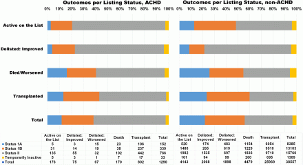
Article Information
vol. 132 no. Suppl 3 A15875
Published By:
American Heart Association, Inc.
Online ISSN:
History:
- Originally published November 6, 2015.
Copyright & Usage:
© 2015 by American Heart Association, Inc.
Author Information
- Laith Alshawabkeh1;
- Nan Hu2;
- Knute D Carter2;
- KellyAnn Light-McGroary1;
- Joseph E Cavanaugh2;
- Heather L Bartlett3
- 1Cardiology, Univ of Iowa Hosps & Clinics, Iowa City, IA
- 2Biostatistics, Univ of Iowa, Iowa City, IA
- 3Cardiology, Univ of Wisconsin Sch of Medicine and Public Health, Madison, WI
Abstract 10919: Contemporary Life Expectancy and Standardized Mortality of Adult Survivors With Congenital Heart Lesions
Jose M Oliver, Pastora Gallego, Ana E Gonzalez Garcia, Diego Garcia Hamilton, Raquel Yotti, Ignacio Ferreira Gonzalez, Francisco Fernandez Aviles
Circulation. 2015;132:A10919
Abstract
Background: Survival beyond the age of 18 years in patients born with congenital heart disease (CHD) is now near 90% but contemporary estimates of outcome in adult survivors are lacking.
Methods: In a prospective cohort of 3,334 adults with CHD followed up to 24 years in a single tertiary centre median age of survival (MAS) was estimated by computing left-truncated and right-censored Kaplan-Meier curves with age as time scale. Standardized mortality ratio (SMR) was also determined using one-sample log-rank test from age at diagnosis-, sex- and time of follow-up-adjusted death rates of general population in Spain. Patients were classified into 3 groups according to CHD complexity: I, simple (1,647); II, moderate (1,283); and III, severe (404). For mortality analysis, data provided by the Spanish national death index were used.
Results: There were 1,688 males and 1,646 females. Median age at first examination was 22 years (IQR 18-39) and median follow-up time 10.6 years (1-18). At the end of the study 336 patients had died (prevalence 10%; annual incidence 0.89%). Estimated MAS was 78.0 years (95% CI 76-82) for group I; 72,1 years (68-78) for group II; and 51.1 years (48-53) for group III (p<0.001). MAS was older than 75 years in all diagnostic categories in group I; between 60 and 75 years (moderate reduction) in subvalvular aortic stenosis, Ebstein anomaly, coarctation of the aorta, tetralogy of Fallot and complete transposition; and < 60 years (severe reduction) in Eisenmenger syndrome, single ventricle, AV discordance and pulmonary atresia The SMR was 1.66 (1.4-2.5) for the group I; 3.27 (2.6-4.1) for the group II; and 20.4 (10-26) for the group III (p<0.001 in all, compared to reference population). The SMRs for main individual diagnostic categories are shown in figure.
Conclusion: Life expectancy for CHD adults is reduced proportionally to complexity of the heart defect. Data for individual diagnostic categories might be used as a prognostic index.
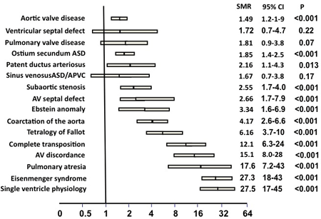
Article Information
vol. 132 no. Suppl 3 A10919
Published By:
American Heart Association, Inc.
Online ISSN:
History:
- Originally published November 6, 2015.
Copyright & Usage:
© 2015 by American Heart Association, Inc.
Author Information
- Jose M Oliver1;
- Pastora Gallego2;
- Ana E Gonzalez Garcia3;
- Diego Garcia Hamilton4;
- Raquel Yotti5;
- Ignacio Ferreira Gonzalez6;
- Francisco Fernandez Aviles5
- 1Cardiology, Instituto Investigacion Gregorio Marañon, Madrid, Spain
- 2Intercentre Adult Congeneital Heart Disease Unit, Virgen Macarena Univ Hosp, Heart Area, Sevilla, Spain
- 3Cardiology, La Paz Univ Hosp, Madrid, Spain
- 4Cardiology, La Paz Univ Hspital, Madrid, Spain
- 5Cardiology, Gregorio Marañon Univ Hosp, Madrid, Spain
- 6Cardiology, Vall d’Hebron Univ Hosp, Barcelona, Spain
Abstract 19626: Cardio-renal Syndrome in Single Ventricle Patients
Scott I Aydin, Kristi Glotzbach, Amy Skversky, Daphne Hsu
Circulation. 2015;132:A19626
Abstract
Introduction: Cardio-renal syndrome (CRS) in single ventricle patients undergoing stage I or II palliation has not been described. CRS in other high-risk pediatric populations has been associated with greater risk of morbidity and mortality. We aim to describe CRS in single ventricle patients undergoing stage I or II palliation and their associations with clinical outcomes.
Methods: Data was collected from the Pediatric Heart Network (PHN) Infant with Single Ventricle trial. Demographics, survival, and data pertaining to stage I and II palliations were obtained. CRS was determined for each patient based on the estimated creatinine clearance (eCCL) as follows; age 0-days to 12-months eCCL < 40 ml/min/1.73m2 and > 12-months eCCL < 90 ml/min/1.73m2. Descriptive statistics and univariate analyses using the student t-test, Mann-Whitney U, chi-square test or Fisher’s exact test were performed to determine the association between CRS and clinical outcomes as well as mortality, respectively.
Results: Two hundred twenty-nine patients, with median age 6-days [4-7], underwent stage I palliation during the study period, of which 86 (38%) had CRS after their palliation. The most common diagnosis was hypoplastic left heart syndrome (HLHS) with 144 patients (63%) with 169 patients (74%) undergoing a Norwood procedure with a modified Blalock-Taussig (MBT) or Sano shunt. Of the 229 patients, 167 (73%), with median age 151-days [123-176], underwent stage II palliation during the study period, of which 60 (48%) had CRS after their palliation. Of the 60-patients, 25 (42%) also had CRS after their stage I palliation. Stage I palliation patients with CRS had significantly lower baseline eCCL as compared to patients with no CRS (36 vs. 56, p < 0.001), as well as significantly longer ICU and hospital length of stays (14-days vs. 11-days, p < 0.001 and 29-days vs. 11-days, p < 0.001). Twenty patients (9%) died prior to stage II palliation; of the 20 patients, 14 patients (70%) had CRS. CRS was significantly associated with mortality prior to stage II palliation (odds ratio 2.6; 95% CI 1.03, 6.7; p = 0.03).
Conclusion: CRS in single ventricle patients is common and associated with lower pre-operative eCCL, longer lengths of ICU and hospital stay, and mortality prior to stage II palliation.
Article Information
vol. 132 no. Suppl 3 A19626
Published By:
American Heart Association, Inc.
Online ISSN:
History:
- Originally published November 6, 2015.
Copyright & Usage:
© 2015 by American Heart Association, Inc.
Author Information
- Scott I Aydin;
- Kristi Glotzbach;
- Amy Skversky;
- Daphne Hsu
- Cardiology and Critical Care, The Children’s Hosp at Montefiore, Bronx, NY
Abstract 18452: Importance of “Dynamic” Rather Than “Static” Central Venous Pressure and Venous Capacitance in Fontan Circulation During Exercise
Clara Kurishima, Seiko Kuwata, Yoichi Iwamoto, Hirotaka Ishido, Satoshi Masutani, Hideaki Senzaki
Circulation. 2015;132:A18452
Abstract
Background: High central venous pressure (CVP) is a key driver of developing late complications in patients after Fontan operation. However, we often realize diversity of clinical outcome among patients with similar levels of CVP measured at resting condition during catheterization. We hypothesized that CVP variably increases with exercise and that degree of the increase is associated with clinical outcomes. We also hypothesized that venous capacitance (VC) is an important determinant of the CVP elevation.
Methods: Dynamic changes in CVP estimated from continuous monitoring of peripheral venous pressure were measured during treadmill exercise (TM) in 21 Fontan patients and 10 age-matched control patients with biventricular (BV) circulation. The VC was calculated during temporal occlusion of inferior vena cava (IVC) at the time of catheterization.
Results: Fontan patients exhibited significantly shorter duration of exercise with limited increases in heart rate and cardiac output. CVP significantly increased during exercise in both groups, but the degree of CVP elevation was much higher in Fontan patients than in controls (Fontan: from 10.8 to 18.4 mmHg vs. control: from 4.3 to 8.8 mmHg, P<0.05). Importantly, maximum CVP during exercise in Fontan group varied considerably among patients with similar resting CVP, and is significantly and positively correlated with a liver fibrosis marker of type 4 collagen 7S (P <0.01). Furthermore, maximum CVP during exercise in Fontan group negatively correlated with the VC, but not with pulmonary resistance.
Conclusions: CVP of the Fontan circulation easily rises during exercise in proportion to the reduced VC, and the degree of CVP rise may be a contributor of hepatic complication. These results suggest the importance of assessing dynamic CVP in patients after Fontan operation. The results also suggest potential importance of daily life modification and/or medical treatment to minimize the dynamic changes in CVP to improve the prognosis of Fontan patients. VC may be a viable target for this purpose.
Article Information
vol. 132 no. Suppl 3 A18452
Published By:
American Heart Association, Inc.
Online ISSN:
History:
- Originally published November 6, 2015.
Copyright & Usage:
© 2015 by American Heart Association, Inc.
Author Information
- Clara Kurishima;
- Seiko Kuwata;
- Yoichi Iwamoto;
- Hirotaka Ishido;
- Satoshi Masutani;
- Hideaki Senzaki
- 1Pediatric Cardiology, Saitama Med Cntr, Saitama Med Univ, Kawagoe, Japan
Abstract 17372: Comparison of Resting Energy Expenditure With Energy Intake in Neonates With Hypoplastic Left Heart Syndrome Following Stage 1 Palliation
Christine L French, Kimberly I Mills, Brian K Walsh, Aditya K Kaza, Alexis Cole, Catherine Baird, Hilary Bond, Nilesh M Mehta, Christopher P Duggan, John N Kheir
Circulation. 2015;132:A17372
Abstract
Introduction: Nutrition provision and fluid administration following cardiac surgery in neonates impacts ventilator-dependent days, intensive care unit length of stay and diuretic administration. Resting energy expenditure (REE) in neonates following stage 1 palliation (S1P) for hypoplastic left heart syndrome (HLHS) has been poorly characterized and is essential to the provision of optimal nutrition.
Methods: We continuously measured REE from postoperative day (POD) 0 through 7 following S1P in n=22 infants using an in-line, indirect calorimetric device (E-COVX moduleTM, GE Healthcare). Artifactual REE values were identified and filtered according to a predefined algorithm. Energy intake (EI) was abstracted from a detailed nutrition record.
Results: There were no complications related to REE measurement, and 7.4% of measurements were found to be artifactual and excluded. Mean REE was 35.9 ± 10.1 kcal/kg/day on POD 0 and increased by an average of 3.1 ± 0.2 kcal/kg/day each day between POD 0 and 7 (A). Current recommended dietary allowance (RDA) estimates propose energy needs to be 108 kcal/kg/day in these patients.
From POD 0-1, EI was less than REE by 18.5±4.3 kcal/kg/day, as fluid intake was primarily comprised of blood products (46.8±7.4%) and flushes (24.5±1.9%, B). From POD 2-7, EI exceeded REE by 22.6±4.4 kcal/kg/day (cumulative amount of 111 ± 7.7 kcal/kg/day over a 5 day period, C). EI from carbohydrates was disproportionately high (69.1±6.2%), and that from protein remained low (7.6±1.2% of calories, 1.53±0.3 g/kg/day, C).
Conclusions: Following S1P, EI correlates poorly with measured REE, which is significantly lower than RDA estimates. This may result in excessive fluid and calorie administration in this fluid-sensitive population. Continuous measurement of REE is safe to perform and should be considered in neonates following cardiac surgery. A future trial comparing restricted EI to estimated EI in postoperative neonates is necessary.
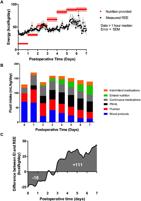
Article Information
vol. 132 no. Suppl 3 A17372
Published By:
American Heart Association, Inc.
Online ISSN:
History:
- Originally published November 6, 2015.
Copyright & Usage:
© 2015 by American Heart Association, Inc.
Author Information
- Christine L French;
- Kimberly I Mills;
- Brian K Walsh;
- Aditya K Kaza;
- Alexis Cole;
- Catherine Baird;
- Hilary Bond;
- Nilesh M Mehta;
- Christopher P Duggan;
- John N Kheir
- Cardiology, Boston Children’s Hosp, Boston, MA
Abstract 17532: Impact of High Hepatic Wedge Pressure, Portal Hypertension, on Hemodynamics and Liver Function in Patients After the Fontan Operation
Jun Negishi, Hideo Ohuchi, Kanae Noritake, Yosuke Hayama, Osamu Sasaki, Tohru Iwasa, Aya Miyazaki, Osamu Sasaki
Circulation. 2015;132:A17532
Abstract
Background: Fontan circulation may be, in part, characterized by portal hypertension that can be estimated by hepatic vein wedge pressure (HWP).
Purpose: To evaluate associations of HWP in cosecutive 318 Fontan patients (18±7 years old) during catheterization and compared the results with the hemodynamics, liver function and the related plasma biomarkers. HWP significantly correlated positively with inferior vena cava pressure (IVCP), pulmonary artery resistance (Rp), ration of Rp to systemic artery resistance (Rs) (Rp/Rs), and cardiac index and negatively with Rs and arterial and mixed venous oxgen saturations. Of those, high IVCP, low Rp and high Rp/Rs were independently correlated with high HWP (p<0.05 for all). As for the liver function, HWP significantly correlated positively with plasma levels of total bilirubin (TB), hyaluronic acid, and type IV collagen (TyIV) and negatively with plasma levels of albumin and cholinesterase (ChE). Of those, ChE, TB and TyIV were independently correlated with high HWP (p<0.01 for all). High HWP, low ChE, and high TyIV significantly correlated with high plasma nitric oxide products (p<0.01).
Conclusion: High HWP associates with a significant reduction of Rp and Rs with preserved cardiac output. This unique Fontan circulation with Rs-dominant reduction may be due to both portal hypertension and overproduction of nitric oxide secondary to liver dysfunction.
Article Information
vol. 132 no. Suppl 3 A17532
Published By:
American Heart Association, Inc.
Online ISSN:
History:
- Originally published November 6, 2015.
Copyright & Usage:
© 2015 by American Heart Association, Inc.
Author Information
- Jun Negishi;
- Hideo Ohuchi;
- Kanae Noritake;
- Yosuke Hayama;
- Osamu Sasaki;
- Tohru Iwasa;
- Aya Miyazaki;
- Osamu Sasaki
- Pediatric Cardiology, National Cerebral and Cardiovascular Cntr, Osaka, Japan
Abstract 17543: The Chicken or the Eggs? Examining the Causal Relationship Between the Development of Atrioventricular Valve Regurgitation and Ventricular Dysfunction in Patients With Functionally Single-Ventricle Physiology
Yasuhiro Kotani, Devin Chetan, Selvi Senthilnathan, Arezou Saedi, Christopher A Caldarone, Glen S Van Arsdell, Luc Mertens, Osami Honjo
Circulation. 2015;132:A17543
Abstract
Introduction: Development of atrioventricular valve regurgitation (AVVR) with or without ventricular dysfunction (VD) often occurs during the first 6 months of life and has a significant impact on outcomes for single-ventricle patients. Yet, it is not well known whether AVVR causes VD or vice versa. Thus, we sought to identify the timing and causal relationship between AVVR and VD.
Methods: Among 156 consecutive single-ventricle patients who had staged palliation (2005- 2012), 28 who had AVV repair at the time of stage II (n=24, 86%) or inter-stage (n=4, 14%) were reviewed. Diagnosis included HLHS in 17 (61%) patients, tricuspid atresia in 2 (7%), and others in 9 (32%). Ventricular morphology was left-dominant in 6 (21%) patients and right-dominant in 22 (79%). AVV morphology included mitral in 6 (21%) patients, tricuspid in 18 (64%), and common in 4 (14%). Serial echocardiograms were reviewed to identify the timing of development of AVVR and/or VD.
Results: After stage I palliation, ventricular end-diastolic dimension (VEDD) z-score significantly increased from 4.01 to 5.69 (p<0.01) AVVR (Figure). By the time of stage II palliation, VEDD further increased and subsequent AVV annular dilation occurred, resulting in 23 patients with significant AVVR. None of the patients, however, had significant VD before stage II palliation/AVV repair, but 9 patients developed significant VD after AVV repair.
Conclusions: Ventricular dilation occurred immediately after stage I palliation and continued until stage II palliation. Secondary annular dilation occurred inter-stage and this further triggered the development of AVVR. Tangible ventricular dysfunction was not seen before AVV repair, however, important ventricular dysfunction was unmasked after volume unloading surgery. Heart failure management and early intervention to significant AVVR may reduce the incidence of ventricular dysfunction following AVV repair.
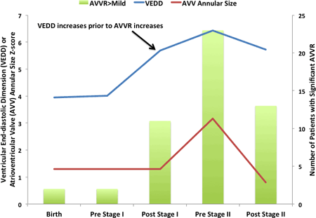
Article Information
vol. 132 no. Suppl 3 A17543
Published By:
American Heart Association, Inc.
Online ISSN:
History:
- Originally published November 6, 2015.
Copyright & Usage:
© 2015 by American Heart Association, Inc.
Author Information
- Yasuhiro Kotani1;
- Devin Chetan2;
- Selvi Senthilnathan3;
- Arezou Saedi2;
- Christopher A Caldarone2;
- Glen S Van Arsdell2;
- Luc Mertens3;
- Osami Honjo2
- 1Cardiovascular Surgery, Okayama Univ, Okayama, Japan
- 2Cardiovascular Surgery, The Hosp for Sick Children, Toronto, Canada
- 3Cardiology, The Hosp for Sick Children, Toronto, Canada
Abstract 14160: Rest and Reserve Functions in Fontan Patients With Right Ventricular Morphology are Worse Than Those With Left Ventricular Morphology
Satoshi Masutani, Seiko Kuwata, Clara Kurishima, Yoichi Iwamoto, Hirofumi Saiki, Hirotaka Ishido, Masanori Tamura, Hideaki Senzaki
Circulation. 2015;132:A14160
Abstract
Introduction: Fontan patients with right ventricular morphology (RV-F) are associated with worse outcome and exercise capacity compared with those with left ventricular morphology (LV-F). Diminished cardiac reserve is one of major mechanisms of impaired exercise capacity in heart failure patients. However, it remains unclear whether and how rest and reserve functions differ between RV-F and LV-F.
Hypothesis: We assessed the hypothesis that rest and reserve functions in RV-F may be worse than those in LV-F.
Methods: This study included 28 RV-F and 17 LV-F (6.0 vs 6.2 years. p=N.S.). Ventricular pressure-area relationships were determined during cardiac catheterization, both before and after β-adrenergic stimulation with dobutamine (5 microg/kg/min) and increased heart rates by atrial pacing.
Results: There were no significant differences in heart rate, central venous pressure, and pulmonary vascular resistance between RV-F and LV-F. End-systolic pressure in RV-F was lower than that in LV-F (89 vs 97mmHg), while end-systolic and end-diastolic volume in RV-F were larger than those in LV-F. Consistently, contractile function in RV-F was worse than that in LV-F (20.1±10.5 vs 30.8±19.7 mmHg/cm2 хm2 in Ees index; and 978±237 vs 1186±297 mmHg/s in dp/dt max). The worse contractile function in RV-F was persisted after dobutamine (24.9 vs 45.1 mmHg/cm2 хm2 in Ees index; and 1721 vs 2241 mmHg/s in dp/dt max). Furthermore, the response of Ees index to faster heart rate in RV-F was blunted, which was in striking contrast to positive chronotropic response in LV-F (Figure).
Conclusions: Compared with LV-F, RV-F has worse systolic function at rest and markedly attenuated chronotropic reserve in systolic function, which can be responsible for worse outcome in RV-F. Given the lack of definitive therapy for Fontan failure, development of medical therapy to improve rest and reserve function should be pursued for better prognosis in Fontan patients.
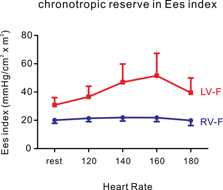
Article Information
vol. 132 no. Suppl 3 A14160
Published By:
American Heart Association, Inc.
Online ISSN:
History:
- Originally published November 6, 2015.
Copyright & Usage:
© 2015 by American Heart Association, Inc.
Author Information
- Satoshi Masutani1;
- Seiko Kuwata1;
- Clara Kurishima1;
- Yoichi Iwamoto1;
- Hirofumi Saiki1;
- Hirotaka Ishido1;
- Masanori Tamura2;
- Hideaki Senzaki1
- 1Pediatric Cardiology, Saitama Med Univ Saitama Med Cntr, Kawagoe, Japan
- 2Pediatrics, Saitama Med Univ Saitama Med Cntr, Kawagoe, Japan
Abstract 13536: Vascular Function is Predictive of Functional Outcomes in Adolescent and Young Adult Fontan Survivors
Bryan H Goldstein, Elaine M Urbina, Philip R Khoury, Zhiqian Gao, Michelle A Amos, Wayne A Mays, Andrew Redington, Bradley S Marino
Circulation. 2015;132:A13536
Abstract
Introduction: Fontan survivors demonstrate diminished vascular function and functional outcomes. The relationships between measures of vascular function and functional outcomes have not been established.
Methods: Cross-sectional single-institution study of Fontan survivors ages 8-25 years. Multimodality assessment of endothelial function (reactive hyperemia index [RHI] and flow mediated dilation [FMD]) and arterial stiffness (augmentation index [AI] and baseline pulse amplitude [BPA]) was performed with peripheral arterial tonometry (PAT), brachial FMD and pulse wave analysis (PWA). Aerobic capacity was determined using cardiopulmonary exercise testing with cycle ergometry and a ramp protocol. Quality of life (QOL) was assessed using the pediatric QOL inventory (PedsQL) and physical activity (PA) was assessed using the PA Questionnaire (PAQ). Vascular measures served as predictor variables while functional outcome measures served as outcome variables. Bivariate predictors (p<0.1) were candidates for inclusion in multivariable models. Model development utilized linear regression and ordered logistic regression techniques.
Results: Sixty patients (52% male) completed the protocol with a mean age of 13.9 ± 4.1 years and duration of Fontan circulation of 9.9 ± 4.2 years. Maximal (max) exercise was achieved in 54 subjects with mean peak and %-predicted oxygen consumption (VO2) of 27.8 ± 7.6 ml/kg/min and 71.0 ± 21.2%, respectively. FMD-RHI (p<0.05) was associated with VO2at anaerobic threshold (AT, R2 0.33). PAT-BPA (p<0.05) was associated with the ratio of minute ventilation to carbon dioxide at AT (R2 0.40). FMD-RHI, PAT-AI and PWA-AI (p<0.05) were associated with max VO2 (R2 0.36-0.53), %-predicted max VO2 (R2 0.40-0.56) and max O2 pulse (R2 0.26-0.31). PAT-AI and PAT-BPA (p<0.05) were associated with PedsQL total (R2 0.19-0.34) and physical heath summary score (R2 0.36-0.41). FMD max and PAT-BPA (p<0.05) were associated with PAQ score (R2 0.33-0.38).
Conclusions: Increased arterial stiffness and decreased endothelial function are associated with lower aerobic capacity, QOL and physical activity in adolescent and young adult Fontan survivors. Vascular dysfunction may be an important target for future therapeutic trials.
Article Information
vol. 132 no. Suppl 3 A13536
Published By:
American Heart Association, Inc.
Online ISSN:
History:
- Originally published November 6, 2015.
Copyright & Usage:
© 2015 by American Heart Association, Inc.
Author Information
- Bryan H Goldstein1;
- Elaine M Urbina1;
- Philip R Khoury1;
- Zhiqian Gao1;
- Michelle A Amos1;
- Wayne A Mays1;
- Andrew Redington1;
- Bradley S Marino2
- 1The Heart Institute, Cincinnati Children’s Hosp Med Cntr, Cincinnati, OH
- 2Cardiology, Ann and Robert H. Lurie Children’s Hosp of Chicago, Chicago, IL
Abstract 10756: Chronic Kidney Disease in Long-term Survivors After Fontan Palliation
Sheena Sharma, Rebecca L Ruebner, Susan L Furth, Kathryn M Dodds, Jack Rychik, David J Goldberg
Circulation. 2015;132:A10756
Abstract
Background: The Fontan operation is a palliative procedure for children with congenital single ventricle heart disease. With advances in prenatal diagnosis and surgical techniques, more children are surviving into adulthood with unique cardiovascular physiology. Little is currently known about long-term kidney function in these patients.
Hypothesis: We hypothesize that long-term survivors after Fontan palliation will have a higher prevalence of chronic kidney disease (CKD) compared to healthy controls.
Methods: We performed a retrospective cohort study of subjects evaluated through the Single Ventricle Survivorship Program (SVSP) at the Children’s Hospital of Philadelphia between July 1, 2010 and December 5, 2014 and healthy children similar in sex and age. The primary outcome was CKD, defined as eGFR <90 ml/min/1.73m2 calculated with age-appropriate estimating equations using creatinine and cystatin C. Secondary outcomes included proteinuria and hyperparathyroidism.
Results: The Fontan cohort included 68 subjects with mean age of 13.9 years (SD 5.8) at SVSP visit who were 11.2 years (SD 5.7) from Fontan operation. The healthy cohort included 70 patients with mean age of 15.9 years (SD 3.9). Mean eGFR was 102.6 versus 101.9ml/min/1.73m2 (p=0.89) in pediatric Fontan versus healthy subjects using the complete CKiD equation, and 128.5 versus 129.7ml/min/1.73m2 (p=0.56) in adult Fontan versus healthy subjects using the CKD-EPI creatinine and cystatin formula. 10% of Fontan subjects had an eGFR<90 ml/min/1.73m2. Mean intact parathyroid hormone level was higher at 68.0pg/mL (SD 35.4) in the Fontan group compared to 26.0pg/mL (SD 13.6) in the healthy group. Proteinuria was found within 34% of the Fontan group compared to 4.6% within the control group.
Conclusion: We found that 10% of subjects have eGFR <90ml/min/1.73m2 after Fontan palliation which would indicate CKD if this remained persistent over time. Although the majority of the cohort had normal kidney function by eGFR, we found a higher proportion with proteinuria and increased parathyroid hormone levels which may indicate early kidney disease. Future studies will focus on evaluating changes in kidney function over time in long-term survivors after Fontan palliation.
Article Information
vol. 132 no. Suppl 3 A10756
Published By:
American Heart Association, Inc.
Online ISSN:
History:
- Originally published November 6, 2015.
Copyright & Usage:
© 2015 by American Heart Association, Inc.
Author Information
- Sheena Sharma1;
- Rebecca L Ruebner1;
- Susan L Furth1;
- Kathryn M Dodds3;
- Jack Rychik3;
- David J Goldberg3
- 1Nephrology, The Children’s Hosp of Philadelphia, Philadelphia, PA
- 2Nephrology, Children’s Hosp of Philadelphia, Philadelphia, PA
- 3Cardiology, The Children’s Hosp of Philadelphia, Philadelphia, PA
Abstract 9747: Truth in Advertising: Are We Providing Consumers With the Proper Information to Participate in Decision Making About Hypoplastic Left Heart Therapies?
Richard A Friedman
Circulation. 2015;132:A9747
Abstract
Introduction: Consumer Health Informatics has become an integral part of healthcare in 2015. The purpose of this survey study is to examine the information available on the internet to a parent(s) of a fetus diagnosed in utero with hypoplastic left heart disease (HLHD) and compare those to scientific, peer reviewed published data. Specifically, is the the provider data complete enough for prospective parent(s) to make a rational decision as to whether to proceed with a 3 stage repair or choose another option.
Hypothesis: The formally untested hypothesis is that there is a signifcant “gap” between what is actually known to providers and what is presented to prospective parent(s)on provider websites.
Methods: Using the list of the top 10 rated Pediatric Cardiovascular programs as ranked by the U.S. News World Report (USNWR) we collected the published information with respect to: # cases of HLHD operated on for Stage 1 Norwood at the hospital/program in question, Stage 2 Glenn shunt and Stage 3 Fontan; mortality at each stage ; interstage mortality ; mortality data for patients completing all 3 stages ; neurodevelopemntal outcomes and other morbidities. For comparison, we used published data on outcomes of HLHD surgery and follow-up from the Society for Thoracic Surgery as it encompasses the largest collection of datasets from many U.S. programs. We compared the STS data set to the publsihed information from the websites..
Results: Websites did not present the statistical data presented by the STS. In addition, no website listed the exact incidence of their interstage mortality, neurodevelopemntal outcomes nor morbidities at their site.
Conclusions: 1. Information published on websites of the top 10 USNWR programs provide information that tell only part of the story of outcomes for HLHD. 2. Critical pieces of information such as interstage mortality and significant morbidities, are not reported. 3.We must reasses the amount and type of data presented on hospital/program websites

Article Information
vol. 132 no. Suppl 3 A9747
Published By:
American Heart Association, Inc.
Online ISSN:
History:
- Originally published November 6, 2015.
Copyright & Usage:
© 2015 by American Heart Association, Inc.
Author Information
- Richard A Friedman
- Pediatrics, Cohen Children’s Med Cntr, New Hyde Park, NY
Abstract 20100: Parental Presence at Cardiac Intensive Care Unit Bedside Transfer Rounds Reduces Parental Anxiety: Results of a Randomized Controlled Trial
Vijay Anand, Elina Williams, Mohamed Elgendi, Leanne Meakins, Chentel Cunningham, Heather McCrady, Gerda Tawfiq, Nicola Devlin, Kumar Shine, Bodil Larsen, Ivan Rebeyka, Ian Adatia
Circulation. 2015;132:A20100
Abstract
Introduction: The transfer of children from the pediatric cardiac intensive care unit (PCICU) to the ward is a time of great anxiety for the parents of children and medical vulnerability for children who are receiving complex therapies.
Hypothesis: We assessed the hypothesis that parental presence at bedside transfer rounds would reduce parental anxiety and improve patient safety following transfer of children from PCICU to the ward.
Methods: We undertook a randomized controlled trial of children discharged from the PCICU to the ward. Consenting parents were randomized to be absent (control group) or present (intervention group) at multidisciplinary face to face bedside transfer rounds. The primary outcome measure was parental stress measured by the validated Spielberger’s State -Trait Anxiety Inventory (STAI) pre and post transfer. Secondary outcome measures included unplanned readmission to the PCICU, medication errors and emergency calls to the ward. We excluded patients being transferred between intensive care units.
Results: We enrolled 230 subjects (control group n=93, intervention group n=91, failed to complete study n= 46). The 2 groups were matched with respect to gender (male 46% control vs 54% intervention), age (median age control 1.9 yrs (range 0.02 to 16.3) vs intervention 0.9 (0.02 to 17), parental age 32 yrs (18-64) vs 33 (20-60), parental years of schooling 15.5 years ( 7-26) vs 15 (9-24), presence of medical co-morbidities (33% each group). There was significantly greater reduction in trait (p=0.004, state (p=0.01) and total anxiety (p=0.0012) pre and post transfer in the intervention group vs the control group. There were no differences in minor medication errors (36 vs 33), unplanned PCICU re-admissions (11 vs 12) and emergency ward calls(7 vs 8)
Conclusions: Parental presence at face to face multidisciplinary transfer rounds from the PCICU is associated with reduced parental anxiety without change in medication errors, readmission rates or emergency calls to the ward. Reduced parental anxiety may improve parental satisfaction with their child’s care.
Article Information
vol. 132 no. Suppl 3 A20100
Published By:
American Heart Association, Inc.
Online ISSN:
History:
- Originally published November 6, 2015.
Copyright & Usage:
© 2015 by American Heart Association, Inc.
Author Information
- Vijay Anand1;
- Elina Williams1;
- Mohamed Elgendi2;
- Leanne Meakins3;
- Chentel Cunningham3;
- Heather McCrady3;
- Gerda Tawfiq4;
- Nicola Devlin3;
- Kumar Shine1;
- Bodil Larsen1;
- Ivan Rebeyka5;
- Ian Adatia1
- 1Pediatrics, Univ of Alberta, Edmonton, Canada
- 2Computer Sciences, Univ of Alberta, Edmonton, Canada
- 3Pediatrics, Stollery Children’s Hosp, Edmonton, Canada
- 4Pharmacy, Stollery Children’s Hosp, Edmonton, Canada
- 5Cardiac Surgery, Univ of Alberta, Edmonton, Canada
Abstract 19722: Cardiac Output Estimation by Inert Gas Rebreathing is Accurate in Mechanically Ventilated Children With Heart Disease
Amanda Marma, Alexander R Opotowsky, Brian K Walsh, Jesse J Esch, James A Dinardo, Barry D Kussman, Jonathan Rhodes
Circulation. 2015;132:A19722
Abstract
Introduction: Real time estimates of pulmonary (Qp) and systemic blood flow (Qs) could improve pediatric cardiac care. Current generation inert gas rebreathing (IGR) devices are safe, portable, and easy to use; can estimate Qp and Qs rapidly at the bedside; and can now be adapted for use in intubated patients. We compared IGR and two types of Fick Qp estimates, those using measured (FickM) and assumed (FickA) oxygen consumption (VO2) values, in mechanically ventilated pediatric cardiac patients with no shunt lesion or exclusive right-to-left shunt. Secondarily, we compared measured VO2 with assumed values and values back-calculated from the Fick equation (using measured saturations, hemoglobin and IGR Qp).
Methods: In 18 intubated patients in the pediatric catheterization laboratory, the modified ventilator-compatible InnocorTM device was used to measure IGR Qp and breath-by-breath VO2; assumed VO2 was taken from LaFarge tables. Sampled pulmonary arterial and venous saturations were used to calculate FickM and FickA. Bland-Altman agreement and Spearman correlation were assessed for IGR Qp with FickA Qp and FickM Qp. Secondarily, agreement was analyzed between measured VO2, assumed VO2, and VO2 calculated from the Fick equation.
Results: Subjects were aged 4-23 years, with a range of cardiac diagnoses. The figure shows Bland-Altman plots for Qp. Compared with FickA, IGR Qp had mean bias -0.9 L/min, 95% limits of agreement (=±1.96 SD) -2.8 to +1.0 L/min, and r=0.75. Compared with FickM, IGR Qp had mean bias -0.2 L/min, 95% limits of agreement -1.3 to +1.0 L/min, and r=0.90. Agreement of assumed and measured VO2 was poor (bias +23%, ±55%); measured VO2 agreed better with Fick calculated VO2 than assumed VO2.
Conclusions: IGR Qp estimates agree well with Fick Qp estimates. This agreement improves when VO2 is measured, as assumed VO2 agrees poorly with measured VO2. IGR is an attractive option for bedside monitoring of Qp in intubated children.
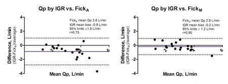
Article Information
vol. 132 no. Suppl 3 A19722
Published By:
American Heart Association, Inc.
Online ISSN:
History:
- Originally published November 6, 2015.
Copyright & Usage:
© 2015 by American Heart Association, Inc.
Author Information
- Amanda Marma1;
- Alexander R Opotowsky1;
- Brian K Walsh2;
- Jesse J Esch1;
- James A Dinardo3;
- Barry D Kussman3;
- Jonathan Rhodes1
- 1Dept of Cardiology, Boston Children’s Hosp, Boston, MA
- 2Div of Critical Care, Boston Children’s Hosp, Boston, MA
- 3Div of Cardiac Anesthesia, Boston Children’s Hosp, Boston, MA
Abstract 17889: A Randomized Controlled Trial of Moderate Glucose Control After Complex Pediatric Cardiac Surgery
Xu Wang, Shoujun Li, Xia Li, Kai Ma, Juxian Yang, Min Zeng, Shengli Li, Leilei Duan
Circulation. 2015;132:A17889
Abstract
Introduction: Infants and children after cardiac surgery may develop hyper- or hypoglycemia, associating with higher nosocomial infection or neurological morbidities.
Hypothesis: The hypothesis of this perspective randomized control study is moderate glucose control may improve clinical outcomes.
Methods: We randomly assigned children(≤3 years of age) who were admitted to the pediatric cardiac intensive care unit (PICU) after cardiopulmonary bypass surgery into either moderate glucose control group (target blood glucose: 110-143mg per deciliter) or conventional glucose control group (target level below 200mg per deciliter). The primary outcome was the rate of nosocomial infection in PICU. Second outcomes include hospital mortality, duration of mechanical ventilation and PICU stay, and a composite morbidity variable included events of requiring extracorporeal membrane oxygenation, delayed sternal closure, dialysis-dependent renal failure and hypoglycemia.
Results: A total of 593 patients underwent randomization: 293 to moderate glucose control group and 300 to conventional glucose control group. The baseline data were balanced between these two groups. Mean 72 hours time-weighted blood glucose average was lower in the moderate control group than in the conventional group (129.9±22.1 mg per deciliter vs. 138.8±28.6 mg per deciliter, p<0.001). Although no statistically significance reached, there is a trend that nosocomial infection occurred less in moderate glucose group (17 (5.80%) vs. 29 (9.67%), P=0.054). Duration of mechanical ventilation was shorter in the moderate control group (18 (range, 4-300) hours vs 22 (range 3-771) hours, p=0.046). Hypoglycaemia (blood glucose ≤65mg per deciliter) which occurred in 7(2.39%) patients in the moderate group versus 8 (2.67%) in the conventional group, was statistically similar between groups. Other secondary outcomes did not differ significantly between groups.
Conclusions: With moderate glucose control, although the major clinical outcomes did not change, this randomized control study showed shorter ventilation time after pediatric cardiac surgery, substantially benefit for patients with complex congenital cardiac anomalies.
Article Information
vol. 132 no. Suppl 3 A17889
Published By:
American Heart Association, Inc.
Online ISSN:
History:
- Originally published November 6, 2015.
Copyright & Usage:
© 2015 by American Heart Association, Inc.
Author Information
- Xu Wang;
- Shoujun Li;
- Xia Li;
- Kai Ma;
- Juxian Yang;
- Min Zeng;
- Shengli Li;
- Leilei Duan
- Dept of Pediatric Intensive Care Unit, Fuwai Hosp, Chinese Academy of Med Science, Peking Union Med College, Beijing, China
Abstract 17967: Systemic Hypertension in Children After Superior Cavo-pulmonary Shunt is Associated With Cerebrovascular Dysautoregulation
Antonio G Cabrera, Ronald B Easley, Rachel Dugan, Michelle Goldsworthy, Katherine K Kibler, Emmett D McKenzie, Charles D Fraser, Jeffrey S Heinle, Dean B Andropoulos, Lara S Shekerdemian, Daniel J Penny, Kenneth M Brady
Circulation. 2015;132:A17967
Abstract
Introduction: Hypertension is frequently seen after superior cavo-pulmonary shunt. It is unknown if hypertension is necessary to maintain cerebral blood flow due to increased cerebral venous pressure. We sought to determine the range of arterial blood pressures (ABP) associated with intact and impaired autoregulation after superior cavo-pulmonary shunt.
Hypothesis: Hypertension (mean ABP>60 mmHg) is associated with cerebrovascular dysautoregulation after superior cavo-pulmonary shunt.
Methods: All patients < 12 months undergoing superior cavo-pulmonary shunt from 10/2014 were eligible. Subjects underwent continuous 100 Hz monitoring of ABP, pulmonary arterial pressure (PAP), and near-infrared spectroscopy measurements of cerebral oximetry (rSO2) and cerebral blood volume (CBV). Cerebrovascular autoregulation was measured by the hemoglobin volume index (HVx). ABP and CBV were low-pass filtered as 10 sec average values. Pearson’s correlation coefficient was performed over 300 sec windows. The associations between HVx changes relative to ABP and PAP were tested using linear regression with generalized estimation of equations. Optimal ABP and PAP defined by lowest HVx was determined using a curve-fit algorithm. The relationship between PAP and ABP was tested by piecewise regression.
Results: Ten patients were enrolled. Median age and weight were 6.5 months and 6.2 kg. Optimal ABP and PAP were obtained in 7/10. HVx became impaired with increased ABP (top panel) and increased PAP (middle panel), indicating worse cerebrovascular dysautoregulation. PAP increased with increasing ABP (r = 0.55, p<0.0001) with an intercept of 72 mmHg above which ΔPAP/ΔABP doubled from 0.23 [0.22- 0.24] to 0.46 [0.43 – 0.49] (bottom panel). Elevations of ABP above optimal for HVx did not improve rSO2 (p>0.05).
Conclusion: Hypertension after superior cavo-pulmonary shunt is associated with elevated PAP, no improvement in rSO2, and cerebrovascular dysautoregulation.
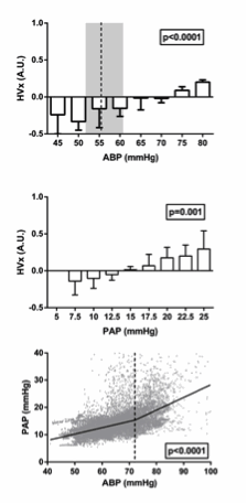
Article Information
vol. 132 no. Suppl 3 A17967
Published By:
American Heart Association, Inc.
Online ISSN:
History:
- Originally published November 6, 2015.
Copyright & Usage:
© 2015 by American Heart Association, Inc.
Author Information
- Antonio G Cabrera;
- Ronald B Easley;
- Rachel Dugan;
- Michelle Goldsworthy;
- Katherine K Kibler;
- Emmett D McKenzie;
- Charles D Fraser;
- Jeffrey S Heinle;
- Dean B Andropoulos;
- Lara S Shekerdemian;
- Daniel J Penny;
- Kenneth M Brady
- Pediatrics, Baylor College of Medicine, Houston, TX
Abstract 16687: A Genetic Contribution to Postoperative Complete Heart Block Following Congenital Heart Surgery
Andrew H Smith, English C Flack, Kimberly Crum, Prince J Kannankeril
Circulation. 2015;132:A16687
Abstract
Background: A familial variant of complete heart block (CHB) is described in association with a substitution mutation in Connexin-40 (GJA5 Q58L) resulting in impaired gap junction formation. We sought to identify genomic predictors that may independently predispose children to the development of early postoperative complete atrioventricular (AV) block following congenital heart surgery.
Methods: Subjects undergoing congenital heart surgery at our institution were consecutively recruited and enrolled from September 2007 through December 2014. In addition to DNA, demographic and perioperative clinical data were obtained from all subjects.
Results: Over the course of the study period, there were 1466 subjects enrolled who underwent 1986 operative procedures; DNA was available for analysis in 1756 cases (1424 subjects). The incidence of at least one episode of postoperative complete AV block was 3.7% (n=65). Genotyping of the common missense polymorphism rs10465885 in GJA5 among subjects yielded allele frequencies in Hardy Weinberg equilibrium (p=0.73), and analysis demonstrated a gene-dose effect association between T allele presence and incidence of postoperative complete AV block (linear by linear association, p=0.009). Multivariate logistic regression analysis demonstrates that TT homozygosity is associated with a two-fold increased odds of new onset complete AV block (OR 2.1, 95%CI 1.2-3.5, p=0.006), independent of other significant univariate predictors including patient weight (p<0.001), operative procedure including closure of a ventricular septal defect (p<0.001), chromosomal anomaly (p=0.016), cross clamp time (p<0.001), ionized calcium and serum lactate on admission (both p<0.001), inotrope use on admission (p<0.001), and preoperative digoxin administration (p<0.001).
Conclusions: A common missense single nucleotide polymorphism within a gene encoding Connexin-40 is associated with the development of early postoperative complete heart block, independent of established risk factors. A refined understanding of genotype-phenotype relationships may serve to guide early postoperative care following congenital heart surgery.
Article Information
vol. 132 no. Suppl 3 A16687
Published By:
American Heart Association, Inc.
Online ISSN:
History:
- Originally published November 6, 2015.
Copyright & Usage:
© 2015 by American Heart Association, Inc.
Author Information
- Andrew H Smith;
- English C Flack;
- Kimberly Crum;
- Prince J Kannankeril
- Pediatrics, Vanderbilt Univ Sch of Medicine, Nashville, TN
Abstract 15022: Alkaline Phosphatase Activity After Infant Cardiac Surgery: Kinetics and Association With Post-operative Cardiovascular Support
Jesse Davidson, Tracy T Urban, Suhong Tong, Mark Twite, Christine Baird, Ludmila Khailova, James Jaggers, Paul Wischmeyer
Circulation. 2015;132:A15022
Abstract
Introduction: Alkaline phosphatase (AP) may be protective after CV surgery through dephosphorylation of endotoxin and extracellular adenine nucleotides. AP activity falls after infant CV surgery and low activity is associated with increased support requirements. However, the kinetics of AP loss and recovery, the isoenzymes involved, and the temporal relation of low AP activity to increased support are unknown. We hypothesized loss of all isoenzymes during surgery leads to decreased AP activity and that early low AP activity is associated with increased cardiovascular support at 6-48hr.
Methods: Prospective study of 93 infants <120 d/o undergoing CV surgery with bypass. Total/isoenzyme specific AP activity and vasoactive inotropic score (VIS) were obtained pre-operation, pre-separation and at 6, 24, 48 and 72hr after ICU admission. AP activities were determined by commercial assay and concentration by multiplex immunoassay. Low AP activity was defined as ≤80 U/L.
Results: Median AP activity decreased through 6hr due to decreased bone and liver 2 isoenzyme activities (fig 1). Liver 1 isoenzyme increased post-bypass then decreased through 72hr. Activity was highly correlated with concentration at all time points (r=0.83-0.89; p<0.0001). Nadir AP activity coincided with peak VIS. Infants with low AP activity at 6hr had significantly higher VIS through 48hr (6hr 12.1 vs 8.1, p<0.0005; 24hr 11.2 vs 5.3, p<0.0001; 48hr 6.9 vs 3.0, p<0.0005). Similar findings occurred with low pre-separation and 24hr AP activity. On multivariate analysis, low AP activity at 6hr was independently associated with higher VIS at 6, 24 and 48hr.
Conclusions: Loss of bone and liver 2 isoenzymes leads to decreased AP activity after infant CV surgery. Low early AP activity is associated with increased cardiovascular support requirements 6-48hr later. AP activity may serve as an early biomarker of post-operative hemodynamic instability and a potential therapeutic target in this population.
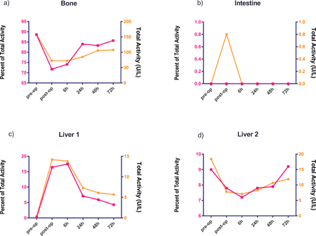
Article Information
vol. 132 no. Suppl 3 A15022
Published By:
American Heart Association, Inc.
Online ISSN:
History:
- Originally published November 6, 2015.
Copyright & Usage:
© 2015 by American Heart Association, Inc.
Author Information
- Jesse Davidson1;
- Tracy T Urban2;
- Suhong Tong3;
- Mark Twite4;
- Christine Baird5;
- Ludmila Khailova5;
- James Jaggers6;
- Paul Wischmeyer5
- 1Pediatric Cardiology/Critical Care, Children’s Hosp Colorado, Aurora, CO
- 2CCRO, Children’s Hosp Colorado, Aurora, CO
- 3Biostatistics, Children’s Hosp Colorado, Aurora, CO
- 4Pediatric Cardiac Anesthesia, Children’s Hosp Colorado, Aurora, CO
- 5Anesthesia, Univ of Colorado, Aurora, CO
- 6Surgery, Univ of Colorado, Aurora, CO
Abstract 13540: Peritoneal Dialysis vs. Furosemide for the Treatment of Oliguria in Infants After Cardiopulmonary Bypass
David M Kwiatkowski, Stuart L Goldstein, David S Cooper, David P Nelson, David L Morales, Catherine D Krawczeski
Circulation. 2015;132:A13540
Abstract
Objectives: Acute kidney injury (AKI) is a frequent and serious complication in infants after cardiac surgery with cardiopulmonary bypass (CPB). Often the earliest sign is oliguria, which can lead to fluid overload, prolonged mechanical ventilation and ICU stay, abnormal electrolytes and increased mortality. We hypothesized that the use of peritoneal dialysis (PD) compared to furosemide use would mitigate these complications.
Methods: This is a single-center, surgical complexity-stratified, randomized controlled trial performed within a cohort of patients younger than 6mo undergoing cardiac surgery with a preoperative plan of PD catheter placement due to risk of post-CPB kidney injury. If enrolled patients had urine output < 1mL/kg/hour for 4 hours during the first postoperative day, the patient was randomized to either a standardized regimen of furosemide or PD. If the patient demonstrated poor response to furosemide, PD could be initiated on postoperative day 2.
Results: A total of 73 patients were randomized and completed the trial, including 2 patients who were randomized to furosemide and subsequently received PD. Using intention-to-treat analysis, the PD group was less likely to have fluid overload, had a lower delayed-extubation rate and fewer electrolyte replacements. There were no differences in mortality or ICU/hospital stay. No serious PD related complications were observed. PD was discontinued early in 9 patients due to pleural-peritoneal communication.
Conclusions: Our study demonstrates that the use of PD in neonates with oliguria after CPB is associated with reduced morbidity but not differences in mortality or ICU/hospital stay. A multi-center study is necessary to further support these findings as well as determine association with mortality.
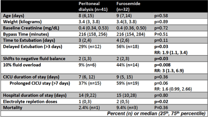
Article Information
vol. 132 no. Suppl 3 A13540
Published By:
American Heart Association, Inc.
Online ISSN:
History:
- Originally published November 6, 2015.
Copyright & Usage:
© 2015 by American Heart Association, Inc.
Author Information
- David M Kwiatkowski1;
- Stuart L Goldstein2;
- David S Cooper3;
- David P Nelson3;
- David L Morales3;
- Catherine D Krawczeski1
- 1Pediatric Cardiology, Lucile Packard Children’s Hosp Stanford, Palo Alto, CA
- 2Pediatric Nephrology, Cincinnati Children’s Hosp Med Cntr, Cincinnati, OH
- 3Heart Cntr, Cincinnati Children’s Hosp Med Cntr, Cincinnati, OH
Abstract 13639: Time to Extubation May Indicate Quality of Care Following Neonatal Cardiac Surgery
Joshua Blinder, Ravi Thiagarajan, Kathryn Williams, Meena Nathan, John Mayer, Thomas Kulik
Circulation. 2015;132:A13639
Abstract
Background: Most neonates survive cardiac surgery, but assessing quality of care is challenging. We aimed to determine: a) minimum time to extubation (TTE) assuming “perfect” care (i.e. no complications and adequate repair) and b) how potentially avoidable events influence TTE.
Methods: Neonates <30 days old undergoing RACHS-1 classifiable surgery from 1/09-1/13 were reviewed using existing data. Technical performance score (TPS) measures adequacy of repair using imaging and clinical criteria prior to hospital discharge: class 1 (trivial/no residua), class 2(minor residua) and class 3 (major residua/re-intervention). Minimum TTE was determined for RACHS-1 class 2, 3-4 and 5-6. Classes 3 and 4 were collapsed as TTE was similar and 5-6 due to too few class 5 pts. Multiple linear regression was used to identify the variables associated with TTE days.
Results: 601 neonates underwent surgery, 58% were male, median age (IQR) was 5.6 (3.6, 9.4) days, median (IQR) BSA was 0.21 (0.19, 0.22) m2; 27% had single ventricle palliation. STS defined-preop factors occurred in 35% (e.g. preop ventilation, genetic anomalies, shock, need for CPR, and sepsis) while postop complications in 72%. 95% of patients with class 3 TPS and 67% of pts with class 1-2 TPS had postoperative complications (p<0.001). Preop (p=0.015) and postop (p<0.001) complications, RACHS-1 class (p<0.001) and class 3 TPS (p<0.001) were associated with increased TTE. Table 1 indicates that with class 3 TPS, preop factors and postop complications, TTE increases across each RACHS-1 class, with the lowest TTE occurring in patients with an class 1 or 2 TPS and no preop factors or postop complications.
Conclusion: Class 3 (major residua/re-intervention) surgical performance, preop factors and postop complications affect TTE. Recognizing that not all postop complications are avoidable, TTE may be a surrogate measurement of the quality of care.
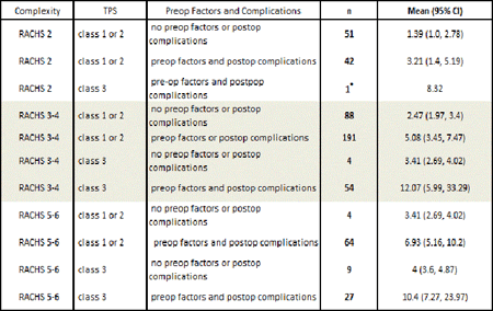
Article Information
vol. 132 no. Suppl 3 A13639
Published By:
American Heart Association, Inc.
Online ISSN:
History:
- Originally published November 6, 2015.
Copyright & Usage:
© 2015 by American Heart Association, Inc.
Author Information
- 1Pediatrics and Anesthesia, Children’s Hosp of Philadelphia, Philadelphia, PA
- 2Cardiology, Boston Children’s Hosp, Boston, MA
- 3Cardiovascular Surgery, Boston Children’s Hosp, Boston, MA
Abstract 11665: Oral Triiodothyronine Supplementation Decreases Time to Extubation After Pediatric Open Heart Surgery in Indonesia
Eva M Marwali, Novik Budiwardhana, Radityo Prakoso, Dicky Fakhri, Cindy E Boom, Mulyadi M Djer, Sudigdo Sastroasmoro, Michael A Portman
Circulation. 2015;132:A11665
Abstract
Background: In Indonesia, children with congenital heart disease (CHD) usually present later for corrective surgery than in developed countries. These children show chronic malnutrition associated with low thyroid hormone levels, which decline further immediately after open-heart surgery.
Objectives: To test the hypothesis that oral triiodothyronine (T3) supplementation improves clinical outcome indexed by time to extubation (TTE) after aortic cross-clamp release.
Methods: The study was a single center, randomized, double blind, and placebo controlled trial in children with CHD younger than 2 years of age undergoing open-heart surgery. Oral T3 or placebo was administered every 6 hours for 60 hours. TTE served as the primary endpoint. Hemodynamics, echocardiography parameters, inotropic score, fluid balance, diuresis, and sepsis complication were serially recorded and evaluated as secondary endpoints
Results: A total of 78 patients were allocated to T3 group and 74 patients to the control group. Oral T3 prevented the declination in serum T3 noted in control group. The T3 group had a shorter time to extubation than placebo with a mean (SD) 25.99(24.30) and 40.71(56.29) hours, respectively and a hazard ratio for chance of extubation (95% CI) 1.49 (1.06-2.11), p=0.02. The median (IQ) cardiac index increased significantly after surgery in T3 (day 1 vs. day 2, 3.28 (2.46 – 4.46) to. 3.65 (2.65 – 4.77) L/min/m2, p=0.01), but not in the placebo group. More diuresis and a significantly more negative fluid balance (p=0.03) were found in T3 group during the first 72 hours post surgery. No difference in the use of inotropic agents or loop diuretics. Sepsis was significantly lower in T3 group with incidence (%) of sepsis in T3 and placebo group 1(1) vs. 10(13) patients, (p=0.004), respectively. Otherwise there was no difference in adverse events including arrhythmia.
Conclusion: Oral T3 supplementation is safe and provides multiple clinical benefits in an extremely vulnerable population of children in a developing country. Any reduction in TTE could improve sparse resources by increasing ventilator and ICU availability.
Article Information
vol. 132 no. Suppl 3 A11665
Published By:
American Heart Association, Inc.
Online ISSN:
History:
- Originally published November 6, 2015.
Copyright & Usage:
© 2015 by American Heart Association, Inc.
Author Information
- Eva M Marwali1;
- Novik Budiwardhana1;
- Radityo Prakoso2;
- Dicky Fakhri1;
- Cindy E Boom1;
- Mulyadi M Djer3;
- Sudigdo Sastroasmoro3;
- Michael A Portman4
- 1Pediatric Cardiac ICU, National Cardiovascular Cntr Harapan Kita Jakarta, Indonesia, Jakarta, Indonesia
- 2Pediatric Cardiology, National Cardiovascular Cntr Harapan Kita Jakarta, Indonesia, Jakarta, Indonesia
- 3Pediatric, Cipto Mangunkusumo Hosp and Faculty of Medicine, Univ of Indonesia, Jakarta, Indonesia
- 4Pediatric, Seattle Children’s Hosp and Univ of Washington, Seattle, WA
Abstract 9997: Utilizing a Collaborative Learning Model to Decrease Length of Postoperative Ventilation Following Infant Heart Surgery
William T Mahle, Susan Nicolson, Lara S Shekerdemian, Michael G Gaies, Madolin K Witte, Eva K Lee, Michelle Goldsworthy, Paul Stark, Kristin M Burns, Marc A Scheurer, David S Cooper, Ravi Thiagarajan, Ben Sivarajan, Danielle Hollenbeck-Pringle, Steven Colan
Circulation. 2015;132:A9997
Abstract
Introduction: Collaborative learning utilizes shared experience and expertise to rapidly disseminate knowledge across systems. We employed this strategy to reduce the duration of mechanical ventilation following infant heart surgery.
Hypothesis: A collaborative learning derived clinical practice guideline (CPG) will result in a significant increase in the rate of early extubation (EE) following surgical repair of tetralogy of Fallot (TOF) or isolated coarctation (Coarct) in infants.
Methods: A collaborative learning initiative that included site visits was employed at five centers (active sites) in the Pediatric Heart Network (PHN). Participants developed a CPG to achieve EE (within 6 hours of surgery) for infants following 2 index operations: 1) repair of isolated Coarct and 2) repair of TOF (30-365 days). EE rates were calculated at the five active sites during the 12 months before and after CPG implementation, and at five other centers in the PHN not participating in collaborative learning or using the CPG (control sites). A differences analysis was performed to compare changes in EE after CPG implementation.
Results: Among the five active sites, there were 322 subjects that met eligibility for the CPG (163 pre- and 159 post-implementation). Among the five control centers, there were 259 subjects that would meet eligibility for the CPG (120 pre- and 139 post-implementation). With CPG implementation the rate of EE increased by 44.8% among active sites, from 28.8% to 73.6% (p<0.001). No change in the rate of EE was found in the control sites 11.7% to 13.7% (p=0.58). At active sites, there was no change in the rate of reintubation within 48 hours (3.1% v 3.1%, p=0.97). At active sites there was no statistically significant impact on median intensive care unit (3.0 days pre- vs. 2.9 post-implementation, p=0.6) or total postoperative length of stay (PLOS)(6 days pre- vs. 5 post-implementation, p=0.5) with CPG implementation.
Conclusions: A collaborative learning strategy designed to shorten postoperative mechanical ventilation led to a significant increase in the rates of EE with no change in the rate of reintubation. The EE CPG did not significantly change intensive care or total PLOS.
Article Information
vol. 132 no. Suppl 3 A9997
Published By:
American Heart Association, Inc.
Online ISSN:
History:
- Originally published November 6, 2015.
Copyright & Usage:
© 2015 by American Heart Association, Inc.
Author Information
- William T Mahle1;
- Susan Nicolson2;
- Lara S Shekerdemian3;
- Michael G Gaies4;
- Madolin K Witte5;
- Eva K Lee6;
- Michelle Goldsworthy7;
- Paul Stark8;
- Kristin M Burns9;
- Marc A Scheurer10;
- David S Cooper11;
- Ravi Thiagarajan12;
- Ben Sivarajan13;
- Danielle Hollenbeck-Pringle8;
- Steven Colan14,
- Pediatric Heart Network Investigators
- 1Pediatrics, Emory Univ Sch of Medicine, Atlanta, GA
- 2Anesthesia, Children’s Hosp of Philadelphia, Philadelphia, PA
- 3Pediatrics, Baylor College of Medicine, Houston, TX
- 4Pediatrics, Univ of Michigan, Ann Arbor, MI
- 5Pediatrics, Univ of Utah, Salt Lake City, UT
- 6Industrial and Systems Engineering, Georgia Institute of Technology, Atlanta, GA
- 7Pediatrics, Baylor College of Medcine, Houston, TX
- 8Rsch, New England Rsch Institutes, Watertown, MA
- 9Cardiovascular Sciences, National Institute of Health, Bethesda, MD
- 10Pediatrics, Med Univ of South Carolina, Charleston, SC
- 11Pediatrics, Cincinnati Children’s Hosp, Cincinnati, OH
- 12Cardiology, Boston Children’s Hosp, Boston, MA
- 13Pediatrics, Hosp for Sick Children, Toronto, Canada
- 14Pediatrics, Boston Children’s Hosp, Boston, MA
Abstract 19440: Reintervention Rates Following Balloon Aortic Valvuloplasty versus Surgical Valve Repair in Isolated Congenital Aortic Valve Stenosis – A Meta-analysis
May T Saung, Courtney McCracken, Ritu Sachdeva, Christopher J Petit
Circulation. 2015;132:A19440
Abstract
Introduction:The optimal treatment for congenital aortic stenosis (AS) is debated despite decades of experience with both balloon aortic valvuloplasty (BAV) and surgical aortic valve repair (SAV). While BAV has been the mainstay of therapy for AS, recent single-center reports suggest optimal results following SAV.
Hypothesis: We propose that reintervention rates following SAV and BAV are equivalent.
Methods: We queried Medline, EMBASE and Web of Science for eligible studies using the keywords: “congenital aortic stenosis”, “balloon valvotomy”, “aortic valve stenosis surgery” and “treatment outcome or reintervention”. Studies were excluded when cohort size was <20 pts, when follow-up was < 2.5 yrs from primary intervention, and when primary indication was not AS (e.g. SAV in the setting of aortic valve regurgitation (AR)). Outcomes analyzed included death, reintervention and moderate or severe AR. Analysis was performed using Comprehensive Meta Analysis v3 using random effects models.
Results: A total of 20 studies were included in our meta-analysis: SAV alone (n=3), BAV alone (n=12), and both (n=5). The mean age at BAV was 3.1 years (range, 4 days – 7 years) with a mean follow-up duration of 6.8 years, while mean age at SAV was 2.8 years (range, 14.2 days – 7.1 years) with a mean follow-up duration of 9.1 years. Mortality rates following BAV and SAV were 12.3% (95% CI: 7.7 – 19.1) and 10.2% (95% CI: 7.0 – 14.5), respectively (p=0.27). Reintervention following initial procedure for treatment of AS was higher following BAV (35.7% [95% CI: 29 – 43.1]) compared to SAV (25.2% [95% CI: 19.9 – 31.3])(p=0.012). Long-term and mid-term follow-up in these studies showed moderate to severe AR was present in 24.1% and 28.1% of BAV and SAV patients, respectively.
Conclusions: Notwithstanding publication bias, both survival rates and development of late AR following BAV and SAV are similar. However, reintervention rates are significantly higher following BAV compared to SAV.
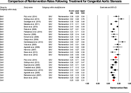
Article Information
vol. 132 no. Suppl 3 A19440
Published By:
American Heart Association, Inc.
Online ISSN:
History:
- Originally published November 6, 2015.
Copyright & Usage:
© 2015 by American Heart Association, Inc.
Author Information
- 1Pediatrics, Emory Univ Sch of Medicine, Atlanta, GA
- 2Biostatistics, Emory Univ Sch of Medicine, Atlanta, GA
Abstract 19002: Predictive Value of the Intraoperative Pulmonary Artery Flow Study for Early and Late Outcome After Surgical Treatment of Pulmonary Atresia With Ventricular Septal Defect and Major Aortopulmonary Collateral Arteries (MAPCAs)
Matteo Trezzi, Antonio Albano, Sonia B Albanese, Enrico Cetrano, Adriano Carotti
Circulation. 2015;132:A19002
Abstract
Introduction & Hypothesis: 1. To evaluate the accuracy of the intraoperative pulmonary artery (PA) flow study in determining the feasibility of concomitant VSD closure after complete one-stage unifocalization in patients with pulmonary atresia with VSD and MAPCAs. 2. To determine the ability of the PA flow study to predict long-term outcomes.
Methods: Between October 1996 and May 2015, a PA flow study was obtained in 92 patients undergoing complete one-stage unifocalization. The study was performed by cannulating the unifocalized central PA. With the heart beating, incremental volumes of blood were pumped through the PAs. The ability to achieve 100% flow (2.5 l/min/m2) into the pulmonary bed at a mean pressure (mPAP) ≤ 30 mmHg was used as an indicator for acceptability of VSD closure.
Results: Among the 92 patients who had a PA flow study, 23 had a mPAP > 30 mmHg and the VSD was left open. VSD closure was performed in 69 patients (mPAP 24 ± 5 mmHg, range 11-34 mmHg). However, four (5.8%) required patch fenestration due to suprasystemic RV pressure. The recorded mPAPs of these 4 patients were 29, 24, 22 and 28 mmHg, respectively. In the whole cohort, median weight at operation was 7.9 kg (range, 2.5-54 kg). The PA flow study accurately predicted successful VSD closure (area under the curve 0.852, Figure 1A). VSD closure at the time of unifocalization resulted in a clear survival advantage (p = 0.0363). mPAP correlated with RVP to LVP ratio (rho 0.460, p = 0.0001) and better long-term results were observed with intraoperative mPAP ≤ 30 mmHg (p = 0.24, Figure 1B).
Conclusions: Successful VSD closure in patients with MAPCAs can be reliably defined by an intraoperative PA flow study with a cut-off of 30 mmHg. The PA flow study predictive value is particularly useful to select patients in which the VSD should be left open. mPAP strongly correlates with the intraoperative RVP/LVP ratio and lower values of mPAP at the time of the unifocalization are associated with improved long-term outcomes.
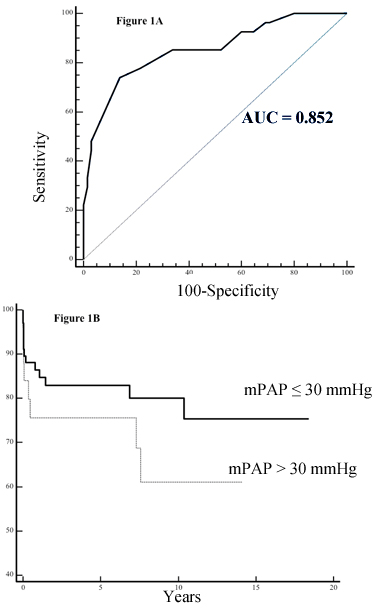
Article Information
vol. 132 no. Suppl 3 A19002
Published By:
American Heart Association, Inc.
Online ISSN:
History:
- Originally published November 6, 2015.
Copyright & Usage:
© 2015 by American Heart Association, Inc.
Author Information
- Matteo Trezzi;
- Antonio Albano;
- Sonia B Albanese;
- Enrico Cetrano;
- Adriano Carotti
- Pediatric Cardiac Surgery, Bambino Gesù Children’s Hosp IRCCS, Rome, Italy
Abstract 18061: Supplementation to Treat Antithrombin Deficiency Improves Sensitivity to Heparin, Anticoagulation and Decreased Thrombogenecity in Neonates and Infants Undergoing Cardiac Surgery With Cardiopulmonary Bypass
Brian W McCrindle, Cedric Manlhiot, Helen M Holtby, Anthony K Chan, Leonardo R Brandao, Martha Rolland, Lia Stenyk, Lynn Crawford-Lean, Celeste Foreman, Glen S Van Arsdell, Colleen E Gruenwald
Circulation. 2015;132:A18061
Abstract
Introduction: Preoperative antithrombin (AT) deficiency is common in infants undergoing cardiac surgery with cardiopulmonary bypass (CPB), and is associated with heparin resistance, difficulties achieving optimal intraoperative anticoagulation and post-operative thrombosis.
Methods: We performed a pilot randomized trial of pooled human AT supplementation for children <1 year with preoperative AT <0.85U/ml. Subjects received a split (patient and CPB prime) dose of AT before heparinization. AT was dosed to reach a predicted AT activity of 1.2U/ml during CPB calculated from preoperative AT level and patient weight.
Results: We randomized 18 subjects; 17 completed the study (9 controls, 8 AT). Mean preoperative AT level was similar between groups (Control: 0.65±0.10 vs. AT: 0.68±0.15 U/mL, p=0.66). AT group subjects had higher AT than controls immediately after start of CPB (Control: 0.44±0.10 vs. AT: 1.15±0.20 U/mL, p<0.001) and at the end of CPB (Control: 0.67±0.14 vs. AT: 1.19±0.16 U/mL, p<0.001). AT supplementation was associated with increased activated clotting time (ACT) post-heparinization (Control: 500±72 vs. AT: 639±172 seconds, p=0.02), increased heparin sensitivity (Control: 89±23 vs. AT: 114±22 ACT seconds per 100U/kg heparin, p=0.02), and decreased heparin dose given during CPB (Control: 1126±344 vs. AT: 845±165 U/kg, p=0.03). After primary heparinization, 56% of control subjects did not achieve ACT target vs. 25% of AT subjects. At the end of CPB, AT group subjects had lower prothrombin levels (Control: 746±372 vs. AT: 460±102 ng/mL, p=0.02) and lower levels of inflammatory cytokines (IL-1α, IL-1β, IL-6, IL-8 and TNFα). Supplementation with AT was not associated with increased chest tube volume loss or blood product requirements. One subject in each group had severe bleeding, and one in each group had post-operative infection. Thrombosis was noted for 3 controls and 1 AT subjects.
Conclusions: AT supplementation to treat preoperative AT deficiency in young children is associated with decreased heparin resistance, improved anticoagulation and decreased thrombogenecity. Safety profile regarding bleeding and infections appears favorable. Equipoise exists for a larger definitive outcome trial.
Article Information
vol. 132 no. Suppl 3 A18061
Published By:
American Heart Association, Inc.
Online ISSN:
History:
- Originally published November 6, 2015.
Copyright & Usage:
© 2015 by American Heart Association, Inc.
Author Information
- Brian W McCrindle1;
- Cedric Manlhiot1;
- Helen M Holtby1;
- Anthony K Chan2;
- Leonardo R Brandao1;
- Martha Rolland1;
- Lia Stenyk1;
- Lynn Crawford-Lean1;
- Celeste Foreman1;
- Glen S Van Arsdell1;
- Colleen E Gruenwald1
- 1Labatt Family Heart Cntr, The Hosp for Sick Children, Toronto, Canada
- 2Pediatric Hematology-Oncology, McMaster Univ, Hamilton, Canada
Abstract 18301: Intra-operative Left Coronary Doppler Profile Independently Predicts Need for Ecmo or Late Revision After Arterial Switch
Fredrik H Halvorsen, Deane Yim, Lynne Nield, Christopher A Caldarone, Glen S Van Arsdell, Eric Pham-Hung, An Duong, Michael Gritti, Edward J Hickey
Circulation. 2015;132:A18301
Abstract
Objective: We hypothesized that intraoperative coronary Doppler profiles after button implantation would help predict survival and morbidity after arterial switch.
Methods: Children (N=319) undergoing arterial switch (2000-2012; TGA 190, TGA-VSD 95, TGA-VSD-PS 10, Taussig-Bing 14, others 10) underwent intraoperative echocardiographic assessment of coronary buttons. Studies were blindly reviewed to delineate Doppler gradient, peak flow velocity, velocity-time integral (VTI) and late diastolic flow reversal, which were explored as predictors of death, ICU morbidity and need for late coronary revision via parametric risk-adjusted regression and bootstrap reliability.
Results: Outcomes included: death=9 (3%), ECMO=13 (4%), surgical coronary revision=9 (3%). Higher left button Doppler values were predictors of chest open duration, post-op JET, need for revision or ECMO. Mean left button gradient was the most reliable – table. Right button Doppler values were not strong predictors of morbidity.
Higher left Doppler gradients confer disproportionate impact – figure. Mean gradient ≥ 1.5 had late revision risk 27%, versus 1.6% for < 1.5 (P=.0016).
Intraoperative strategies to improve button geometry included release of fascia (16, 5%) or button revision (9, 3%). Such strategies reduced gradients, velocities and VTI (P between .004 and .03). Left Doppler values retained their predictive value even in 296 (93%) children who had no such coronary manipulation – table.
Conclusions: Aiming for mean left coronary gradient < 1.5-2 and peak <4 mmHg through routine Doppler assessment will help lower risk of ECMO and late coronary problems.
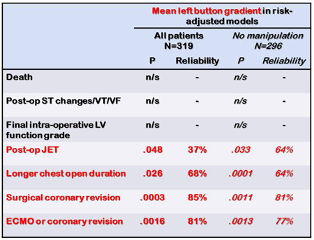
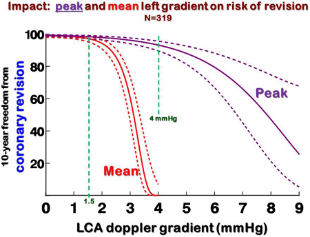
Article Information
vol. 132 no. Suppl 3 A18301
Published By:
American Heart Association, Inc.
Online ISSN:
History:
- Originally published November 6, 2015.
Copyright & Usage:
© 2015 by American Heart Association, Inc.
Author Information
- Fredrik H Halvorsen1;
- Deane Yim2;
- Lynne Nield2;
- Christopher A Caldarone1;
- Glen S Van Arsdell1;
- Eric Pham-Hung1;
- An Duong1;
- Michael Gritti1;
- Edward J Hickey1
- 1Cardiovascular Surgery, The Hosp for Sick Children, Toronto, Canada
- 2Cardiology, The Hosp for Sick Children, Toronto, Canada
Abstract 17272: Regionalization of Pediatric Cardiac Surgery Lowers Mortality, Re-operation, and Healthcare Costs
Michael F Swartz, Joseph Orie, Matthew Egan, Francisco Gensini, George M Alfieris
Circulation. 2015;132:A17272
Abstract
Introduction: The mortality of infants requiring cardiac surgery is related to center and surgeon case volume. One solution for increasing case volume is to regionalize pediatric cardiac surgical care.
Hypothesis: We hypothesized that the regionalization of pediatric cardiac surgical care from two medical centers would lower mortality, rate of re-operation, and subsequent health care costs.
Methods: Infants undergoing a biventricular repair were divided into two groups: pre-regionalization when operations were performed in two separate hospitals (1991-2000) and post-regionalization when all operations were performed in one regional hospital (2001-2010). Operative survival, midterm mortality, need for re-operation, and subsequent healthcare costs were compared between the groups.
Results: There were 508 infants in the pre-regionalization group, and 479 infants in the post-regionalization group. There were no significant differences in pre-operative age, weight, or gender between groups. Pre-regionalization operative survival was significantly lower than the state average. Post-regionalization operative survival was significantly greater than pre-regionalization and was similar to the state average (Figure 1). Five and 10 year survival remained significantly greater in the post-regionalization group (5 year: 96.8% vs 90.6%, 10 year: 96.8% vs. 89.9%, p<0.01). Five and ten year freedom from re-operation was significantly higher in the post-regionalization group (5 year: 88.8 vs 79.5 %, 10 year: 82.7 vs 73.3%, p=0.001). Subsequent health care costs were significantly lower in the post-regionalization group ($20,233 ± 53596 vs. 37,516 ± 82,947, p<0.001).Conclusions: Regionalization of pediatric cardiac surgical care lowered operative and 10 year mortality, rate of re-operation, and subsequent costs of health care.
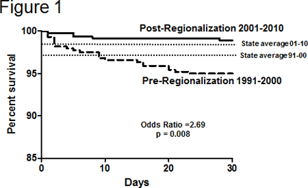
Article Information
vol. 132 no. Suppl 3 A17272
Published By:
American Heart Association, Inc.
Online ISSN:
History:
- Originally published November 6, 2015.
Copyright & Usage:
© 2015 by American Heart Association, Inc.
Author Information
- 1Surgery, Univ of Rochester, Rochester, NY
- 2Pediatrics, Univ of Rochester, Rochester, NY
Abstract 19540: US Center Variability in the Incidence of Stroke for the Berlin Heart EXCOR Pediatric Ventricular Assist Device
Christopher S Almond, Angela Lorts, Joseph W Rossano, Lori C Jordan, Christina VanderPluym, Jack F Price, Kevin P Daly, Katsuhide Maeda, Andrew Y Shin, David N Rosenthal
Circulation. 2015;132:A19540
Abstract
Background: Ventricular assist device (VAD)-related stroke is an important performance benchmark for adult VAD programs. No benchmarks are available for pediatric VAD programs where the stroke rate is higher. We sought to describe center variability in the stroke rate for the Berlin Heart EXCOR® Pediatric VAD, the most commonly used durable VAD in US children, to support pediatric VAD quality improvement efforts.
Methods: All children age <18 years supported with a Berlin Heart EXCOR Pediatric VAD at a site participating in the EXCOR IDE or FDA Post-Approval Study were identified using the Sponsor’s database. The incidence of neurological dysfunction, defined by INTERMACS criteria, was estimated for patients at centers with at least 300 support days.
Results: Between 2007 and March 2014, a total of 320 children underwent implantation of a Berlin Heart EXCOR Pediatric VAD at 42 centers, of which 271 patients at 25 centers met the study inclusion criteria. The baseline characteristics of the cohort have been reported previously. Overall, there were a total of 64 neurological dysfunction episodes in 53 unique patients over 20,875 days of support, yielding an overall stroke incidence of 0.31 events per 100 days of support and a prevalence of 19%. Centers varied considerably (ten-fold) in their incidence rate of stroke ranging from 0 events to 1.7 events per 100 days of support (Figure). Among 13 centers with at least 10 total implants (totaling 205 patients over 14,405 support days), the overall stroke prevalence was 20% (range 6 to 46%) (Incidence rate 0.33 events per 100 days of support). Centers with ≥10 implants did not have fewer strokes than lower volume centers (P=0.30).
Conclusion: The incidence of stroke among pediatric VAD centers using the Berlin Heart EXCOR Pediatric VAD was 0.31 events per day of support and varied considerably across pediatric centers. These data may be useful for developing performance benchmarks for neurological dysfunction in pediatric VAD support.
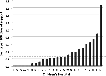
Article Information
vol. 132 no. Suppl 3 A19540
Published By:
American Heart Association, Inc.
Online ISSN:
History:
- Originally published November 6, 2015.
Copyright & Usage:
© 2015 by American Heart Association, Inc.
Author Information
- Christopher S Almond1;
- Angela Lorts2;
- Joseph W Rossano3;
- Lori C Jordan4;
- Christina VanderPluym5;
- Jack F Price6;
- Kevin P Daly5;
- Katsuhide Maeda7;
- Andrew Y Shin1;
- David N Rosenthal1
- 1Pediatrics (Cardiology), Stanford Univ, Palo Alto, CA
- 2Pediatric Cardiology, Cincinnati Children’s Hosp, Cincinnati, OH
- 3Cardiology, Children’s Hosp of Philadelphia, Philadelphia, PA
- 4Neurology, Vanderbilt Univ, Nashville, TN
- 5Cardiology, Boston Children’s Hosp, Boston, MA
- 6Cardiology, Texas Children’s Hosp, Houston, TX
- 7Cardiac Surgery, Stanford Univ, Palo Alto, CA
Abstract 17469: Expanding the Donor Pool in Pediatric Heart Transplant With a Novel Three Dimensional Technique
Jonathan Plasencia, Justin Ryan, Jacob Lindquist, Susan Sajadi, Micheal VanAuker, Randy Richardson, Erik Ellsworth, Susan Park, Robyn Augustyn, Richard Southard, John Nigro, Stephen Pophal, David Frakes, Steven Zangwill
Circulation. 2015;132:A17469
Abstract
Introduction: Heart transplant (HTx) outcomes continue to improve although organs remain limited. Donor-recipient (DR) size matching is critical in pediatrics. Usually, centers apply a subjective donor weight (wt) range based on recipient chest x-ray. DR wt ratios may vary from 0.7–4.
Hypothesis: A novel 3D tool can be used to predict ideal donor size.
Methods: A library of 3D reconstructed normal hearts was created from CT/MRI data. A linear fit of normal heart volume vs. body wt was determined for wts up to 22.5kgs (R2=0.983). The linear fit was used to predict donor heart volumes and ideal donor wt. Similarly, pre and post HTx hearts were reconstructed from 3 infant pts undergoing HTx.
Results: [Table] The library was used to predict ideal donor wt targeting 1:1 DR heart volume ratio. Pt A had cardiomegaly and an ideal DR wt ratio of 3.5, which may be outside a center’s usual range. While all 3 Pts had DR wt ratios > 1, Pt A had a downsized graft while Pt B and C had upsized grafts (based purely on DR heart volumes). 3D reconstructions > 1 week post HTx suggest the downsized graft became larger and upsized grafts smaller.
Conclusion: 3D reconstruction has tremendous potential to improve DR size matching in HTx. As the virtual library grows, the ability to accurately predict DR heart volumes will improve. Further, retrospective analysis of transplant cases, wherein 3D matching was used, will allow us to predict the limits of under and oversizing. Finally, this technique has potential to allow “virtual transplantations” wherein we can model a particular donor (green) into a patient-specific recipient (red) and assess fit along with potential for mismatch related morbidity.

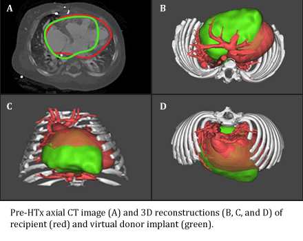
Article Information
vol. 132 no. Suppl 3 A17469
Published By:
American Heart Association, Inc.
Online ISSN:
History:
- Originally published November 6, 2015.
Copyright & Usage:
© 2015 by American Heart Association, Inc.
Author Information
- Jonathan Plasencia1;
- Justin Ryan2;
- Jacob Lindquist1;
- Susan Sajadi1;
- Micheal VanAuker1;
- Randy Richardson3;
- Erik Ellsworth2;
- Susan Park2;
- Robyn Augustyn4;
- Richard Southard4;
- John Nigro2;
- Stephen Pophal2;
- David Frakes1;
- Steven Zangwill2
- 1Sch of Biological and Health Systems Engineering, Arizona State Univ, Tempe, AZ
- 2Cardiology, Phoenix Children’s Hosp, Phoenix, AZ
- 3Radiology, St. Joseph’s Hosp and Med Cntr, Phoenix, AZ
- 4Radiology, Phoenix Children’s Hosp, Phoenix, AZ
Abstract 15011: Intravenous Immunoglobulin for Acute Myocarditis is Associated With Increased Resource Utilization but Equivalent Survival: An Outcome Analysis Amongst U.S. Children’s Hospitals
Pirouz Shamszad, Deipanjan Nandi, Matthew J O’Connor, Kimberly Y Lin, Andrew T Costarino, Robert E Shaddy, Joseph W Rossano
Circulation. 2015;132:A15011
Abstract
Introduction: Intravenous immunoglobulin (IVIg) is frequently utilized in the treatment of pediatric acute myocarditis (AM) though the data surrounding the efficacy of intravenous immunoglobulin (IVIg) are equivocal.
Hypothesis: IVIg utilization does not improve survival in pediatric AM but is associated with increased resource utilization.
Methods: A retrospective review of the Pediatric Health Information System database was performed for children <18 years hospitalized with AM between 2004 and 2014. A 1:1 propensity matched case control design was used to compare children who received IVIg (IVIg-group) to those who did not receive IVIg (no IVIg-group). The propensity score was based on a model including treatment with mechanical ventilation (MV), inotropes, or mechanical circulatory support (MCS). High- and low-use hospitals were defined by quartiles of percent usage of IVIg.
Results: A total of 1802 children with AM were identified (median age 8y [IQR 1 – 15]; 60% male, 60% IVIg given, 65% inotrope use, 46% MV use, 16% MCS use). After matching, 552 children were included in both the IVIg-group and no IVIg-group. Overall median follow-up of the matched cohort was 1.4y [IQR 0.4-3.4]. Unadjusted in-patient mortality was similar between groups (6% vs. 9%, p=0.09). On multivariable Cox regression, there was no association between IVIg and follow-up mortality (OR 1.0, 95%CI 0.3- 2.9). Although in-patient cardiac transplantation occurred more frequently in the IVIg-group (2% vs. 0%, p<0.001), there was no association between IVIg and cardiac transplantation during the follow up period (OR 0.5, 95%CI 0.2-1.1). Mortality was similar between high- and low-use centers (6% vs. 8%, p=0.316) as was length of stay (7d [IQR 8-18] vs. 7d [4-18], p=0.771). Total charges, as well as pharmacy charges, were significantly higher in the IVIg-group ($125k vs. $77k, $32k vs. $2.8k; p<0.001 for both) and high-use centers ($125k vs. $112k, p=0.025; $27k vs. $5.1k, p<0.001).
Conclusions: IVIg is frequently utilized in children for the treatment of AM. Overall survival was similar in patients treated and not treated with IVIg, though resource utilization was significantly increased. Further study is needed to assess the most optimal, cost-effective treatment for AM in children.
Article Information
vol. 132 no. Suppl 3 A15011
Published By:
American Heart Association, Inc.
Online ISSN:
History:
- Originally published November 6, 2015.
Copyright & Usage:
© 2015 by American Heart Association, Inc.
Author Information
- Pirouz Shamszad1;
- Deipanjan Nandi1;
- Matthew J O’Connor1;
- Kimberly Y Lin1;
- Andrew T Costarino2;
- Robert E Shaddy1;
- Joseph W Rossano1
- 1Div of Cardiology, Children’s Hosp of Philadelphia, Philadelphia, PA
- 2Div of Critical Care, Children’s Hosp of Philadelphia, Philadelphia, PA
Abstract 14706: Cardiac Phenotype in Children Undergoing Screening for Familial Hypertrophic Cardiomyopathy: Is it Time to Change Screening Guidelines?
Laura Zahavich, Jacob Mathew, Judith Wilson, Kristen George, Sarah Bowdin, Leland N Benson, Seema Mital
Circulation. 2015;132:A14706
Abstract
Introduction: Current hypertrophic cardiomyopathy (HCM) screening guidelines recommend that clinical and/or genetic screening of children with family history of HCM be initiated from age 10 years (European Society of Cardiology 2014) or 12 years (American Heart Association 2011) onward and earlier only in the presence of malignant family history, presence of symptoms and/or participation in competitive sports. We assessed the proportion of asymptomatic children undergoing HCM screening that developed HCM phenotype or major adverse cardiac events (MACE) prior to age 12 years.
Methods: This was a single centre retrospective study of HCM kindreds referred for screening to our cardiomyopathy clinic. Index cases with HCM, and cases with secondary cardiomyopathy (metabolic, neuromuscular, syndromic) were excluded. Genotype and phenotype status at initial screening and last follow-up were analyzed. MACE was defined as occurrence of death, transplant, resuscitated cardiac arrest, implantation of ICD or myectomy.
Results: From 2005-2014, 427 children were referred for cardiac screening secondary to a family history of HCM. Of these, 379 (88.8%) had first-degree relatives with HCM and 146 (34.1%) had a family history of sudden cardiac death. The median age at first screening was 7.4 years (range 1 day-17 years), and median follow-up duration was 3 years. 45% (13/29 tested) phenotype-positive children, and 53% (32/60 tested) phenotype-negative children has positive genetic testing. 27/427 (6.3%) were phenotype-positive (maximal LV wall thickness Z-score >2.0) at initial screening and an additional 13 (3%) became phenotype-positive during follow-up. The median age at positive phenotypic diagnosis of HCM was 8 years (range 5 days-17 years) with 24 children being <12 years old (5.6%). 11/427 (2.6%) suffered a MACE during follow-up. Median age at MACE occurrence was 10.9 years (range 6-15 years) with 6 children being <12 years old at occurrence of MACE (1.4%).
Conclusion: Overall, more than 5% children being screened for HCM were phenotype-positive and 1.4% suffered a MACE before 12 years of age. These data suggest the importance of revising existing guidelines to recommend clinical and/or genetic screening of HCM of all family members independent of age.
Article Information
vol. 132 no. Suppl 3 A14706
Published By:
American Heart Association, Inc.
Online ISSN:
History:
- Originally published November 6, 2015.
Copyright & Usage:
© 2015 by American Heart Association, Inc.
Author Information
- Laura Zahavich1;
- Jacob Mathew2;
- Judith Wilson1;
- Kristen George1;
- Sarah Bowdin1;
- Leland N Benson1;
- Seema Mital1
- 1PEDIATRICS, Hosp for Sick Children, Toronto, Canada
- 2DIVISION OF CARDIOLOGY, Cincinnati Children’s Hosp, Cincinnati, OH
Abstract 20041: Single Ventricle Outcome Following Neonatal Palliation of Severe Ebstein’s Anomaly With Modified Starnes Procedure
- Ram Kumar, Nathan Noh, Novell Castillo, Brian Fagan, Grace Kung, Winfield J Wells, Vaughn A Starnes
Circulation. 2015;132:A20041
Abstract
Background: We have previously shown that neonates in profound cardiogenic shock due to severe Ebstein’s anomaly can be successfully salvaged with fenestrated right ventricular (RV) exclusion and systemic to pulmonary shunt (modified Starnes procedure). The long-term outcome of single ventricle management in these patients is not known.
Methods: We retrospectively reviewed the records of 26 patients who underwent neonatal Starnes procedure between 1989 and 2011. Patient demographics, clinical variables and outcome data were collected. Data is presented as mean ± standard errors or median (interquartile ranges).
Results: 26 patients (12, 46% boys) underwent Starnes procedure at 7 (5-9) days of life. All were intubated and on prostacyclin infusion, 24 (92%) were inotrope-dependent and 23 (88%) had no antegrade flow from the RV. Two patients had had prior intervention (one tricuspid annuloplasty and one shunt alone). Three patients underwent non-fenestrated RV exclusion, two (67%) of whom died. Of the remaining 23, 3 (13%) died during the same hospitalization. The 21 neonatal survivors have been followed for 7 (6-8) years. One patient died after Glenn. The remaining 20 have successfully undergone Fontan completion with an indexed pulmonary resistance of 1.8 (1.2-2.3) W/m2 and mean pulmonary pressure of 12 (9-18) mm Hg. At last follow-up, all patients have normal left ventricular function, and all but one patient are in NYHA Class I symptoms. Two patients have required pacemaker implantation, while the rest are in sinus rhythm. Survival at 1, 5 and 10 years are 81±4%, 77±3% and 77±3%, respectively.
Conclusion: Long-term single ventricle outcomes amongst neonatal survivors of modified Starnes procedure are excellent. There is reliable remodeling of the excluded RV and excellent function of the left ventricle.

Article Information
vol. 132 no. Suppl 3 A20041
Published By:
American Heart Association, Inc.
Online ISSN:
History:
- Originally published November 6, 2015.
Copyright & Usage:
© 2015 by American Heart Association, Inc.
Author Information
- S. Ram Kumar1;
- Nathan Noh1;
- Novell Castillo2;
- Brian Fagan2;
- Grace Kung3;
- Winfield J Wells1;
- Vaughn A Starnes1
- 1Surgery, Univ of Southern California, Los Angeles, CA
- 2Heart Institute, Children’s Hosp, Los Angeles, Los Angeles, CA
- 3Pediatrics, Univ of Southern California, Los Angeles, CA
Abstract 18747: Young Infants With Severe Tetralogy of Fallot: Early Primary Surgery versus Trans-catheter Palliation
Travis J Wilder, Glen S Van Arsdell, Lee Benson, An Duong, Eric Pham-Hung, Michael Gritti, Alexandra Page, Christopher A Caldarone, Edward J Hickey
Circulation. 2015;132:A18747
Abstract
Introduction: Infants with severe tetralogy of Fallot (TOF) may undergo: 1) early primary surgical repair (EARLY) or 2) early catheter palliation (CATH) prior to delayed surgical repair. We compared these two strategies to 3) usual elective single stage TOF repair (USUAL).
Methods: We studied 453 TOF repairs (2000-2012, excluding BT shunts). USUAL strategy at our institution is repair ≥3 months. Risk adjusted hazard analysis compared freedom from surgical or catheter reintervention. Somatic size, branch PA size and RV systolic pressure were modeled using 2543 echo reports via mixed model regression.
Results: Table: group characteristics for USUAL (383), EARLY (42) and CATH (28). CATH involved: RVOT stent=18, RVOT balloon=9, ductal stent=1.
Risk adjusted freedom from surgical reoperation was 88%, 87% and 85% for USUAL, EARLY and CATH respectively, at 10 years. EARLY and CATH had similar reoperation rates, except for very young children (<1 month) where EARLY primary repair conferred increased risk of further surgery (Figure).
Risk adjusted freedom from catheter reintervention was higher for EARLY (76%) and especially so for CATH (53%) at 10 years, versus USUAL (83%; Figure).
Somatic growth and progression of RVSP (37-40mmHg) was similar among groups at 8 years. EARLY (P=.02) and CATH (P=.09) tend to have smaller bPAs initially. The CATH group tend to remain smaller in the long-term, whereas growth in EARLY matches USUAL.
Conclusions: Early primary repair at very young ages comes with a cost of increased late surgical reoperation. Early transcatheter palliation tends to come with a cost of increased late transcatheter intervention – possibly related to slower branch PA growth.
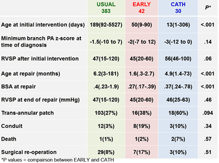
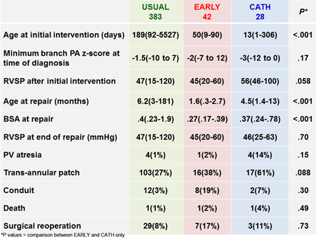
Article Information
vol. 132 no. Suppl 3 A18747
Published By:
American Heart Association, Inc.
Online ISSN:
History:
- Originally published November 6, 2015.
Copyright & Usage:
© 2015 by American Heart Association, Inc.
Author Information
- Travis J Wilder1;
- Glen S Van Arsdell2;
- Lee Benson3;
- An Duong2;
- Eric Pham-Hung2;
- Michael Gritti2;
- Alexandra Page2;
- Christopher A Caldarone2;
- Edward J Hickey2
- 1CHSS Data Cntr, The Hosp For Sick Children, Toronto, Canada
- 2Dept of Cardiovascular Surgery, The Hosp For Sick Children, Toronto, Canada
- 3Dept of Pediatric Cardiology, The Hosp For Sick Children, Toronto, Canada
Abstract 18478: Balloon Angioplasty of Branch Pulmonary Arteries in Children: A Long Term Outlook
Yinn Khurn Ooi, Sung In Kim, Scott E Gillespie, Zhi Geng, Dennis W Kim, Robert N Vincent, Petit J Christopher
Circulation. 2015;132:A18478
Abstract
Introduction: Balloon angioplasty (BA) is widely employed for branch pulmonary artery stenosis (BPAS) in children. Vessel recoil, and hence need for repeat intervention, is common.
Hypothesis: BA of BPAS has poor long term outcomes.
Methods: We reviewed our cath database over an 11-year period ending in 2013 and collected data for all pts undergoing BA for BPAS. Primary endpoint was repeat intervention (RI), with other endpoints including acute results, and need for stent or surgical PA plasty (S/S). Central branch pulmonary artery (cPA) was defined as pre-hilum, while peripheral BPA (pPA) was define as post-hilum.
Results: 175 pts underwent 371 BA procedures for BPAS. Primary diagnosis included major aorto-pulmonary collateral arteries (MAPCAs) in 18% (n=32) pts, and single ventricle in 11% (n=20) pts. Median age and weight were 23.6 (9.9 – 65.8) months and 10.7 kg (7.8 – 18.2 kg). Pt follow up was 4.1 (1.1 – 6.5) years. 69% (n=256) of BA was performed on cPA and 31% (n=115) on pPA. Acutely, there was a 14.9% (7.6 – 26.7%) improvement in stenosis in cPA, and 18.5% (9.8 – 28.6%) in pPA (p=0.053). Improvement of pressure gradient was 34.3% (10.4 – 63.4%) in cPA, and 31.6% (0 – 60.7%) in pPA (p=0.324). Higher rates of RI was seen in pPA (55% RI, p=0.035).
For cPA, freedom from RI was 69% at 1 year and 44% at 5 years. Freedom from S/S was 81% at 1 year and 62% at 5 years. Factors associated with higher rate of RI include MAPCAs (65% RI, p=0.039), younger pt age (p=0.042), and lower pt weight (p=0.047). Lower rates of RI were seen in pts with discrete (p=0.007) and severe stenosis (p<0.001). For pPA, freedom from RI was 59% at 1 year and 38% at 5 years. Freedom from S/S was 98% at 1 year and 92% at 5 years.
Conclusions: Long term outcomes of BA for BPAS are disappointing. BA for BPAS is associated with a high rate of RI with rates exceeding 50% by 5 years. Patients with pre-hilar BPAS and with discrete, severe stenosis appear to respond more favorably. Peripheral BPAS and MAPCAs in particular have a high rate of recurrence.
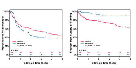
Article Information
vol. 132 no. Suppl 3 A18478
Published By:
American Heart Association, Inc.
Online ISSN:
History:
- Originally published November 6, 2015.
Copyright & Usage:
© 2015 by American Heart Association, Inc.
Author Information
- Yinn Khurn Ooi1;
- Sung In Kim2;
- Scott E Gillespie1;
- Zhi Geng2;
- Dennis W Kim1;
- Robert N Vincent1;
- Petit J Christopher1
- 1Dept of Pediatrics, Emory Univ Sch of Medicine, Atlanta, GA
- 2Div of Cardiology, Emory Univ Rollins Sch of Public Health, Atlanta, GA
Abstract 18359: Long Term Follow-up of Aortic Coarctation Repair Using Leg-arm Blood Pressure Difference to Assess Hemodynamic Profiles
Daniel F Labuz, James A Berry, Lee A Pyles
Circulation. 2015;132:A18359
Abstract
Introduction: Patients with successfully repaired coarctation (CoA) need long term follow up due to the risk of abnormal arterial elasticity and complications of hypertension. We hypothesize that even subtle hemodynamic abnormalities are marked by lack of a blood pressure standing wave effect to the lower extremities (arm ≥ leg systolic BP: ARM Group) and is associated with elevated aortic flow velocities, increased left ventricular posterior wall thickness (LVPWd), higher arm BP and a greater requirement for interventions compared to subjects with the normal standing wave effect (leg > arm SBP: LEG Group).
Methods: CoAs repaired between 1990 and 2008 with at least two documented right arm and leg SBPs at clinic visits along with M-mode, 2D and Doppler echocardiography were selected. The largest recorded peak instantaneous (PeakV) and mean velocities (MeanV) in the descending aorta were reviewed, as were measures of the proximal transverse arch (TA) and LVPWd. Measurements were indexed by Z-score. Patient records were also reviewed for reintervention and antihypertensive medical therapy.
Results: Eighty patients met our criteria with 29 ARM (median age 12 years) and 51 LEG (median 13 years) Group members. Compared to LEG, ARM had significantly increased arm SBP Z-score (p <0.001), PeakV (p <10-4), MeanV (p <10-6), and LVPWd (p <0.01) (Figure). There was no difference in arm diastolic BP (p =0.7) or TA diameter (p =0.3). Even without overt markers such as hypertension or medication, otherwise healthy ARM subjects were still found to have the same relationships. Furthermore, 11/29 (38%) ARM compared to 39/51 (76%) LEG were normotensive with no previous intervention and no BP medicine (Fisher’s p <0.001).
Conclusions: Higher leg SBP after CoA repair is a marker for salutary hemodynamic and echo parameters as well as freedom from interventions post repair. Follow-up tailored by recognizing good outcome in LEG Group could be accomplished with less expense and obtrusive testing.
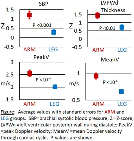
Article Information
vol. 132 no. Suppl 3 A18359
Published By:
American Heart Association, Inc.
Online ISSN:
History:
- Originally published November 6, 2015.
Copyright & Usage:
© 2015 by American Heart Association, Inc.
Author Information
- 1Surgery, Univ of Minnesota Med Sch – Twin Cities, Minneapolis, MN
- 2Pediatrics, Univ of Minnesota Med Sch – Twin Cities, Minneapolis, MN
- 3Pediatrics, West Virginia Univ Sch of Medicine, Morgantown, WV
Abstract 17678: Earlier Stage I Operation in Neonates With Hypoplastic Left Heart Syndrome is Not Associated With Worse Short-term Outcomes
Ann L McCarthy, Madeline E Winters, Elizabeth Bixler, Jennifer M Lynch, John J Newland, David R Busch, Tiffany Ko, Rui Xiao, Susan C Nicolson, Lisa M Montenegro, Christopher E Mascio, Stephanie Fuller, William J Gaynor, Thomas L Spray, Daniel J Licht, Maryam Y Naim
Circulation. 2015;132:A17678
Abstract
Introduction: We have previously reported that earlier stage I palliation (S1P) is protective against White Matter Injury (WMI) for term infants with hypoplastic left heart syndrome (HLHS). The present study compares clinical outcomes from the acute hospitalization between neonates who had early (≤ 4 days) versus late surgery (> 4 days).
Hypothesis: Earlier S1P intervention is associated with greater post-operative complications rates.
Methods: A retrospective chart review was performed on all neonates with HLHS or variants who underwent S1P at a single institution between 10/2008 – 03/2013. Excluded were a preterm (<37 weeks gestation), left-ventricle physiology, prior operations and international referral. Anthropometric data, pre- post- and intra-operative factors, post-operative complications, and both intensive care unit (ICU) and hospital length of stay were evaluated for differences between early vs. late surgery. Analysis was performed excluding patients who had either early, or late surgery for non-elective diagnoses.
Results: A total of 145 infants, 114 (73 with early surgery, 41 late) met inclusion criteria. The mean day of surgery was 4.2 days (early 2.7±0.1 days, late 6.9±3.7 days). There were no significant differences between the groups’ post-operative complications or operative factors (see Table 1). In addition exclusion of patients with non-elective change to their surgical date did not change results significantly. Neonates who had earlier surgery had significantly longer post-operative ICU stay (median 9 days vs. median 13 days, p = 0.02).
Conclusion: Early surgery for neonates with HLHS was not associated with either protection against or greater risk for post-operative complications. Neonates who have earlier surgery have a longer initial post-operative ICU stay, but total ICU and hospital stay was not different than that for infants with surgery after day four.
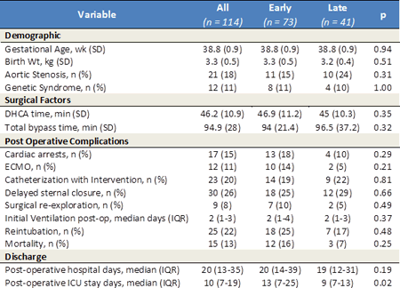
Article Information
vol. 132 no. Suppl 3 A17678
Published By:
American Heart Association, Inc.
Online ISSN:
History:
- Originally published November 6, 2015.
Copyright & Usage:
© 2015 by American Heart Association, Inc.
Author Information
- Ann L McCarthy1;
- Madeline E Winters2;
- Elizabeth Bixler3;
- Jennifer M Lynch4;
- John J Newland1;
- David R Busch1;
- Tiffany Ko5;
- Rui Xiao6;
- Susan C Nicolson7;
- Lisa M Montenegro7;
- Christopher E Mascio8;
- Stephanie Fuller8;
- William J Gaynor8;
- Thomas L Spray8;
- Daniel J Licht9;
- Maryam Y Naim10
- 1Dept of Neurology, Children’s Hosp of Philadelphia, Philadelphia, PA
- 2Div of Neurology, Children’s Hosp of Philadelphia, Philadelphia, PA
- 3Neurology, Children’s Hosp of Philadelphia, Philadelphia, PA
- 4Dept of Physics and Astronomy, Univ of Pennsylvania, Philadelphia, PA
- 5Dept of Bioengineering, Univ of Pennsylvania, Philadelphia, PA
- 6Biostatistics, Univ of Pennsylvania, Philadelphia, PA
- 7Div of Cardiac Anesthesiology, Children’s Hosp of Philadelphia, Philadelphia, PA
- 8Div of Cardiothoracic Surgery, Children’s Hosp of Philadelphia, Philadelphia, PA
- 9Divs of Neurology, Children’s Hosp of Philadelphia, Philadelphia, PA
- 10Div of Critical Care Medicine, Children’s Hosp of Philadelphia, Philadelphia, PA
Abstract 17203: Postnatal Management of Fetuses With Ebstein Anomaly or Tricuspid Valve Dysplasia in the Current Era: A Multi-center Study
Lindsay R Freud, Brian T Kalish, Maria C Escobar-Diaz, Rukmini Komarlu, Michael D Puchalski, Edgar T Jaeggi, Anita L Szwast, Grace Freire, Stephanie M Levasseur, Ann Kavanaugh-McHugh, Erik C Michelfelder, Anita J Moon-Grady, Mary T Donofrio, Lisa W Howley, Elif Seda Selamet Tierney, Bettina F Cuneo, Shaine A Morris, Jay D Pruetz, Mary E van der Velde, John P Kovalchin, Catherine M Ikemba, Margaret M Vernon, Cyrus Samai, Gary M Satou, Nina L Gotteiner, Colin K Phoon, Norman H Silverman, Doff B McElhinney, Wayne Tworetzky
Circulation. 2015;132:A17203
Abstract
Background: A recent multi-center study of perinatal outcome in fetuses with Ebstein anomaly or tricuspid valve dysplasia (EA/TVD) found that 1/3rd of live-born patients (pts) died prior to hospital discharge. The purpose of this study was to explore differences in postnatal management and the relationship to outcome.
Methods: This 23-center, retrospective study included 243 fetuses with EA/TVD from 2005 to 2011. Neonatal procedure (NP) was defined as surgery or interventional catheterization (cath) prior to discharge. Associations between postnatal management and outcome at discharge were explored.
Results: Of 176 live-born pts, 7 received comfort care only, 11 died <24 hrs of life, and 4 had insufficient data. Among 154 remaining pts, 38 (25%) did not survive to discharge. Pts who required ECMO at any point (n=18) had 83% mortality. More than half of pts (54%) did not have an NP, 34% had surgery, 8% had interventional cath, and 4% had both. The median age at 1st NP was 6 days (quartiles: 1-11). Survival did not differ between pts who had an NP and those who did not (70% vs. 80%; p=0.19) or between pts who had surgery and those who did not (68% vs. 80%; p=0.09). However, mortality differed by NP performed and whether pulmonary regurgitation, an indicator of high risk, was present prenatally (Figure). No pts with a right ventricular exclusion (RVE) died. Of 49 surviving neonates with ≥1 procedure, 28 (57%) were palliated with a shunt or RVE and 21 (43%) had a biventricular circulation. Thus, in total, 86 of 154 live-born pts (56%) survived with a biventricular circulation: 65 with medical management only and 21 with ≥1 NP.
Conclusion: Among live-born pts diagnosed with EA/TVD in utero, a variety of postnatal management strategies were employed with overall poor outcomes. If surgery beyond PDA ligation is necessary, then RVE or other palliative procedure may need to be considered. A prospective, multi-center study utilizing a management algorithm would help elucidate the optimal strategy.
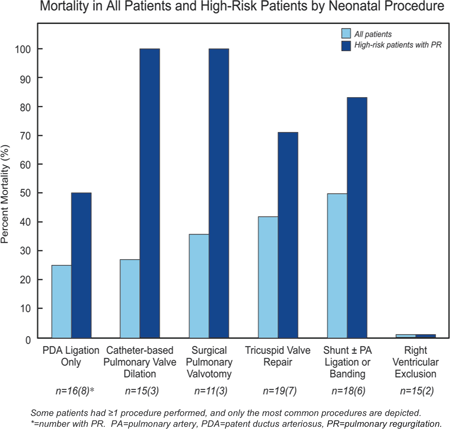
Article Information
vol. 132 no. Suppl 3 A17203
Published By:
American Heart Association, Inc.
Online ISSN:
History:
- Originally published November 6, 2015.
Copyright & Usage:
© 2015 by American Heart Association, Inc.
Author Information
- Lindsay R Freud1;
- Brian T Kalish2;
- Maria C Escobar-Diaz1;
- Rukmini Komarlu1;
- Michael D Puchalski3;
- Edgar T Jaeggi4;
- Anita L Szwast5;
- Grace Freire6;
- Stephanie M Levasseur7;
- Ann Kavanaugh-McHugh8;
- Erik C Michelfelder9;
- Anita J Moon-Grady10;
- Mary T Donofrio11;
- Lisa W Howley12;
- Elif Seda Selamet Tierney13;
- Bettina F Cuneo14;
- Shaine A Morris15;
- Jay D Pruetz16;
- Mary E van der Velde17;
- John P Kovalchin18;
- Catherine M Ikemba19;
- Margaret M Vernon20;
- Cyrus Samai21;
- Gary M Satou22;
- Nina L Gotteiner23;
- Colin K Phoon24;
- Norman H Silverman13;
- Doff B McElhinney13;
- Wayne Tworetzky1
- 1Dept of Cardiology, Boston Children’s Hosp, Boston, MA
- 2Dept of Medicine, Boston Children’s Hosp, Boston, MA
- 3Dept of Pediatrics, Div of Cardiology, Primary Children’s Hosp, Salt Lake City, UT
- 4Dept of Pediatrics, Div of Cardiology, Hosp for Sick Children, Toronto, Canada
- 5Dept of Pediatrics, Div of Cardiology, Children’s Hosp of Philadelphia, Philadelphia, PA
- 6Dept of Pediatrics, Div of Cardiology, All Children’s Hosp, Johns Hopkins Medicine, St. Petersburg, FL
- 7Dept of Pediatrics, Div of Cardiology, Morgan Stanley Children’s Hosp of New York-Presbyterian, New York, NY
- 8Dept of Pediatrics, Div of Cardiology, Monroe Carell Jr. Children’s Hosp, Nashville, TN
- 9Dept of Pediatrics, Div of Cardiology, Cincinnati Children’s Hosp Med Cntr, Cincinnati, OH
- 10Dept of Pediatrics, Div of Cardiology, UCSF Benioff Children’s Hosp, San Francisco, CA
- 11Dept of Pediatrics, Div of Cardiology, Children’s National Med Cntr, Washington, DC
- 12Dept of Pediatrics, Div of Cardiology, Children’s Hosp Colorado, Aurora, CO
- 13Dept of Pediatrics, Div of Cardiology, Lucile Packard Children’s Hosp, Palo Alto, CA
- 14Dept of Pediatrics, Div of Cardiology, Advocate Children’s Hosp, Oak Lawn, IL
- 15Dept of Pediatrics, Div of Cardiology, Texas Children’s Hosp, Houston, TX
- 16Dept of Pediatrics, Div of Cardiology, Children’s Hosp Los Angeles, Los Angeles, CA
- 17Dept of Pediatrics, Div of Cardiology, C.S. Mott Children’s Hosp, Ann Arbor, MI
- 18Dept of Pediatrics, Div of Cardiology, Nationwide Children’s Hosp, Columbus, OH
- 19Dept of Pediatrics, Div of Cardiology, Children’s Med Cntr of Dallas, Dallas, TX
- 20Dept of Pediatrics, Div of Cardiology, Seattle Children’s Hosp, Seattle, WA
- 21Dept of Pediatrics, Div of Cardiology, Children’s Healthcare of Atlanta, Atlanta, GA
- 22Dept of Pediatrics, Div of Cardiology, Mattel Children’s Hosp, Los Angeles, CA
- 23Dept of Pediatrics, Div of Cardiology, Ann & Robert H. Lurie Children’s Hosp of Chicago, Chicago, IL
- 24Dept of Pediatrics, Div of Cardiology, Hassenfeld Children’s Hosp of NYU Langone Med Cntr, New York, NY
Abstract 14197: Industrial Developmental Toxicant Emissions and Congenital Heart Disease in Urban and Rural Alberta, Canada
Deliwe P Ngwezi, Lisa K Hornberger, Jesus Serrano-Lomelin, Deborah Fruitman, Alvaro Osornio-Vargas
Circulation. 2015;132:A14197
Abstract
Background: We have previously demonstrated a downward temporal association between mixtures of organic solvents released into air from industrial sources and congenital heart disease (CHD) in Alberta.
Hypothesis: In the current study we hypothesized that the downward temporal associations between developmental toxicants (DTs) and CHD would have a different industrial sector participation in the urban and rural areas of Alberta.
Methods: We extracted yearly emissions of DTs (tonnes) from Canada’s National Pollutant Release Inventory as released to air between 2003-2010. We identified at the postal code level, all CHD cases born between 2004-2011 through our provincial-wide echocardiography database. We calculated yearly urban and rural CHD crude rates. Urban and rural were defined according to Forward Sortation Areas. Principal Component Analysis was undertaken to reduce the dimensionality of the number of DTs and the yearly solution was applied to rural and urban areas to test correlations with corresponding yearly CHD rates.
Results: Three principal components (PCs) were identified: PC1 consisted of a mixture of organics and gases, PC2 consisted of a mixture of organics only and PC3 consisted of metals. There were strong positive correlations between CHD rates and urban PC1 emissions from mining and manufacturing, (r=0.76, p=0.03; 0.74, p=0.04); whereas in rural areas, PC1 emissions from mining and utilities and PC2 emissions from mining and manufacturing were associated with rates of CHD, (r=0.71, p=0.05; r=0.74, p=0.04 and r=0.86, p=0.01; r=0.74, p=0.04, respectively). PC3 showed no positive correlations (Table 1).
Conclusions: CHD rates are consistently strongly correlated with mixtures of organic compounds and gases, with different patterns of chemicals and sectors involved in the urban and rural settings. This provides an opportunity to further investigate spatial correlations between DTs and CHD rates at a higher resolution scale.
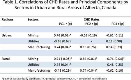
Article Information
vol. 132 no. Suppl 3 A14197
Published By:
American Heart Association, Inc.
Online ISSN:
History:
- Originally published November 6, 2015.
Copyright & Usage:
© 2015 by American Heart Association, Inc.
Author Information
- Deliwe P Ngwezi1;
- Lisa K Hornberger1;
- Jesus Serrano-Lomelin2;
- Deborah Fruitman3;
- Alvaro Osornio-Vargas1
- 1Pediatrics, Univ of Alberta, Edmonton, Canada
- 2Sch of Public Health, Univ of Alberta, Edmonton, Canada
- 3Pediatrics, Univ of Calgary, Calgary, Canada
Abstract 12635: Nationwide Long-term Follow-up of Pulmonary Atresia With Ventricular Septal Defect
Anu Kaskinen, Juha-Matti Happonen, Ilkka P Mattila, Olli M Pitkänen
Circulation. 2015;132:A12635
Abstract
Introduction: The naturally poor survival of pulmonary atresia with ventricular septal defect (PA+VSD) has improved due to evolved perioperative and surgical treatment. Studies including PA+VSD patients, both with and without major aortopulmonary collateral arteries (MAPCAs), with extensive follow-up are scarce. This nationwide study aimed to investigate survival and surgical treatment in PA+VSD patients with and without MAPCAs.
Methods: Study comprised 109 PA+VSD patients born in Finland between 1970 and 2007. We reviewed retrospectively medical records and operative reports through December 2011, as well as first available angiograms and preoperative angiograms prior to repair attempt.
Results: The median follow-up time for the total study population, including also patients who died during the follow-up, was 11.4 years (IQR 0.8 – 21.1). The incidence of PA+VSD, which could be determined reliably from 1995 to 2007, was 6.1 per 100 000 live births.
Although the patients with (n = 43) or without MAPCAs (n = 66) showed no difference in survival (p = 0.74), the patients without MAPCAs had better probability to achieve repair (64% vs. 28%, p < 0.0001). The bigger size of true central pulmonary arteries assessed by McGoon index at first angiogram [HR 0.66 (CI95% 0.49 – 0.88) per 0.5 McGoon index units, p = 0.006] and achievement of repair [HR 0.07 (CI95% 0.03 – 0.17), p < 0.0001] improved the overall survival.
After successful repair survival was 93% at 1 year and 91% from 2 years on. Palliated patients, instead, had survival at 1, 5, 10, and 20 years of age of 55%, 42%, 34%, and 20% respectively. However, patients with right ventricle – pulmonary artery connection and septal fenestration had better survival than rest of the palliated patients (p = 0.001). Palliation with a systemic-pulmonary artery shunt increased McGoon index by 41% (p < 0.0001).
Conclusions: The patients with MAPCAs had higher risk to remain palliated than patients without, although their survival was similar. Survival of PA+VSD was influenced by the initial size of true central pulmonary arteries and whether repair was achieved. Although palliative procedures may not improve the final outcome of PA+VSD, palliative surgery may have a role in its treatment.
Article Information
vol. 132 no. Suppl 3 A12635
Published By:
American Heart Association, Inc.
Online ISSN:
History:
- Originally published November 6, 2015.
Copyright & Usage:
© 2015 by American Heart Association, Inc.
Author Information
- Anu Kaskinen;
- Juha-Matti Happonen;
- Ilkka P Mattila;
- Olli M Pitkänen
- 1Children’ s Hosp, Div of Pediatric Cardiology and Cardiac and Transplantation Surgery, Univ of Helsinki and Helsinki Univ Hosp, Helsinki, Finland
Abstract 11416: The “Natural History” of Intra-operative Pulmonary Artery Stents: A 20-year Perspective
Jeffrey D Zampi, Emefah Loccoh, Aimee K Armstrong, Sunkyung Yu, Ray Lowery, Albert Rocchini, Jennifer C Hirsch-Romano
Circulation. 2015;132:A11416
Abstract
Background: Branch pulmonary artery (PA) stenoses are typically treated with catheter-based percutaneous therapies or surgical arterioplasty. Intra-operative (intra-op) PA stent placement is an alternative strategy. In this study, we sought to describe our 20-year experience with intra-op PA stent placement and to evaluate long-term patient outcomes, specifically the need and risk factors for re-intervention.
Methods: We performed a retrospective review of all intra-op PA stents placed at our institution from 1994-2013. Patient and stent characteristics and outcome data were collected. Risk factors associated with re-intervention were identified using univariate cox regression analysis.
Results: Eighty-one PA stents were placed in 68 patients, mostly to treat proximal PA stenoses (85%). Stent implantation was acutely successful in 67 patients (98.5%) with a procedural complication rate of 4.4%. Stents capable of being dilated to large diameters (“adult-size”) were used in 97% of patients. At hospital discharge, 93% of patients had no or mild residual PA stenosis by echocardiogram. During a median follow-up period of 6 years (interquartile range 0.9-12.7), 31 patients (46%) underwent PA re-intervention with a median time to first re-intervention of 2.5 years. Risk factors for re-intervention included age < 18 months (Hazard ratio [HR] 3.26, p=0.002) and body surface area < 0.47 m2 (HR 3.47, p=0.001) at the time of stent implantation, and the presence of multiple aorto-pulmonary collaterals in patients with tetralogy of Fallot (HR 3.19, p=0.02).
Conclusions: Intra-op PA stent implantation is technically feasible with low procedural complications. It offers several advantages over transcatheter stenting, including the ability to implant adult-size stents in small patients while avoiding injury to percutaneous vessels, to position and flare stents to facilitate future percutaneous stent re-dilation, and to access the PAs directly, which shortens procedural time and eliminates radiation exposure. It also may prevent re-stenosis from scar tissue caused by patch arterioplasty. A future direct comparison of intra-op PA stenting to catheter-based stenting and surgical arterioplasty for PA stenosis is necessary.
Article Information
vol. 132 no. Suppl 3 A11416
Published By:
American Heart Association, Inc.
Online ISSN:
History:
- Originally published November 6, 2015.
Copyright & Usage:
© 2015 by American Heart Association, Inc.
Author Information
- Jeffrey D Zampi;
- Emefah Loccoh;
- Aimee K Armstrong;
- Sunkyung Yu;
- Ray Lowery;
- Albert Rocchini;
- Jennifer C Hirsch-Romano
- Congenital Heart Cntr, Univ of Michigan, Ann Arbor, MI
Abstract 19801: Obesity, Insulin Resistance and Loss of Dipping are Associated With Pulse Wave Velocity in Prepubertal Children
Ana Luísa Costa, Liane Correia-Costa, Alberto Caldas Afonso, Franz Schaefer, António Guerra, Cláudia Moura, Cláudia Mota, Henrique Barros, José Carlos Areias, Ana Azevedo
Circulation. 2015;132:A19801
Abstract
Introduction: Arterial stiffness is a dynamic property of the vessels which reflects changes in their structure and function and pulse wave velocity (PWV) is a noninvasive and high-resolution technique to indirectly evaluate it. Childhood obesity is known to be associated with the development of several cardiovascular comorbidities and to the progression of atherosclerosis.
Hypothesis: We aimed to compare carotid-femoral PWV between normal weight and overweight/obese prepubertal children and to evaluate its association with other cardiovascular risk factors.
Methods: We conducted a cross-sectional study of 315 children aged 8-9 years that have been followed in the population-based birth cohort Generation XXI (Portugal). Anthropometrics, 24-hour ambulatory blood pressure (BP) and PWV were measured. Classification of obesity was according to World Health Organization body mass index (BMI)-for-age reference values.
Results: Compared to normal weight children, overweight and obese children presented significantly higher levels of PWV (4.95 (P25-P75: 4.61-5.23), 5.00 (4.71-5.33), 5.10 (4.82-5.50) m/sec, respectively; p for trend<0.001). Significant positive correlations were found between PWV and total cholesterol, LDL cholesterol, triglycerides, fasting insulin and insulin resistance levels (estimated by homeostasis model assessment of insulin resistance – HOMA-IR) and with high-sensitivity C-reactive protein. In a multivariate linear regression model adjusted for sex, age, height and 24-hour systolic BP z-score, the independent determinants of PWV were BMI, HOMA-IR and the absence of dipping pattern.
Conclusions: The association between PWV and the loss of dipping and insulin resistance levels, independently of the BMI, reinforces the contribution of these comorbidities to vascular injury in early life. Longitudinal studies are needed to strengthen the role of PWV as a valid tool for risk stratification in children.
Article Information
vol. 132 no. Suppl 3 A19801
Published By:
American Heart Association, Inc.
Online ISSN:
History:
- Originally published November 6, 2015.
Copyright & Usage:
© 2015 by American Heart Association, Inc.
Author Information
- Ana Luísa Costa1;
- Liane Correia-Costa2;
- Alberto Caldas Afonso2;
- Franz Schaefer3;
- António Guerra4;
- Cláudia Moura1;
- Cláudia Mota1;
- Henrique Barros5;
- José Carlos Areias1;
- Ana Azevedo5
- 1Dept of Pediatric Cardiology, Cntr Hospar São João, Porto, Portugal
- 2Dept of Pediatric Nephrology, Cntr Hospar São João, Porto, Portugal
- 3Div of Pediatric Nephrology, Cntr for Pediatrics and Adolescent Medicine, Univ of Heidelberg, Heidelberg, Germany, Heidelberg, Germany
- 4Dept of Pediatric Nutrition, Cntr Hospar São João, Porto, Portugal
- 5Dept of Clinical Epidemiology, Predictive Medicine and Public Health, EPIUnit – Institute of Public Health, Univ of Porto, Portugal; Dept of Clinical Epidemiology, Predictive Medicine and Public Health, Faculty of Medicine of Univ of Porto, Portugal, Porto, Portugal
Abstract 19301: Blood Pressure Trajectories From Age 7 To 31 are Associated With Augmentation Index at Age 31 in Young Adults: The Kangwha Study
Ju-Mi Lee, Norrina B Allen, Yuichiro Yano, Hyeon Chang Kim, Joo Young Lee, Philip Greenland, Il Suh
Circulation. 2015;132:A19301
Abstract
Introduction: Longterm blood pressure (BP) trajectories have been associated with subsequent subclinical atherosclerosis in early adulthood. However the association of BP trajectories at earlier age with subsequent arterial stiffness have not been studied.
Objectives: Are BP trajectories from early childhood to adulthood associated with augmentation index (AIx) in young adults?
Methods: The Kangwha Study is a community-based prospective cohort study that started in 1986 in Kangwha County, South Korea. Study participants were recruited at age 7 years, and 15 follow-up examinations have been conducted until age 31 (every year from age 7 to 18, and at age 20, 26, and 31 years). The current analysis includes 239 (male 110, female 129) participants whose BP were measured at least 3 times (on average 10 times) over 25 years. Augmentation index (AIx) as a measure of arterial stiffness was determined using radial artery tonometry by (Second peak systolic BP- diastolic BP) / (first peak SBP- DBP)*100 at the age 31 examination and adjusted for heart rate 75 were used. Latent mixture modeling was used to identify trajectories in mid-BP ((SBP+DBP)/2) over time. The associations of BP trajectories with AIx were assessed by linear regression.
Results: We identified 5 mid-BP trajectories: low-stable (Group 1; 18.9%), continuously increasing (Group 2; 27.3%), increased slow and remain lower (Group 3; 29.9%), increasing moderately and remain lower (Group 4; 18.6%), and increasing fast and remain (Group 5; 5.3%). When we adjusted for sex and age, group 2 (β=6.05, p=0.01) and group 4 (β=6.72, p<0.01) showed higher AIx than group 1. With additional adjustment for obesity, high fasting glucose, high cholesterol, cigarette smoking, alcoholic drinks, and exercise, group 2 (β=6.53, p=0.01), and group 4 (β=7.47, p<0.01) showed higher AIx than group1.
Conclusions: Longterm BP trajectories from early childhood had increased associations with arterial stiffness in early adulthood.
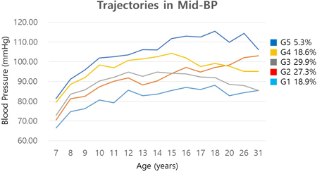
Article Information
vol. 132 no. Suppl 3 A19301
Published By:
American Heart Association, Inc.
Online ISSN:
History:
- Originally published November 6, 2015.
Copyright & Usage:
© 2015 by American Heart Association, Inc.
Author Information
- Ju-Mi Lee1;
- Norrina B Allen2;
- Yuichiro Yano2;
- Hyeon Chang Kim1;
- Joo Young Lee1;
- Philip Greenland2;
- Il Suh1
- 1Preventive medicine, Yonsei university college of medicine, Seoul, Korea, Republic of
- 2Preventive medicine, Northwestern Univ, Chicago, IL
Abstract 17084: Clinical Experience With the Subcutaneous Implantable Cardioverter Defibrillator in Paediatric Patients
Reinoud Knops, Tom Brouwer
Circulation. 2015;132:A17084
Abstract
Introduction: In pediatric patients prevention of Sudden Cardiac Death (SCD) using Implantable Cardioverter Defibrillators (ICD) is associated with a higher risk of complications than in the adult population. Surgical re-interventions due to infection, lead dislocation or migration and inappropriate shocks on intra and extracardiac oversensing are the most prevalent complications. The Subcutaneous ICD (S-ICD) has been introduces as a less invasive and extravascular alternative to overcome some of these complications. The scientific data that has led to the approval of the S-ICD did not include pediatric patients. Contradicting reports in small cohorts have been published on the effectiveness of S-ICD therapy in the pediatric population. We here report the largest pediatric cohort with the longest follow up to date where the S-ICD has been used for SCD prevention.
Hypothesis: The S-ICD is safe and effective for the prevention of SCD in a diverse pediatric population.
Methods: We retrospectively studied all consecutive pediatric patients (age<18 yrs)that were implanted with a S-ICD in our center.
Results: Fifteen S-ICDs were implanted in 14 patients (mean age 14, range 8-17), 71% were male, mean BMI was 18.8 (range 17.2-28.8), 57% had a secondary prevention indication, 77% had a genetic arrhythmia diagnosis and the median follow up was 21 months (range 1- 57).
The first shock efficacy of induced and spontaneous episodes was 100% (n=13 and n =2 respectively). Inappropriate shocks due to intracardiac oversensing occurred in 3 (21%) patients. No lead failures or dislocations were observed. Two patients (14%) experienced a complication requiring re-intervention: one infection and one pocket erosion. After the pocket erosion the S-ICD has been successfully re-implanted. In one patient the S-ICD was removed after cardiac transplantation. None of the patients died during follow up.
Conclusions: The S-ICD is safe and effective in a diverse pediatric populations. There was a very high efficacy of defibrillation therapy (100%) and a relative low and mild occurrence of complications. The S-ICD should be considered as a viable alternative for transvenous ICD’s in pediatric patients.
Article Information
vol. 132 no. Suppl 3 A17084
Published By:
American Heart Association, Inc.
Online ISSN:
History:
- Originally published November 6, 2015.
Copyright & Usage:
© 2015 by American Heart Association, Inc.
Author Information
- 1Cardiology, Academic Med Cntr Amsterdam, Hilversum, Netherlands
- 2Cardiology, Academic Med Cntr Amsterdam, Amsterdam, Netherlands
Abstract 17014: Getting to Zero: Impact of Electroanatomical Mapping on Fluoroscopy Use in Pediatric Catheter Ablation
Bradley C Clark, Kohei Sumihara, Robert McCarter, Charles I Berul, Jeffrey P Moak
Circulation. 2015;132:A17014
Abstract
Introduction: Over the past several years, alternative imaging techniques including electroanatomic mapping systems such as CARTO®3 (C3) have been developed to improve anatomic resolution and potentially limit radiation exposure in electrophysiology (EP) procedures. We retrospectively examined the effect of the introduction of C3 on patient radiation exposure during EP studies and ablation procedures at a children’s hospital.
Methods: All patients that underwent EP and ablation procedures between January 2012 and November 2014 were included; demographic information, fluoroscopy time in minutes (FT), total radiation dose in mGy (RAD), and dose-area product in μGy/m2 (DAP) were collected. Patients were stratified by time period (before vs. after C3 introduction), structural group (normal heart, congenital heart disease (CHD), and those with normal cardiac anatomy requiring trans-septal (TS) access), and arrhythmia diagnosis (Accessory Pathway (AP), AV Nodal Reentry Tachycardia (AVNRT), atrial, or ventricular arrhythmia). Mean values were compared using a single sample t-test, as well as analysis of covariance to control for age, weight, and arrhythmia diagnosis.
Results: Mean FT decreased after the introduction of C3 in patients with normal hearts (p<0.001), AP (p<0.001), AVNRT (p=0.002), and CHD (p=0.007). After controlling for age, weight, and arrhythmia diagnosis, there was a statistically significant decrease in FT in all three groups (normal heart, CHD and TS), in RAD in the TS group, and in DAP in both the normal heart and TS groups. In all other groups, there was a trend towards decreased RAD and DAP, but they did not reach statistical significance. After the introduction of C3, zero fluoroscopy was achieved in 18/66 (27%) and ≤ 1 minute of FT in 28/66 (42%) of ablation procedures in patients with normal hearts.
Conclusions: We have shown a decrease in all metrics that measure radiation exposure when comparing the time periods before and after the introduction of C3, secondary to reducing fluoroscopy time, fluoroscopic pulse rate and radiation dose per pulse. Further refinements are still needed to decrease radiation exposure towards the goal of zero fluoroscopy, but this cannot be achieved without thinking beyond fluoroscopy time.
Article Information
vol. 132 no. Suppl 3 A17014
Published By:
American Heart Association, Inc.
Online ISSN:
History:
- Originally published November 6, 2015.
Copyright & Usage:
© 2015 by American Heart Association, Inc.
Author Information
- 1Cardiology, Children’s National Health System, Washington, DC
- 2Biostatistics and Informatics, Children’s National Health System, Washington, DC
Abstract 16584: Association of Lower Height and Higher LDL-c in a Statewide Schoolchild Cholesterol Screening Program
Lee A Pyles, Christa Lilly, Eloise Elliott, William A Neal
Circulation. 2015;132:A16584
Abstract
Introduction: The Coronary Artery Risk Detection in Appalachian Communities (CARDIAC) Project has screened West Virginia 5th graders since 1998 to facilitate primordial prevention of coronary heart disease (CHD) in WV. LDL-c levels above 175 mg/dl in children suggest Familial Hyperlipidemia (FH) in the child’s family and a level above 160 mg/dl with history of CHD in relatives can also establish a diagnosis.
Hypothesis: Based on previous adult literature, the association of lower height with higher LDL level observed in adults begins in childhood and is prominent in children with LDL in FH range.
Methods: Fifth graders are screened yearly in WV schools with parental consent for Body Mass Index and lipid panel. Lipids were analyzed with respect to either short stature < 2 SD for height or comparing 1st (shortest) and 4th quartiles of the population. Statistical analysis for age- and gender-adjusted height percentiles was performed in SAS.
Results: 59,386 children had lipid and height data. Mean LDL-c for 1st vs. 4th quartile of height was 94.08 mg/dl (95% Confidence Interval-CI 93.66-94.51) vs. 90.03 mg/dl (CI 89.65-90.42). First quartile of height students had average 4.05 mg/dl higher LDL-c (95% CI 3.48 -4.62 mg/dl). 4398 children had an LDL level above 130 g/dl, 632 above 160 mg/dl and 247 above 175 mg/dl. The Chi square analysis of short stature (height 130 g/dl was also significant (p=0.013) with increased odds of LDL-c above 130 g/dl compared to non-short stature (OR= 1.37, CL 1.07-1.75). Table 1 shows odds ratio for varying levels of elevated LDL-c for the first (shortest) vs. 4th (tallest) quartile of students.
Conclusions: Shorter stature is associated with higher LDL-c level in WV 5th graders generally and in those children with increased risk for genetic dyslipidemia. The trend to increasing odds ratio in strata of higher LDL-c supports a recent report of association of single nucleotide polymorphisms selecting for lower genetic height and higher LDL-c.

Article Information
vol. 132 no. Suppl 3 A16584
Published By:
American Heart Association, Inc.
Online ISSN:
History:
- Originally published November 6, 2015.
Copyright & Usage:
© 2015 by American Heart Association, Inc.
Author Information
- 1Pediatrics, West Virginia Univ, Morgantown, WV
- 2Biostatistics, West Virginia Univ, Morgantown, WV
- 3College of Physical Activity and Sport Science, West Virginia Univ, Morgantown, WV
Abstract 16193: Sport Activity and Sudden Cardiac Death in Women
Cristina Basso, Stefania Rizzo, Kalliopi Pilichou, Elisa Carturan, Gaetano Thiene
Circulation. 2015;132:A16193
Abstract
Background: No specific data are available on the prevalence and characteristics of cardiac sudden death (SCD) during sport activities among young women.
Methods and Results: From a prospective 30-year target project on SCD in the young, involving 695 subjects 1-40 years old who died suddenly in the same geographic areas, 649 were due to SCD (196 female, 30%). A total of 76 young adults were competitive athletes and died during or soon after effort (11%). Only 6 (8%) of such events occurred in women (mean age 23±10 yrs, range 12-38), specifically during jogging (2), and swimming, volley, gymnastic and triathlon (one each). Cause of death seemed more likely to be associated with structurally normal heart in women compared with men (50% versus 7%; P<0.01). In particular, while inherited cardiomyopathies (i.e. arrhythmogenic, hypertrophic and dilated) and coronary atherosclerosis are the leading cause in the overall population of athletes (24/76, 32% and 11/76, 14.5% respectively) and in the male sub-group (24/70, 34% and 11/70, 16%, respectively), they were never observed in female athletes.
Conclusions: Sports-related SCD in women is dramatically less common compared with men. In the female athletic population, SCD occurs in the setting of a structurally normal heart in half of cases. In a country with obligatory pre-participation screening for sport activity, inherited cardiomyopathies and atherosclerotic coronary artery diseases are almost exclusive of the male athletic population.
Article Information
vol. 132 no. Suppl 3 A16193
Published By:
American Heart Association, Inc.
Online ISSN:
History:
- Originally published November 6, 2015.
Copyright & Usage:
© 2015 by American Heart Association, Inc.
Author Information
- 1Dept Cardiac, thoracic and vascular sciences, Univ of Padova, Padova, Italy
- 2Cardiac, thoracic and vascular sciences, Univ of Padua, Padova, Italy
Abstract 14231: Effectiveness and Safety of Statin Therapy in Children: A Multi-Center, Real-World Clinical Practice Experience
Rae Ellen W Kavey, Cedric Manlhiot, Tanveer Collins, Samuel S Gidding, Matthew Demczko, Sarah Clauss, Ruth Phillippi, Ashraf Harahsheh, Michele Mietus-Snyder, Brian McCrindle
Circulation. 2015;132:A14231
Abstract
Results of statin use in clinical practice in children are very limited, with effectiveness and safety data based on short-term clinical trials. To address this, we reviewed the results of all children with primary hypercholesterolemia treated with statins for more than 6 months from 4 Pediatric Lipid Clinics. Expert Panel guidelines recommend statin if LDL-cholesterol (LDL-C) exceeds 4.9 mmol/L (190 mg/dL), or 4.1 mmol/L (160 mg/dL) with additional risk factors, after 6 months of lifestyle modification, with hepatic enzyme monitoring plus clinical surveillance and CK measurement for myositis. Patients at all study sites were managed using these guidelines.
Results: There were 246 pts (59% male) who had 1488 clinical assessments. Mean age at statin initiation was 12.5±0.5 yrs. Mean duration of therapy was 2.9 yrs (IQR: 1.9_4.7) with 48% more than 3 yrs. Initial statin prescribed varied over time but 61% of pts were started on atorvastatin and 70% remained on the first statin prescribed. Mean compliance was assessed at 92% and 234 pts (94%) remained on statin therapy at the end of the review. Baseline and follow-up lab values and anthropometry (mean + SD) appear below.
While 13 pts (5%) had transient AST or ALT > 3xULN, or CK >10xULN at some time during follow-up, no pt was diagnosed with myositis. In regression analysis adjusted for repeated measures over time, statin treatment was associated with significant reductions in total cholesterol, LDL-C and non-HDL-C with no change in HDL-C, TG, safety labs or anthropometry. Despite statin, 51% and 70% of pts had LDL-C levels above minimal (<3.35mmol/L; 130 mg/dL) and ideal (<2.85mmol/L; 110 mg/dL) targets at last follow-up.
Conclusions: These findings in a large series of pts from real-world clinical practice show that statin therapy in children with primary hypercholesterolemia is safe and effectively lowers LDL-C on mid-term follow-up. Side effects are rare and discontinuation of treatment is uncommon.
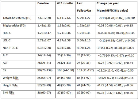
Article Information
vol. 132 no. Suppl 3 A14231
Published By:
American Heart Association, Inc.
Online ISSN:
History:
- Originally published November 6, 2015.
Copyright & Usage:
© 2015 by American Heart Association, Inc.
Author Information
- Rae Ellen W Kavey1;
- Cedric Manlhiot2;
- Tanveer Collins2;
- Samuel S Gidding3;
- Matthew Demczko4;
- Sarah Clauss5;
- Ruth Phillippi6;
- Ashraf Harahsheh7;
- Michele Mietus-Snyder7;
- Brian McCrindle8
- 1Pediatrics, Univ of Rochester Med Cntr, Rochester, NY
- 2Rsch scientist, Hosp for Sick Children, Toronto, Canada
- 3Cardiology, Nemours Alfred I du Pont Children’s Hosp, Wilmington, DE
- 4Pediatrics, Nemours Alfred I du Pont Children’s Hosp, Wilmington, DE
- 5Pediatric Cardiology, Children’s National Med Cntr, Washington, DC
- 6Rsch Coordinator, Children’s National Med Cntr, Washington, DC
- 7Cardiology, Children’s National Med Cntr, Washington, DC
- 8Cardiology, Hosp for Sick Children, Toronto, Canada
Abstract 13716: Pharmacokinetics of Pravastatin in Pediatric Dyslipidemia Patients: Clinical Impact of Genetic Variation on Statin Disposition
Jonathan Wagner, Susan Abdel-Rahman, Leon van Haandel, Geetha Raghuveer, Ralph Kauffman, J. Steven Leeder
Circulation. 2015;132:A13716
Abstract
Background: In adults, genetic variation of the drug transporter, organic anion transporting polypeptide (OATP1B1) encoded by the gene SLCO1B1, decreases hepatic uptake of statins. This leads to increased circulating statin concentrations in plasma, thereby placing patients at higher risk for adverse events and decreased efficacy. Expression of OATP transporters in mice changes during development, raising the question of whether in humans the genotype-phenotype relationship observed in adults is relevant in the developing child.
Methods: Participants (8-21 years) with at least one allelic variant of SLCO1B1 c.521 T>C (521TC; n=8) and wild type controls (521TT; n=5) were enrolled in a single oral dose pharmacokinetics (PK) study. Pravastatin and isomers were quantified by liquid chromatography tandem mass spectrometry. A model-independent PK approach was used to define the peak plasma concentration (Cmax), area under the curve (AUC) during the sample period and AUC to infinity.
Results: 521TC subjects had a nearly 3-fold increase in Cmax (177.62 vs. 56.53 ng/ml; p=0.001) and AUC (389.81 vs. 139.57 ng/ml*h; p=0.004) compared to 521TT controls (Fig. 1). Formation of the 3-alpha-iso-pravastatin acid and lactone isomers in the acidic environment of the stomach prior to absorption was variable in both groups. There was a linear relationship between pravastatin acid AUC and the ratio of pravastatin acid to total analyte (pravastatin acid, 3-alpha-iso-pravastatin, and corresponding lactones) AUC amongst genetic variants (R=0.8961) that does not appear to be present in the control.
Conclusion: The impact of the SLCO1B1 c.521 gene variant on pravastatin PK in adults was replicated in our pediatric cohort. However, other physicochemical factors appear to contribute to variability in the dose-exposure relationship. This data must be replicated in a larger cohort, and the pharmacodynamic consequences assessed, before implementing OATP1B1 driven dosing guidelines.
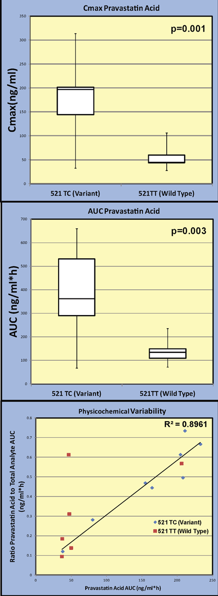
Article Information
vol. 132 no. Suppl 3 A13716
Published By:
American Heart Association, Inc.
Online ISSN:
History:
- Originally published November 6, 2015.
Copyright & Usage:
© 2015 by American Heart Association, Inc.
Author Information
- Jonathan Wagner1;
- Susan Abdel-Rahman2;
- Leon van Haandel2;
- Geetha Raghuveer1;
- Ralph Kauffman2;
- J. Steven Leeder2
- 1Pediatric Cardiology, Children’s Mercy Hosp, Kansas City, MO
- 2Pediatric Clinical Pharmacology, Children’s Mercy Hosp, Kansas City, MO
Abstract 13142: Characteristics of Decremental Accessory Pathways in Children
Allison Hill, Michael Silka, Yaniv Bar-Cohen
Circulation. 2015;132:A13142
Abstract
Introduction: While decremental retrograde accessory pathways (DAPs) classically present clinically as permanent junctional reciprocating tachycardia (PJRT), they may be diagnosed unexpectedly during electrophysiology study (EPS). Although DAPs associated with PJRT have been studied, information about other variants of DAPs in children is limited.
Methods: We retrospectively studied patients 0 – 21 years of age with retrograde DAPs who underwent EPS between 2005 and 2014. Decrement was defined as rate-dependent prolongation of the local ventriculo-atrial (VA) time by > 30 ms during ventricular extrastimulus or rapid ventricular pacing. Clinical presentation, electrophysiologic properties and ablation outcomes of patients with DAPs were compared to an age-matched cohort of patients with non-decremental accessory pathways. In addition, a sub-analysis examined patients with DAPs depending on whether clinical PJRT was diagnosed prior to the EPS.
Results: A total of 29 patients with DAPs and 83 controls were included [mean age at time of EPS 10.3 ± 5.7 years and 10.7 ± 5.2 years, respectively (p = NS)]. Compared to controls, DAPs were more likely to be associated with syncope [6/29 (21%) vs 3/83 (4%), p = 0.01], ventricular dysfunction [6/29 (21%) vs 4/83 (5%), p = 0.04], and treatment with antiarrhythmic medications [20/29 (69%) vs 39/83 (47%) p = 0.04]. In addition, DAPs were more likely to be right-sided [20/29 (69%) vs. 24/83 (29%), p < 0.01] and septal [19/29 (66%) vs 17/83 (21%), p < 0.01]. DAPs and their age-matched controls had similar rates of acute ablation success [26/29 (90%) vs 77/83 (93%), p = NS] and recurrence [2/29 (7%) vs 4/83 (5%), p = NS]. There were no major procedural complications in either group. Of the patients with DAPs, only 12 (41%) had clinical PJRT, and these patients had more syncope [6/12 (50%) vs 0/17 (0%), p < 0.01], slower ORT (mean cycle length 383 ± 89 ms vs 323 ± 60 ms, p = 0.04), and longer maximum VA times (mean 283 ± 116 ms vs 208 ± 42 ms, p = 0.01) compared to those with DAPs without clinical PJRT.
Conclusions: The majority of pediatric patients with DAPs do not present with clinical PJRT. DAPs in general are typically associated with more severe symptoms, but ablation outcomes are similar to age-matched controls with non-decremental pathways.
Article Information
vol. 132 no. Suppl 3 A13142
Published By:
American Heart Association, Inc.
Online ISSN:
History:
- Originally published November 6, 2015.
Copyright & Usage:
© 2015 by American Heart Association, Inc.
Author Information
- Allison Hill;
- Michael Silka;
- Yaniv Bar-Cohen
- Pediatrics, Children’s Hosp Los Angeles, Los Angeles, CA
Abstract 12713: Epidemiology and Mortality of Very and Extremely Preterm Infants With Congenital Heart Defects
Patricia Y Chu, Jennifer S Li, Andrzej S Kosinski, Christoph P Hornik, Kevin D Hill
Circulation. 2015;132:A12713
Abstract
Introduction: Congenital heart disease (CHD) is estimated to occur in 6-10 per 1000 births. Although epidemiology and outcomes for term and near term infants with CHD are well described, data are limited for very and extremely preterm (VEP) infants. We used the Healthcare Cost and Utilization Project Kids’ Inpatient Database (KID), a nationally representative administrative database, to evaluate epidemiology and outcomes for VEP infants (25 to 32 weeks gestational age, GA) with CHD.
Methods: Two separate cohorts were defined from the KID: an epidemiologic cohort including birth hospitalizations in ‘03, ’06, ’09 and ‘12; and an outcomes cohort including hospitalizations at a children’s hospital or pediatric unit for infants < 1 month of age in ‘06 and ‘09. CHD was defined by ICD-9-CM codes with severe CHD defined as those defects expected to be universally diagnosed during a preterm birth hospitalization. Weighted multivariate logistic regression analysis was used to calculate odds ratios (OR) for mortality, adjusted for race, sex, GA, year, small for gestational age and hospital teaching status.
Results: Our epidemiologic and outcomes cohorts included 249,011 and 49,893 VEP infants, respectively. Incidence of CHD (116/1000 VEP births) and severe CHD (7/1000 VEP births) were both higher than previously reported in term infants. Relative risk of severe CHD in VEP vs term infants was 4.80 (95% CI 4.76, 4.84) and decreased with increasing GA (5.7 at 25 weeks GA to 4.3 at 31 weeks, p=0.005). Hospital mortality (Figure) was substantially higher for VEP infants with vs without severe CHD (26% vs 5%; adjusted OR 7.5 [95% CI: 5.9, 9.6]). Overall 16% of VEP infants with severe CHD underwent cardiac surgery during the neonatal hospitalization with mortality after surgery of 16%.
Conclusions: CHD incidence is increased in very and extremely preterm infants and outcomes are poor. These data underscore the need for interventions to decrease preterm delivery when severe CHD is diagnosed in utero.
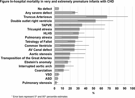
Article Information
vol. 132 no. Suppl 3 A12713
Published By:
American Heart Association, Inc.
Online ISSN:
History:
- Originally published November 6, 2015.
Copyright & Usage:
© 2015 by American Heart Association, Inc.
Author Information
- 1Pediatrics, Duke Clinical Rsch Institute, Duke Univ Sch of Medicine, Durham, NC
- 2Pediatrics, Duke Clinical Rsch Institute, Duke Univ Med Cntr, Durham, NC
- 3Biostatistics and Bioinformatics, Duke Clinical Rsch Institute, Duke Univ Med Cntr, Durham, NC
Abstract 19872: 30-day Readmission Rate and Center Variability for US Children Hospitalized With Heart Failure
Christopher S Almond, David N Rosenthal, Matthew Wood, Joseph W Rossano, Jack F Price, Kevin P Daly, Esther Liu, Chealsea Nather, Kristin Jensen, Andrew Y Shin
Circulation. 2015;132:A19872
Abstract
Background: The 30-day readmission rate is a well-established performance benchmark for adult heart failure programs. Little is known about the 30-day readmission rate for pediatric heart failure programs or the variability among centers. We sought to determine the 30-day readmission rate for children admitted with acute decompensated heart failure (ADHF) to support benchmarking efforts in pediatrics.
Methods: The Pediatric Health Information System (PHIS) database was queried to identify all children ≤21 years of age admitted between January 2009 and March 2014 with a primary or secondary diagnosis of heart failure. All-cause 30-day readmission rates were calculated, as was mean length of stay.
Results: Over the 5-year study period, a total of 39,150 hospitalizations at 47 children’s hospitals met the inclusion criteria. Overall, 56% were infants, 25% age 1-12 years, 9% adolescents, and 9% adults, 48% were female, 72% had congenital heart disease, 28% had cardiomyopathy/myocarditis. The mean hospital length of stay was 25 days (range 11,41 days; SD 7 days). In-hospital mortality was 8.4%. Overall 30-day readmission rate was 20% (range 10%, 30%; SD 5%) and varied considerably by center (Figure).
Conclusion: A substantial portion of children (one in five) admitted to the hospital for heart failure is re-admitted within 30 days. There is considerable center-based variation in re-admission rates. The mean hospital length of stay exceeds 3 week and also varies considerably by center.
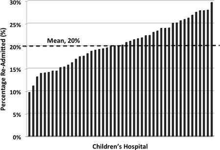
Article Information
vol. 132 no. Suppl 3 A19872
Published By:
American Heart Association, Inc.
Online ISSN:
History:
- Originally published November 6, 2015.
Copyright & Usage:
© 2015 by American Heart Association, Inc.
Author Information
- Christopher S Almond1;
- David N Rosenthal1;
- Matthew Wood2;
- Joseph W Rossano3;
- Jack F Price4;
- Kevin P Daly5;
- Esther Liu1;
- Chealsea Nather2;
- Kristin Jensen1;
- Andrew Y Shin1
- 1Pediatrics (Cardiology), Stanford Univ, Palo Alto, CA
- 2Cntr for Quality and Clinical Effectiveness, Stanford Univ, Palo Alto, CA
- 3Cardiology, Children’s Hosp of Philadelphia, Philadelphia, PA
- 4Cardiology, Texas Children’s Hosp, Houston, TX
- 5Cardiology, Boston Children’s Hosp, Boston, MA
Abstract 19165: Regional Changes in Cellular and Molecular Pathology in Children With End-stage Dilated Cardiomyopathy: Correlation With Cardiac Performance
Ching Kit Chen, Mei Sun, Cameron Slorach, Ryo Ishii, Andrew N Redington, Mark K Friedberg
Circulation. 2015;132:A19165
Abstract
Dilated cardiomyopathy (DCM) carries poor prognosis in children, often needing heart transplant. Regional differences in left ventricular (LV) function suggest inhomogeneous expression of contractility related proteins. However, associations between myocardial performance and histological / molecular findings are poorly defined.
We sought to investigate associations of regional myocardial performance with histological and molecular changes in hearts explanted from children with DCM, undergoing transplantation.
Clinical and echo features (including tissue Doppler and strain) were correlated with histological and molecular findings in hearts explanted from 9 children [median age 2.8 years (range 4.4 months-17 years); 6 had LVAD]. Interstitial fibrosis, myocyte volume, and expression of proteins (western blot) implicated in fibrosis and function (p38, ERK, phospho-JNK, phospho-GSK, SMA, myosin heavy chain, cadherin, ILK, sarcoplasmic reticulum Ca2+-ATPase [SERCA2a], phospho-CamKII, phospholamban [PLN], and phospho-PLN) were correlated with speckle tracking strain from 11 regions per patient (basal and mid regions of anterior septum, anterior, lateral, posterior and inferior LV segments, and right ventricular [RV] free wall).
On average, no significant difference was noted for the percentage of fibrosis and myocyte volume between the various LV segments, and between RV and LV. LV dimension, systolic and diastolic function were not related to interstitial fibrosis, myocyte volume or molecular signaling. Tricuspid annular plane systolic excursion was related to myocyte volume (r=0.89; p<0.01). An inverse relationship between the extent of interstitial fibrosis and segmental longitudinal strain was present for LV basal and mid posterior segments (basal posterior, r=0.96, p<0.01; mid posterior, r=0.74, p=0.05). Global longitudinal strain was related to expression of ILK and SERCA2a (ILK, r=0.78, p=0.02; SERCA2a, r=0.71, p=0.04).
This study demonstrated association of regional cardiac function in children with DCM with expression of calcium-cycling and contractile proteins, as well as interstitial fibrosis. These findings have implications for diagnosis and therapeutic targets in pediatric DCM.
Article Information
vol. 132 no. Suppl 3 A19165
Published By:
American Heart Association, Inc.
Online ISSN:
History:
- Originally published November 6, 2015.
Copyright & Usage:
© 2015 by American Heart Association, Inc.
Author Information
- 1Cardiology Service, Dept of Paediatric Subspecialties, KK Women’s and Children’s Hosp, Singapore, Singapore
- 2Div of Cardiology, the Labatt Family Heart Cntr, The Hosp for Sick Children, Toronto, Canada
- 3Pediatric Cardiology, Dept of Pediatrics, Cincinnati Children’s Hosp, Cincinnati, OH
Abstract 18039: Resource Utilization for Initial Hospitalization in Pediatric Heart Transplantation in the United States
Dana M Boucek, Ashwin K Lal, Nelangi M Pinto, Hsin-Yi Cindy Weng, Jacob F Wilkes, Shaji C Menon
Circulation. 2015;132:A18039
Abstract
Background: Pediatric heart transplantation (HT) is resource intensive. Event driven pediatric HT databases do not capture data on resource use. The objective of this study was to evaluate resource utilization and identify associated factors during initial hospitalization for pediatric HT in a large multi-institutional cohort.
Methods: This is a multicenter retrospective cohort study using the PHIS database (43 US children’s hospitals) of children ≤ 19 years of age who underwent HT between 1/07 and 7/13. Data collected: Demographic variables including site, payer, distance and time to center, clinical pre and post-transplant variables, mortality, cost and charge. Total length of stay (LOS) and charge for initial hospitalization were used as surrogates for resource use. Charges were inflation adjusted to 2013 $. Gamma regression analysis was performed to evaluate factors associated with resource use.
Results: Of 1629 subjects, 54% were male, and the median age at HT was 5 years (IQR 0-13). The median total and ICU LOS were 51 (IQR: 23-98) and 23 (IQR: 9-58) days respectively, and mortality occurred in 82 (5%). Total charge and cost for hospitalization were $852,713 ($464,900-$1,609,300) and $383,600 ($214,900-$681,000) respectively. Factors associated with resource use on multivariate analysis are shown in Table 1. Younger age, lower center volume, southern region, and comorbidities prior to HT were associated with higher resource use. In later years, costs increased despite shorter LOS.
Conclusions: This large multicenter study provides novel insight into factors associated with resource use in pediatric HT that cannot be assessed in alternative event driven transplant databases. Peri-transplant morbidities are associated with increased cost and LOS. Reducing costs in line with LOS will improve health care value. Regional and center volume differences need further investigation for optimizing value-based care and efficient use of scarce resources.
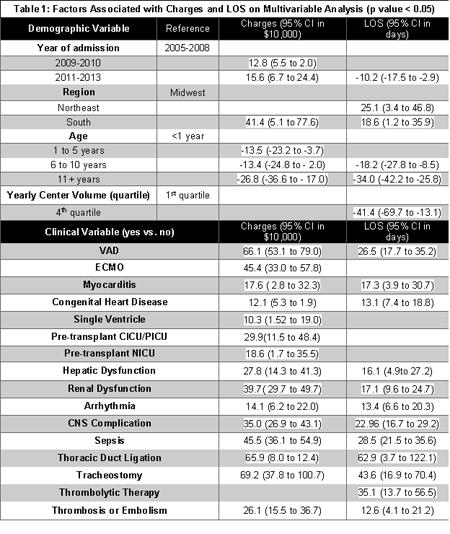
Article Information
vol. 132 no. Suppl 3 A18039
Published By:
American Heart Association, Inc.
Online ISSN:
History:
- Originally published November 6, 2015.
Copyright & Usage:
© 2015 by American Heart Association, Inc.
Author Information
- Dana M Boucek1;
- Ashwin K Lal1;
- Nelangi M Pinto1;
- Hsin-Yi Cindy Weng2;
- Jacob F Wilkes3;
- Shaji C Menon1
- 1Pediatric Cardiology, Univ of Utah, Salt Lake City, UT
- 2Div of Epidemiology, Univ of Utah, Salt Lake City, UT
- 3Pediatrics, Univ of Utah, Salt Lake City, UT
Abstract 18106: 2d Strain is a More Accurate Measure of Systolic Function Than Shortening Fraction and Ejection Fraction in Pediatric Patients Following Orthotopic Heart Transplant
Meghna D Patel, Georgeann K Groh, Justin Varughese, Craig Myers, Tim Sekarski, Diana Hartman, Joshua J Murphy, Gautam K Singh, Shafkat Anwar
Circulation. 2015;132:A18106
Abstract
Introduction: 2D strain by echocardiography is a sensitive technique for assessing LV systolic function in pediatric heart transplant patients, although it is under-utilized clinically.
Methods: We prospectively compared 2D LV 4-chamber Lagrangian longitudinal strain (LS) in 34 echo studies against cardiac output indexed to body surface area (cardiac index, Ci) via cardiac catheterization to assess accuracy of LS. Of those echos, 31/34 were simultaneous with cath, and 3/34 were within 10 days of catheterization. Ci was also compared to LV shortening fraction (SF) and single plane ejection fraction (EF). FS via M-mode in parasternal short-axis and EF via 4 chamber were measured. Strain was obtained in the 4 chamber apical view as an average of 6 segments. Right heart catheterization was performed and evaluated by a cardiac interventionalist blinded to echo data. Pearson’s correlation coefficient was used to assess relationships.
Results: Mean age was 10.2 years (0.3 – 19 years), 11 males. Mean HR was 99 bpm (64-155) for catheterization, and 97 bpm (66-148) for echocardiography, mean frame rate 72 f/sec (47-109). Mean Ci was 3.8 L/min/m2 (95% CI 1.9 to 5.73). LS was diminished for this cohort, mean -13.7 (95% CI – 4 to -23.3), however, EF and SF were normal (mean EF 59.1% (95% CI 32.58%- 85.6%), mean FS 35.6% (95% CI 24%- 47%)).
There was moderate positive correlation between LS and Ci, (r = 0.48, p = 0.002). FS and EF did not correlate with Ci (r = 0.34, p = 0.03, and r = 0.15, p = 0.39 respectively). There was fair positive correlation between LS and EF and LS and SF (r = 0.36, p = 0.03 and r = 0.38, p = 0.02 respectively).
Conclusion: 4 chamber LV LS may be a more sensitive and reliable non-invasive method to assess systolic function in pediatric heart transplant patients. Further investigation in a larger sample using global (18 segment) strain is warranted.

Article Information
vol. 132 no. Suppl 3 A18106
Published By:
American Heart Association, Inc.
Online ISSN:
History:
- Originally published November 6, 2015.
Copyright & Usage:
© 2015 by American Heart Association, Inc.
Author Information
- Meghna D Patel;
- Georgeann K Groh;
- Justin Varughese;
- Craig Myers;
- Tim Sekarski;
- Diana Hartman;
- Joshua J Murphy;
- Gautam K Singh;
- Shafkat Anwar
- Pediatric Cardiology, Washington Univ Sch of Medicine, St. Louis, MO
Abstract 17697: Myocardial Response to Milrinone in Single Ventricle Heart Disease
Stephanie J Nakano, Penny Nelson, Carmen C Sucharov, Shelley D Miyamoto
Circulation. 2015;132:A17697
Abstract
Background: Single ventricle heart disease (SV) is a subset of congenital heart defects characterized by a univentricular circulation. Despite surgical intervention, progressive heart failure (HF) is a common cause of death and indication for heart transplantation in children with SV. The exact mechanisms underlying SV HF are poorly understood, limiting the ability to identify effective therapies. Empiric treatment with milrinone, a phosphodiesterase 3 inhibitor (PDE3i), has become increasingly common in SV HF; however the response to milrinone has yet to be characterized in the pediatric SV population. The purpose of this study was to determine the myocardial response to PDE3i in children with SV.
Methods and Results: Cyclic adenosine monophosphate (cAMP) levels, phosphodeisterase (PDE) activity, and phospholamban phosphorylation (pPLB) were determined in explanted human ventricular myocardium from non-failing pediatric donors (n=10) and pediatric patients transplanted secondary to SV HF (of right ventricular [RV] morphology). SV HF subjects were divided into those who received milrinone therapy (n=13) and those who did not (n=12). In comparison to non-failing RV myocardium, cAMP levels are lower in SV HF patients treated with milrinone (ANOVA, p=0.046). Chronic milrinone treatment does not alter total PDE or PDE3 activity in SV myocardium. Additionally, when compared with non-failing RV myocardium, SV myocardium (both with and without milrinone) demonstrates equivalent pPLB at the protein kinase A site (serine 16 residue).
Conclusions: As evidenced by preserved pPLB, the molecular adaptation associated with SV HF differs significantly from that demonstrated in pediatric HF due to dilated cardiomyopathy. These alterations support a pathophysiologically distinct mechanism of HF in pediatric SV patients, which has direct implications regarding the putative response to PDE3i treatment in this population.
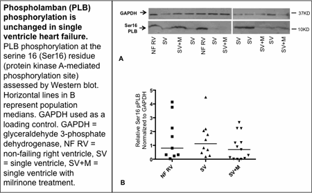
Article Information
vol. 132 no. Suppl 3 A17697
Published By:
American Heart Association, Inc.
Online ISSN:
History:
- Originally published November 6, 2015.
Copyright & Usage:
© 2015 by American Heart Association, Inc.
Author Information
- 1Pediatric Cardiology, Univ of Colorado, Aurora, CO
- 2Medicine, Cardiology, Univ of Colorado, Aurora, CO
Abstract 14382: Outcomes of Heart Transplantation in the Smallest Infants: To List or Let Grow?
Matthew J Bock, Elfriede Pahl, Osama Eltayeb, John M Costello, Jeffrey G Gossett
Circulation. 2015;132:A14382
Abstract
Introduction: Waitlist and post-transplant outcomes for heart transplantation (HTx) in infants below 5 kg are not well defined.
Hypothesis: We hypothesize that lower listing weight leads to higher waitlist and post-HTx mortality in the smallest infants.
Methods: All children <5 kg listed for primary (1°) HTx between 10/87-12/14 were identified in the United Network for Organ Sharing database. Demographic and clinical covariates, along with waitlist and post-HTx survival, were compared between weight groups. Waitlist and post-HTx survival differences were assessed via the Kaplan-Meier method and a Cox proportional hazards model assessed independent risk factors for mortality.
Results: Over the 27 year study period, 2,785 infants <5 kg were listed for 1° HTx. Median waiting time was 25 days [IQR 9-57]. The majority had congenital heart disease (79%) and 205 (7%) were <2.5 kg. Waitlist death or removal due to deterioration was more common in infants <2.5 kg (37%), compared to 31% in those 2.5-5 kg (p <0.01). Waitlist survival was worse for patients with progressively lower listing weight (p <0.01; Fig. 1a). In addition, 30-day and 1-year post-HTx survival were worse in the <2.5 kg cohort [77%, 64% (<2.5 kg) vs. 89%, 79% (2.5-5 kg), respectively] (p <0.01; Fig. 1b). However, 1-year conditional survival after HTx was not different between the groups (p=0.82; Fig. 1c). In the <2.5 kg group, pre-HTx ECMO requirement was an independent risk factor for waitlist and post-HTx mortality [waitlist adjusted HR 4.3 (CL 2.0, 9.2), post-HTx adjusted HR 11.3 (CL 4.0, 31.8); p <0.01].
Conclusions: For small infants, lower listing weight is associated with increasing waitlist mortality. Early post-HTx mortality is worse for infants weighing <2.5 kg and the need for ECMO while listed greatly increases these risks. Despite elevated waitlist and early post-HTx mortality, long term conditional survival for infants <2.5 kg is similar to that achieved in patients weighing 2.5-5 kg.
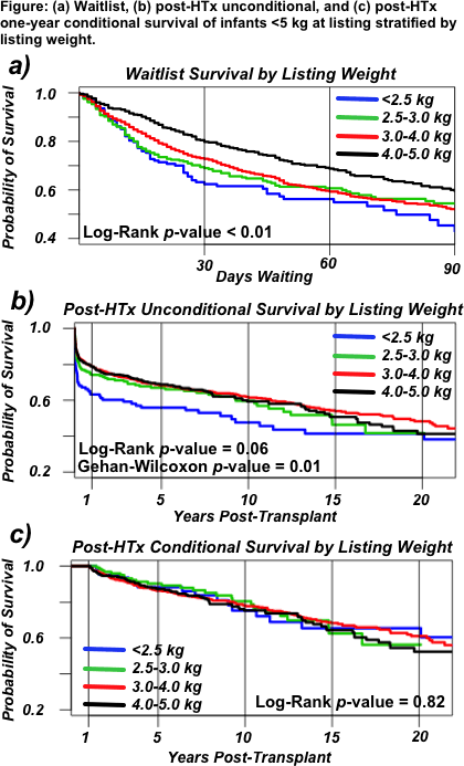
Article Information
vol. 132 no. Suppl 3 A14382
Published By:
American Heart Association, Inc.
Online ISSN:
History:
- Originally published November 6, 2015.
Copyright & Usage:
© 2015 by American Heart Association, Inc.
Author Information
- 1Pediatric Cardiology, Ann & Robert H. Lurie Children’s Hosp of Chicago, Chicago, IL
- 2Cardiovascular Thoracic Surgery, Ann & Robert H. Lurie Children’s Hosp of Chicago, Chicago, IL
Abstract 13879: Clinical Features and Prognosis of Patients With Left Ventricular Noncompaction Cardiomyopathy: A Comparison Between Infantile and Juvenile Types
Asami Takasaki, Sayaka Ozawa, Nariaki Miyao, Hideyuki Nakaoka, Keijiro Ibuki, Keiichi Hirono, Naoki Yoshimura, Fukiko Ichida
Circulation. 2015;132:A13879
Abstract
Introduction: Left ventricular noncompaction cardiomyopathy (LVNC) is characterized by a left ventricle with a prominent trabecular meshwork. Long-term prognosis of LVNC has not been elucidated yet.
Purpose: The aim of this study is to clarify the difference between infantile and juvenile cases and identify the risk factor of poor prognosis of LVNC in the largest series to date.
Methods: Based on nationwide surveys of LVNC in Japanese children, we compared the clinical features and anatomical properties of infantile cases of LVNC (< 2 years: 85 cases) with that of juvenile cases (2-15 years: 73 cases). In addition to the standard noncompacted to compacted layer (N/C) ratio, we developed an echocardiographic criteria which represents an average of N/C ratios in five wall segments to estimate the severity of LVNC.
Results: The duration of follow-up ranged from 15 days to 22 years (median 5 years). Although most patients in the infantile group had clinical signs of heart failure at initial presentation (70.1%), the majority of juvenile cases were asymptomatic and identified when screened for cardiac abnormalities, such as ECG screening (55.8%). WPW syndrome was higher in both groups (infantile group: 9.7%, juvenile group: 10.0%), the incidence of LBBB and VT was lower than those reported in adults. On echocardiography, the maximum N/C ratio was observed at the apex in both groups. Neither noncompaction score nor N/C score was significantly different between groups. Left ventricular ejection fraction (LVEF) at initial presentation was significantly lower in the infantile group than in the juvenile group. Although survival analysis showed poor prognosis in the infantile group, the significant risk factor was LVEF below 50% (p = 0.0004, hazard ratio = 9.0), rather than age of onset.
Conclusions: LVNC in both infantile and juvenile groups showed poor prognosis when correlated with depressed LVEF at initial presentation.
Article Information
vol. 132 no. Suppl 3 A13879
Published By:
American Heart Association, Inc.
Online ISSN:
History:
- Originally published November 6, 2015.
Copyright & Usage:
© 2015 by American Heart Association, Inc.
Author Information
- Asami Takasaki1;
- Sayaka Ozawa1;
- Nariaki Miyao1;
- Hideyuki Nakaoka1;
- Keijiro Ibuki1;
- Keiichi Hirono1;
- Naoki Yoshimura2;
- Fukiko Ichida1
- 1Pediatrics, Univ of Toyama, Toyama, Japan
- 2Thoracic and Cardiovascular Surgery, Univ of Toyama, Toyama, Japan
Abstract 12452: Clinical Practice Patterns are Relatively Uniform Between Pediatric Heart Transplant Centers: A Survey Based Assessment
Chesney Castleberry, Sonja Ziniel, Christopher Almond, Scott Auerbach, Seth Hollander, Lal Ashwin, Matthew Fenton, Joseph Rossano, Melanie Everitt, Kevin Daly
Circulation. 2015;132:A12452
Abstract
Background: Randomized control trials (RCT) of immunosuppression are needed in pediatric heart transplantation. However, variations in clinical practice could confound results. We surveyed centers to describe practice variations and understand willingness to modify protocols for the purpose of a RCT.
Hypothesis: We hypothesized that though variability in practice exists, there would be a willingness to change protocols as part of a RCT.
Methods: Pediatric heart transplant centers were identified based on participation in the Pediatric Heart Transplant Study. One member from each institution was sent an electronic request to complete a survey using a customized Qualtrics tool. Simple descriptive statistics were used.
Results: The response rate was 72% (40 responses from 52 contacted centers, 37 complete). Respondents were from the United States (36, 90%), Great Britain (2, 5%), Canada (1, 2.5%), and Brazil (1, 2.5%). Mean center volume was 10 transplants/year (range of 1 to 25). A majority of centers use tacrolimus (36, 95%) and mycophenolate mofotil (36, 95%) as maintenance therapy. Other immunosuppression was cyclosporine (7, 18%), everolimus or sirolimus (3, 8%), and azathioprine (2, 5%). Responses for most clinical practices were over 70% similar except for steroid use and biopsy frequency (Table). Overall, willingness to change clinical practice was greater than 70%. A majority of respondents, 97% (36/37) responded that they would be willing to participate in a RCT and 100% (37/37) said they would be willing to participate in a RCT investigating everolimus or sirolimus.
Conclusion: Though practice variations exist between pediatric heart transplant centers, most major components of clinical practice protocols are similar. Importantly, a majority of centers are willing to adopt a common protocol for a RCT and practice variations should not be considered a barrier to trial design. There is overwhelming support for a trial involving everolimus or sirolimus.
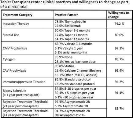
Article Information
vol. 132 no. Suppl 3 A12452
Published By:
American Heart Association, Inc.
Online ISSN:
History:
- Originally published November 6, 2015.
Copyright & Usage:
© 2015 by American Heart Association, Inc.
Author Information
- Chesney Castleberry1;
- Sonja Ziniel2;
- Christopher Almond3;
- Scott Auerbach4;
- Seth Hollander3;
- Lal Ashwin5;
- Matthew Fenton6;
- Joseph Rossano7;
- Melanie Everitt4;
- Kevin Daly8
- 1Pediatric Cardiology, Washington Univ in St. Louis, St. Louis, MO
- 2Pediatrics, Boston Children’s Hosp, Boston, MA
- 3Pediatric Cardiology, Stanford Univ, Palo Alto, CA
- 4Pediatric Cardiology, Children’s Hosp of Colorado, Aurora, CO
- 5Pediatric Cardiology, Primary Children’s Hosp, Salt Lake City, UT
- 6Pediatric Cardiology, Great Ormond Street Hosp for Children, London, United Kingdom
- 7Pediatric Cardiology, Children’s Hosp of Philadelphia, Philadelphia, PA
- 8Pediatric Cardiology, Boston Children’s Hosp, Boston, MA
Abstract 10895: The Association of Growth Patterns and Clinical Outcomes in Children With Dilated Cardiomyopathy: A Report From the Pediatric Cardiomyopathy Registry Study Group
Chesney Castleberry, John L Jefferies, Ling Shi, James D Wilkinson, Jeffrey A Towbin, Ryan W Harrison, Charles Canter, Joseph Rossano, Elfriede Pahl, Teresa Lee, Linda Addonizio, Paolo Rusconi, Justin Godown, Joseph Mahgerefteh, Steven D Colan, Paul F Kantor, Steven E Lipshultz, Tracie Miller
Circulation. 2015;132:A10895
Abstract
Introduction: Although malnourishment is associated with mortality in adult dilated cardiomyopathy (DCM), a paradox exists where obesity is protective. We aim to look at the role of these risk factors in pediatric DCM.
Hypothesis: We hypothesize malnourishment (MN) and obesity are risk factors for death or heart transplantation.
Methods: The NHLBI- funded Pediatric Cardiomyopathy Registry (PCMR) database was used for this analysis of patients (age < 18 years) with idiopathic or familial DCM and myocarditis. Patients were categorized by anthropological measurements: MN (defined as BMI <5% for ≥2years or weight for length <5% for <2 years), obese (BMI> 95% for ages ≥2years or weight for length >95% for <2 years), and normal.
Results: Of 904 DCM patients, 23.7% (214) were MN, 13.3% (120) were obese, and 63.1% (570) were normal (Table). Obese patients were significantly older (p<0.001) and more likely to have a family history of DCM (both p<0.05). MN were more likely to have congestive heart failure, increased cardiac dimensions, and higher ventricular mass (p<0.01). In a Kaplan Meier analysis, there was a difference in time to death or transplant by groups (Figure, p=0.0498). Adjusting for age and myocarditis, MN was associated with increased risk of death (HR: 1.857, 95%CI: 1.159-2.977, p=<0.001), but obesity was not (HR: 1.362, 95%CI: 0.722, 2.566). Competing outcome analysis showed an increased risk of death for MN (p=0.026) but no difference between groups in transplant rate (p=0.159).
Conclusion: Malnourishment is significantly associated with poor clinical outcomes. Unlike adult DCM where obesity is protective, obesity is not associated with improved survival in pediatric DCM.
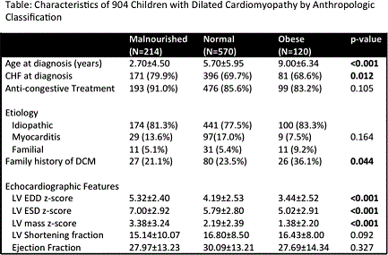
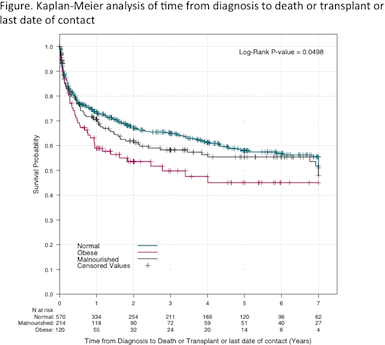
Article Information
vol. 132 no. Suppl 3 A10895
Published By:
American Heart Association, Inc.
Online ISSN:
History:
- Originally published November 6, 2015.
Copyright & Usage:
© 2015 by American Heart Association, Inc.
Author Information
- Chesney Castleberry1;
- John L Jefferies2;
- Ling Shi3;
- James D Wilkinson4;
- Jeffrey A Towbin5;
- Ryan W Harrison3;
- Charles Canter1;
- Joseph Rossano6;
- Elfriede Pahl7;
- Teresa Lee8;
- Linda Addonizio8;
- Paolo Rusconi9;
- Justin Godown10;
- Joseph Mahgerefteh11;
- Steven D Colan12;
- Paul F Kantor13;
- Steven E Lipshultz14;
- Tracie Miller15
- 1Pediatric Cardiology, Washington Univ in St. Louis, St. Louis, MO
- 2Pediatric Cardiology, Cincinnati Children’s Hosp Med Cntr, Cincinnati, OH
- 3Biostatistics, New England Rsch Institute, Watertown, MA
- 4Pediatrics, Wayne State Univ Sch of Medicine, Detroit, MI
- 5Pediatric Cardiology, Le Bonheur Children’s Hosp, Memphis, TN
- 6Pediatric Cardiology, Children’s Hosp of Philadelphia, Philadelphia, PA
- 7Pediatric Cardiology, Ann & Robert H. Lurie Children’s Hosp of Chicago, Chicago, IL
- 8Pediatric Cardiology, Columbia Univ Med Cntr, New York, NY
- 9Pediatric Cardiology, Univ of Miami Miller Sch of Medicine, Miami, FL
- 10Pediatric Cardiology, Vanderbilt Univ Sch of Medicine, Nashville, TN
- 11Pediatric Cardiology, Children’s Hosp at Montefiore, Bronx, NY
- 12Pediatric Cardiology, Boston Children’s Hosp, Bostan, MA
- 13Pediatric Cardiology, Stollery Children’s Hosp, Univ of Alberta, Edmonton, Canada
- 14Pediatric Cardiology, Wayne State Univ, Detroit, MI
- 15Pediatric Gastroenterology, Univ of Miami Health System, Miller Sch of Medicine, Miami, FL
Abstract 10025: Human Umbilical Cord Stem Cells Increase Heart Contractility and Decrease Fibrosis in Congenital Cardiomyopathy
Robert Henning, Ernesto Jimenez
Circulation. 2015;132:A10025
Abstract
Introduction: We have established that human umbilical cord blood stem cells (hUCSC) decrease inflammation and infarct size in acute myocardial infarctions in rats. We hypothesized that hUCSC can limit the progressive LV fibrosis and LV failure that occurs in TO2 hamsters, a model of congenital cardiomyopathy due to sarcoglycan deficiency.
Methods: 22 TO2 1 month old hamsters were treated with intramyocardial (IM) hUCBC, 4х 106 ,in Isolyte®, and 23 TO2 1 month old hamsters were treated with IM Isolyte. 16 1 month old F1B hamsters served as controls and received IM Isolyte. No hamster was given immunosuppressive treatment. Echocardiograms were performed on all hamsters prior to and monthly after treatment for 6 months. Heart tissues were stained with hematoxylin and eosin, Masson’s Trichrome and human nuclear antibody.
Results: In the F1B hamsters, LV fractional shortening (FS) and ejection fractions (EF) did not significantly decrease over 6 months. In contrast, in Isolyte treated TO2 hamsters, FS decreased from 56.2±1.0% to 19.7±3.2% and EF decreased from 89.5±1.4% to 41.9 ±5.9% at 6 months (both p < 0.001). The FS and EF in hUCSC treated TO2 hamsters also progressively decreased over 6 months but changes were more gradual, especially during first month after hUCSC when FS was 52.0±1.5% and EF was 89.5±1.4%, which was not significantly different from the F1B hamsters. In the hUCSC treated hamsters, the FS and EF were 20-30% greater than FS and EF in Isolyte TO2 hamsters at 3 and 5 months (p < 0.01). Injection of hUCBC in a subset of Isolyte treated TO2s at 4 months increased LV EF at age 5 months to 57.5±2.3% compared with a EF of 41.6±3.1% (p< 0.01) in Isolyte treated TO2s. In Isolyte treated TO2s at 6-7 months, fibrosis involved 30.0±5.0% of LV and 35.0±5.0% of LV septum. In contrast, in hUCSC treated hamsters, fibrosis involved only 16.5± 2.3% of LV and 16.3±1.8% of septum (p< 0.05). The average number of blood vessels per myocardial microscopic field in hUCSC treated hearts was 53.5 ± 0.8 versus 46.2 ± 3.0 in Isolyte treated TO2 hearts (p < 0.05).
Conclusion: hUCSC, given as a single intramyocardial injection, can limit fibrosis and increase heart contractility over the short term in TO2 hamsters with congenital cardiomyopathy.
Article Information
vol. 132 no. Suppl 3 A10025
Published By:
American Heart Association, Inc.
Online ISSN:
History:
- Originally published November 6, 2015.
Copyright & Usage:
© 2015 by American Heart Association, Inc.
Author Information
- Robert Henning;
- Ernesto Jimenez
- MEDICINE, James A. Haley Hosp/Univ of South Florida, Tampa, FL
Abstract 18230: Wringing Pump or Roller Pump? Insights From Rotational Mechanics in Children With Left Ventricular Noncompaction Cardiomyopathy
Hythem Nawaytou, Putri Yubbu, Kelley Miller, Anirban Banerjee
Circulation. 2015;132:A18230
Abstract
Introduction: Left ventricular noncompaction (LVNC) is a rare form of cardiomyopathy in children, characterized by abnormal endocardial myofiber arrangement. This may result in an altered pattern of LV rotation depicted by a “roller pump” like motion and described as rigid body rotation (RBR). This pattern, is characterized by the apex and the base rotating in the same direction.
Hypothesis: To describe the LV rotational patterns in children with LVNC using speckle tracking echocardiography and detect the relationship between the different rotational patterns and measures of LV systolic function.
Methods: We prospectively studied 19 children (age 0.9 to 17.8 years) with LVNC and compared them with age-matched controls. Apical and basal rotations, peak twist and peak torsion were analyzed as indices of systolic function. Peak apical recoil rate was measured as an index of diastolic function.
Results: Children with LVNC had significantly lower apical rotation (6.26°± 3.09°, 8.59°± 2.19° p=0.01) than controls. In 7 out of 19 (36.8%) patients a pattern of RBR was detected. Twelve patients exhibited the typical “wringing” motion of the LV, where the apex rotated counterclockwise and the base clockwise. Patients with LVNC and RBR had significantly lower ejection fraction than patients with LVNC without RBR (49.1 ± 9.2% vs 60.3 ± 10.2%, p<0.05). There were no significant differences in the remainder of the systolic and diastolic indices between LVNC patients and controls.
Conclusions: Children with LVNC can exhibit an abnormal LV rotational pattern characterized by RBR that is associated with impaired systolic function.
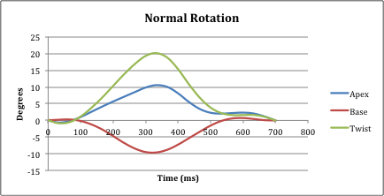
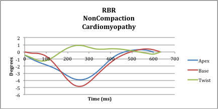
Article Information
vol. 132 no. Suppl 3 A18230
Published By:
American Heart Association, Inc.
Online ISSN:
History:
- Originally published November 6, 2015.
Copyright & Usage:
© 2015 by American Heart Association, Inc.
Author Information
- Hythem Nawaytou;
- Putri Yubbu;
- Kelley Miller;
- Anirban Banerjee
- Pediatrics, Children’s Hosp of Philadelphia, Philadelphia, PA
Abstract 18262: Left Ventricle Strain Rate in Children Demonstrates Heart Rate Dependence and a Force-frequency Relationship
Silvia Alvarez, Mohammed Alhabden, Michal Kantoch, Joseph Atallah, Timothy Colen, Edythe Tham, Nee Khoo
Circulation. 2015;132:A18262
Abstract
Introduction: In adult pigs and human studies, strain rate (SR) is a valid and reproducible marker of contractility. It is also heart rate (HR) independent, hence lacks force-frequency relationship (FFR). Isovolumic acceleration (IVA) is a proven non-invasive load-independent measure of LV contractility used in research settings. This study sought to assess SR behavior during tachycardia and inotropic stimulation in children, compared to IVA.
Methods: Twenty-four patients (median age, 13.9; range 7.8 – 22.5 years) with no structural or functional heart abnormalities were evaluated after a radiofrequency ablation procedure. Echocardiogram was performed at baseline, during atrial pacing and isoprenaline infusion to achieve 130% of baseline HR. Speckle tracking global LV longitudinal SR and tissue Doppler septal IVA were measured. Relationships between HR, SR and IVA were assessed. Percent (%) change and absolute differences between SR and IVA at baseline, pacing and isoprenaline were evaluated. Data are reported as median and interquartile ranges.
Results: SR and IVA showed a moderate correlation with HR at baseline (SR: r=-0.68, p=0.0002; IVA: r=0.46, p=0.01), and during pacing (SR: r=-0.56, p=0.003; IVA: r=0.58, p=0.002). Both SR and IVA increased with pacing and isoprenaline (table 1), however the greatest % change was seen during isoprenaline infusion for IVA (p < 0.006) and SR (p < 0.0001).
Conclusion: SR enhances with increasing HR in children, demonstrating a relative HR dependence and a FFR. This is contrary to findings in adult studies, thus this study highlights that children show a different LV mechanical response to chronotropic effects and therefore caution should be use when extrapolating of adult findings to children.

Article Information
vol. 132 no. Suppl 3 A18262
Published By:
American Heart Association, Inc.
Online ISSN:
History:
- Originally published November 6, 2015.
Copyright & Usage:
© 2015 by American Heart Association, Inc.
Author Information
- Silvia Alvarez1;
- Mohammed Alhabden1;
- Michal Kantoch2;
- Joseph Atallah2;
- Timothy Colen1;
- Edythe Tham1;
- Nee Khoo1
- 1Divs of Pediatric Cardiology and Diagnostic Imaging, Stollery Children’s Hosp, Univ of Alberta, Edmonton, Canada
- 2Div of Pediatric Cardiology, Stollery Children’s Hosp, Univ of Alberta, Edmonton, Canada
Abstract 17892: Strain Rate is Affected by Heart Rate in Younger Heart: A Simultaneous Invasive and Noninvasive in vivo Piglet Model
Etienne Fortin-Pellerin, Lisa K Hornberger, James Y Coe, Lindsay Mills, Jesus Serrano-Lomelin, Po-Yin Cheung, Nee S Khoo
Circulation. 2015;132:A17892
Abstract
Introduction: In adult human and pig hearts, left ventricular (LV) systolic strain rate (SR) has been shown to be independent of heart rate (HR) in atrial tachycardia. It has been hypothesized that any increase in contractility related to the force-frequency relationship is balanced by a decrease in contractility due to reduced filling time and preload at higher HRs. In this study, we explore the impact of atrial tachycardia on SR of the young infant heart using a simultaneous invasive and noninvasive piglet model to determine whether SR of the immature heart is similarly not influenced by increasing HR.
Methods: Under general anesthesia (propofol, isoflurane), 1 – 15 day old piglets were instrumented intravascularly with Millar high-fidelity and pacing catheters in the left ventricle and right atrium, respectively. After stabilization, invasive hemodynamic and echocardiography parameters were acquired at baseline, and at 200, 230 and 260bpm. Basal circumferential SR was analyzed off-line by speckle tracking (frame rates 247±7 Hz). Each animal was its own control and repeated measure ANOVA was used for comparison, data is expressed as mean ± SE.
Results: Fourteen piglets of mean age 8.5±1.8 days, weight 3.6±0.5kg and baseline heart rate of 152±5bpm were assessed. Baseline LV systolic SR was -1.53±0.13 1/s and dP/dt 1656±115mmHg/s. With pacing, LV SR increased significantly (p = 0.002). The increase in SR mirrored the increase in contractility assessed invasively by dP/dt (p<0.001). M-mode LV end diastolic dimension decreased from baseline to 260bpm (73±9.9% of baseline value, p < 0.001) consistent with reduced preload with tachycardia.
Conclusion: Our study suggests that in the younger heart, SR is augmented by atrial tachycardia itself even in the presence of decreased preload. This is in keeping with preservation of the force frequency relationship. Given our findings, HR should be taken into account when assessing contractility using SR in young patients.
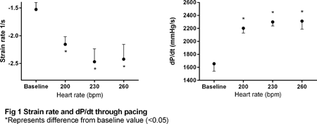
Article Information
vol. 132 no. Suppl 3 A17892
Published By:
American Heart Association, Inc.
Online ISSN:
History:
- Originally published November 6, 2015.
Copyright & Usage:
© 2015 by American Heart Association, Inc.
Author Information
- Etienne Fortin-Pellerin1;
- Lisa K Hornberger2;
- James Y Coe2;
- Lindsay Mills2;
- Jesus Serrano-Lomelin3;
- Po-Yin Cheung4;
- Nee S Khoo2
- 1Pediatrics, Neonatology, CHU Sherbrooke, Sherbrooke, Canada
- 2Pediatric Cardiology, Stollery Children’s Hosp, Edmonton, Canada
- 3Sch of Public Health, Univ of Alberta, Edmonton, Canada
- 4Pediatrics, Neonatology, Stollery Children’s Hosp, Edmonton, Canada
Abstract 16866: Depressed Biventricular Outputs in Fetuses of Diabetic Mothers When Standardized to Estimated Fetal Weight
Jennifer Hickman, Mary Craft, Ling Li, Aparna Kulkarni, Lisa Hornberger, David Danford, Shelby Kutty
Circulation. 2015;132:A16866
Abstract
Background: Although the results of maternal diabetes on the newborn heart are well described, in-utero cardiovascular effects of maternal diabetes and obesity are not as well understood.
Hypothesis: Echocardiographically derived measures of right and left heart flow volumes in fetuses of diabetic mothers (FDM) and fetuses of obese mothers (FOM) differ from those of normal fetuses.
Methods: We prospectively enrolled 264 normal fetuses, 30 FDM and 49 FOM for prenatal research echocardiography. Estimated fetal weight (EFW) based norms for aortic and pulmonary valve velocity time integrals (AVVTI, PVVTI), right and left ventricular outflow tract diameters (LVOTD, RVOTD), aortic and pulmonary valve stroke volume (AVSV, PVSV), and cardiac output (LVCO, RVCO) were derived from the 264 control group fetuses using nonlinear regression to a Gompertz growth function. Z values for each parameter were determined, then analyzed using a non-paired two-tailed t-test to compare the three groups.
Results: The mean ± SD EFW (g) for normal fetuses, FDM and FOM were 1610 ± 1201, 964 ± 694 and 1075 ± 1013; respective mean gestational ages (weeks) were 28.1±1.3, 25.3± 5, and 25 ± 6.6. The table shows mean Z value comparisons of VTI, OTD, SV and CO for the RV and LV in the three groups.
Conclusions: FDM have significantly lower right and left heart stroke volumes and outputs than other fetuses matched for size, while FOM did not have demonstrable right or left heart stroke volume or cardiac output changes relative to normal fetuses. Similar to their relationship with gestational age, normal right and left ventricular stroke volumes and outputs follow a non-linear correlation with estimated fetal weight. Flow volumes in macrosomic fetuses may be better normalized to EFW rather than to gestational age. Reduced placental blood flow and ventricular compliance changes may be contributing to depressed biventricular cardiac output in FDM.
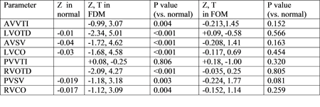
Article Information
vol. 132 no. Suppl 3 A16866
Published By:
American Heart Association, Inc.
Online ISSN:
History:
- Originally published November 6, 2015.
Copyright & Usage:
© 2015 by American Heart Association, Inc.
Author Information
- Jennifer Hickman1;
- Mary Craft1;
- Ling Li1;
- Aparna Kulkarni2;
- Lisa Hornberger3;
- David Danford1;
- Shelby Kutty1
- 1DIVISION OF PEDIATRIC CARDIOLOGY, Univ of Nebraska College of Medicine, Omaha, NE
- 2Div of Cardiology, Albert Einstein College of Medicine, New York, NY
- 3Div of Cardiology, Stollery Childrens Hosp, Edmonton, Canada
Abstract 13554: Left Atrial Function in Children by Volume and Strain Analysis: Impact of Congenital Aortic Stenosis
Divya Shakti, Kevin G Freidman, David M Harrild, Kimberly Gauvreau, Tal Geva, Steven D Colan, David W Brown
Circulation. 2015;132:A13554
Abstract
Background: Left atrial (LA) function by volume and strain analysis in children has been little studied. LA dysfunction related to aortic stenosis has been reported in adults.
Hypotheses: 1. LA volume and strain analysis is feasible and reproducible in normal children.
- Children with isolated congenital aortic stenosis (AS) as compared to matched controls have higher maximal LA volume (LAV) and lower total longitudinal LA strain.
Methods: Retrospective chart review and offline analysis of 2D echocardiogram images for phasic LAVs, volume indices, and longitudinal LA strain (Fig 1) by speckle tracking (TomTec software) in: 1. Healthy children with a normal echocardiogram; and, 2. Children ≥1 years of age with significant (≥ moderate) isolated AS. AS cases were compared with age, gender and BSA-matched normal controls.
Results: There were 67 normal subjects [age: 10.5 (1.3-22.1) yrs; 50 males, 17 females; BSA 1.22 (0.44-2.39) m2] studied. No significant inter-observer and intra-observer differences were noted in normal subjects (Fig 2). Between June 2004 and October 2012, there were 41 eligible AS cases [Age: 11.7 (1.6-26.8) yrs; 32 males, 9 females] with median maximal Doppler aortic stenosis gradient of 63 (43-94) mm Hg. Significantly higher phasic indexed LAVs and indexed active LA stroke volume (pump function) and lower strain values for LA reservoir and conduit function were noted in AS cases (Fig 2).
Conclusions: LA volume and strain analysis is reproducible and feasible in children. Children with significant AS have abnormal LA function with higher phasic LA volumes, higher LA pump function, and lower strain-derived reservoir and conduit LA function.
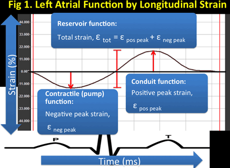
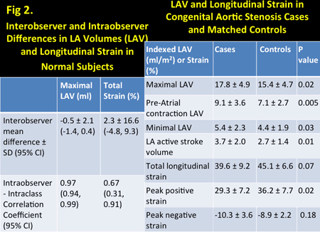
Article Information
vol. 132 no. Suppl 3 A13554
Published By:
American Heart Association, Inc.
Online ISSN:
History:
- Originally published November 6, 2015.
Copyright & Usage:
© 2015 by American Heart Association, Inc.
Author Information
- Divya Shakti1;
- Kevin G Freidman2;
- David M Harrild3;
- Kimberly Gauvreau2;
- Tal Geva4;
- Steven D Colan4;
- David W Brown5
- 1Pediatric Cardiology, Batson Children’s Hosp, Univ of Mississippi Med center, Jackson, MS
- 2Cardiology, Boston Childrens Hosp, Boston, MA
- 3Cardiology, Boston Childrens Hosp, Jackson, MS
- 4Cardiology, Boston Children’s Hosp, Boston, MA
- 5Pediatric Cardiology, Boston Children’s Hosp, Boston, MA
Abstract 12511: Exercise Stress Test and Myocardial Perfusion Imaging Have Limited Diagnostic Reliability in Detecting Coronary Abnormalities in Children After Arterial Switch Operation for Transposition of the Great Arteries
Takeshi Tsuda, Abdul M Bhat, Jeanne M Baffa, Wolfgang Radtke, Bradley Robinson
Circulation. 2015;132:A12511
Abstract
Background: Exercise stress test (EST) and myocardial perfusion imaging (MPI) are common non-invasive modalities to screen for occult myocardial ischemia, but their sensitivity and specificity in children are unknown.
Hypothesis: EST and MPI have limited sensitivity and specificity in detecting occult myocardial ischemia in children.
Patients and Methods: We retrospectively reviewed EST, MPI, and coronary angiogram in asymptomatic patients after arterial switch operation (ASO) for d-transposition of the great arteries (TGA) and examined the sensitivity and specificity of EST/MPI in detecting coronary abnormalities. Positive ECG findings for exercise-induced ischemia included ST-T depression > 2mm and exercise-induced ventricular ectopies. Positive MPI is defined as a perfusion defect. Data are shown as mean ± SD.
Results: Thirty asymptomatic patients (20 male/10 female; 23 with intact ventricular septum and 7 with ventricular septal defect; ages 12.5 ± 4.4 years) had EST and 26 had simultaneous MPI. Respiratory quotient was 1.11 ± 0.07 and maximum oxygen consumption was 80.7 ± 15.0% predicted, suggesting mildly diminished aerobic fitness level. There was no exercise-induced arrhythmia. Twenty two patients had coronary angiogram (Table 1). Four patients with positive EST and/or MPI were confirmed to have coronary abnormalities by coronary angiogram, whereas 4 patients revealed variable degree of coronary obstruction despite negative EST/MPI including 1 patient who later developed life-threatening ventricular fibrillation. Three patients with positive EST or MPI showed no coronary abnormalities.
Conclusions: A combination of EST and MPI has low sensitivity (50%; 4/8) and moderate specificity (79%; 11/14) in detecting coronary abnormalities after ASO. Thus, we recommend performing coronary angiography or equivalent coronary investigation in addition to EST/MPI in children after ASO to detect coronary abnormalities and myocardial ischemia.
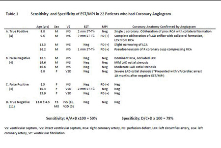
Article Information
vol. 132 no. Suppl 3 A12511
Published By:
American Heart Association, Inc.
Online ISSN:
History:
- Originally published November 6, 2015.
Copyright & Usage:
© 2015 by American Heart Association, Inc.
Author Information
- Takeshi Tsuda;
- Abdul M Bhat;
- Jeanne M Baffa;
- Wolfgang Radtke;
- Bradley Robinson
- NEMOURS CARDIAC CENTER, Alfred I. duPont Hosp for Children, Wilmington, DE
Abstract 11518: Diastolic Left Ventricular Dysfunction is a Common and Early Cardiac Abnormality in Hutchinson-Gilford Progeria Syndrome
Ashwin Prakash, Leslie B Gordon, Monica E Kleinman, Ralph D’Agostino, Joe Massaro, Mark W Kieran, Marie Gerhard-Herman, Leslie B Smoot
Circulation. 2015;132:A11518
Abstract
Introduction: Hutchinson-Gilford progeria syndrome (HGPS) is associated with premature death due to cardiovascular events. However, little is known about the natural history of cardiac disease in this population. We prospectively evaluated children with HGPS to identify echocardiographic abnormalities in ventricular and valve function and to assess the effect of treatment on these abnormalities.
Methods and Results: Transthoracic echocardiography was used to evaluate 51 children with HGPS (45 with classic type, 6 with non-classic type; 55% males) at a median age of 10.7 (2.4-19.8) years. Among the study population, 19 were evaluated prior to entry into an open label drug trial of the farnesyltransferase inhibitor lonafarnib while the other 32 were receiving combination therapy with lonafarnib, pravastatin and zoledronic acid (triple therapy) at the time of evaluation. As seen in the Table, diastolic dysfunction diagnosed using pulsed wave tissue Doppler was the most common cardiac abnormality at all ages, developing early and present universally beyond 12.1 years of age. Other cardiac abnormalities including left ventricular hypertrophy (LVH), LV systolic dysfunction, and mitral or aortic valve disease were less common and appeared later. Using multiple linear regression to control for age and gender, patients receiving triple therapy demonstrated less severe LV diastolic dysfunction evidenced by higher lateral E’ velocities (p=0.01) and lower mitral inflow E velocity/lateral E’ velocity ratio (p=0.006). Other cardiac abnormalities were not associated with triple therapy.
Conclusions: In patients with Hutchinson-Gilford progeria syndrome, LV diastolic dysfunction is common and appears early. Other cardiac abnormalities are less common and appear at a later age. Triple therapy is associated with less severe diastolic dysfunction. Echocardiographic parameters of LV diastolic dysfunction may be useful endpoints in future clinical trials.
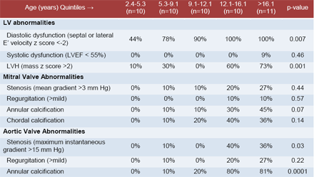
Article Information
vol. 132 no. Suppl 3 A11518
Published By:
American Heart Association, Inc.
Online ISSN:
History:
- Originally published November 6, 2015.
Copyright & Usage:
© 2015 by American Heart Association, Inc.
Author Information
- Ashwin Prakash1;
- Leslie B Gordon2;
- Monica E Kleinman3;
- Ralph D’Agostino4;
- Joe Massaro4;
- Mark W Kieran5;
- Marie Gerhard-Herman6;
- Leslie B Smoot1
- 1Dept of Cardiology, Boston Children’s Hosp, Boston, MA
- 2Dept of Anesthesia, Boston Children’s Hosp, Boston, MA
- 3Div of Critical Care Medicine, Boston Children’s Hosp, Boston, MA
- 4Dept of Mathematics and Statistics, Boston Univ, Boston, MA
- 5Div of Pediatric Oncology, Dana Farber Cancer Institute, Boston, MA
- 6Dept of Medicine, Cardiovascular Div, Brigham and Women’s Hosp, Boston, MA
Abstract 10876: Speckle-tracking vs. Pressure-Volume Loop Measures of Left Ventricular Systolic Function in Children: An Inter-vendor Comparison
Shahryar M Chowdhury, Ryan J Butts, Carolyn L Taylor, Karen S Chessa, Anthony M Hlavacek, Girish S Shirali, Arni Nutting, George H Baker
Circulation. 2015;132:A10876
Abstract
Introduction: The variability in speckle-tracking echocardiography (STE) algorithms between vendors is well recognized. While some are attempting to standardize algorithms across vendors, it is currently unknown which algorithm best compares to gold-standard measures of systolic function. The objective of this study was to compare STE measures of deformation from two vendors vs. gold-standard measures of LV contractility and global ventricular efficiency in children.
Methods: Children with normal loading conditions undergoing routine left heart catheterization were prospectively enrolled. Pressure-volume loops were obtained via conductance catheterization. The gold-standard measure of contractility, end-systolic elastance (Ees), was obtained via balloon occlusion of the vena cavae. Ventriculo-arterial coupling was assessed using the arterial elastance to Ees ratio (Ea/ Ees). STE was performed immediately after PVL analysis under the same anesthetic conditions. Offline STE analysis was perfomed using both QLAB v9.0 (Phillips, Andover, MA) and Cardiac Performance Analysis v3.0 (Tomtec, Hamden, CT) by a single blinded observer. The relationships between PVL and STE measures of systolic function were determined using Spearman’s correlation.
Results: Of 24 patients, 18 patients were s/p heart transplant, 6 patients had a small patent ductus arteriosus or small coronary fistula. Mean age was 9.1 ± 5.6 years. The average invasive Ees was 3.04 ± 1.65 mmHg/mL. The average Ea/Ees was 0.88 ± 0.35. Correlations between invasive Ees and Ea/Ees to STE measures of deformation are reported in the Table.
Conclusion: STE measures of GLS and GLSR derived from Cardiac Performance Analysis have better correlation with gold-standard measures of cardiac performance when compared to QLAB. GCS derived from both vendors has no correlation to invasive measures. Attempts to standardize STE algorithms should take these results into consideration.

Article Information
vol. 132 no. Suppl 3 A10876
Published By:
American Heart Association, Inc.
Online ISSN:
History:
- Originally published November 6, 2015.
Copyright & Usage:
© 2015 by American Heart Association, Inc.
Author Information
- Shahryar M Chowdhury1;
- Ryan J Butts1;
- Carolyn L Taylor1;
- Karen S Chessa1;
- Anthony M Hlavacek1;
- Girish S Shirali2;
- Arni Nutting1;
- George H Baker1
- 1Pediatrics, Med Univ of South Carolina, Charleston, SC
- 2Pediatrics, Children’s Mercy Hosp, Kansas City, MO
Abstract 10878: Validation of Non-invasive Measures of Diastolic Function in Children: A Simultaneous Echocardiography and Conductance Catheterization Study
Shahryar M Chowdhury, Ryan J Butts, Anthony M Hlavacek, Carolyn L Taylor, Varsha M Bandisode, Karen S Chessa, Girish S Shirali, Arni Nutting, George H Baker
Circulation. 2015;132:A10878
Abstract
Introduction: The accuracy of echocardiography in evaluating left ventricular (LV) diastolic function has not been validated in children. The objective of this study was to compare echocardiographic and gold-standard measures of LV diastolic function in children.
Methods: Patients undergoing routine left heart catheterization were prospectively enrolled. Pressure-volume loops (PVL) were obtained via conductance catheters. The end-diastolic pressure-volume relationship was obtained via balloon occlusion of the vena cavae. PVL measures of diastolic function were divided into early active relaxation (the isovolumic relaxation time constant, tau), and ventricular stiffness (the chamber stiffness constant, β). End-diastolic pressure (EDP) was also recorded. Echocardiographic measures of diastolic function were derived from spectral Doppler, tissue Doppler, and 2D speckle-tracking. The relationships between PVL and echocardiographic measures were determined using Spearman’s correlation.
Results: Of 24 patients, 18 patients were s/p heart transplant, 5 patients had a small patent ductus arteriosus or coronary fistula. Mean age was 9.1 ± 5.6 years. The median τ was 24.9 ms (IQR 22.8 – 28.4 ms), median β was 0.094 (IQR 0.035 – 0.154), and median EDP was 9 mmHg (IQR 8 – 13 mmHg). Statistically significant correlations between invasive and echocardiographic measures of diastolic function are reported in the Table. No echocardiographic measures correlated with β.
Conclusion: Early diastolic echocardiographic measures correlate with tau and may accurately represent early active relaxation in children. Modest associations exist between echocardiographic measures and EDP. The use of these non-invasive measures in accurately assessing LV diastolic function appears promising in children. However, no echocardiographic measures correlate with chamber stiffness. The development of such measures merits further study.
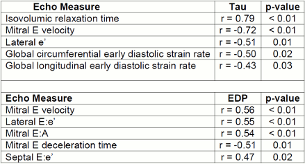
Article Information
vol. 132 no. Suppl 3 A10878
Published By:
American Heart Association, Inc.
Online ISSN:
History:
- Originally published November 6, 2015.
Copyright & Usage:
© 2015 by American Heart Association, Inc.
Author Information
- Shahryar M Chowdhury1;
- Ryan J Butts1;
- Anthony M Hlavacek1;
- Carolyn L Taylor1;
- Varsha M Bandisode1;
- Karen S Chessa1;
- Girish S Shirali2;
- Arni Nutting1;
- George H Baker1
- 1Pediatrics, Med Univ of South Carolina, Charleston, SC
- 2Pediatrics, Children’s Mercy Hosp, Kansas City, MO
Abstract 18025: Systematic Screening is Necessary to Detect Post-operative Thrombosis and Evaluate Risk in Children Undergoing Cardiac Surgery: A Prospective Observational Study
Cedric Manlhiot, Leonardo R Brandao, Helen M Holtby, V. Ben Sivarajan, Jennifer L Russell, Luc Mertens, Margaret L Rand, Collen E Gruenwald, Steven M Schwartz, Seema Mital, Lia Stenyk, Martha Rolland, Sunita O’Shea, Glen S Van Arsdell, Christopher A Caldarone, Anthony K Chan, Brian W McCrindle
Circulation. 2015;132:A18025
Abstract
Introduction: Postoperative thrombosis in children undergoing cardiac surgery with cardiopulmonary bypass (CPB) is a frequent, under-diagnosed and clinically detrimental complication.
Methods: Pediatric patients were prospectively enrolled before surgery with CPB in an observational cohort study. Children <1 year old and cyanotic patients were oversampled. All subjects had serial lab assessment of hemostatic status and standardized vascular ultrasound for surveillance of post-operative thrombosis. Eligible non-enrolled patients were retrospectively reviewed as concurrent controls without study surveillance. All imaging findings underwent blinded adjudication to confirm the presence and clinical importance of identified thrombi.
Results: Of 400 enrolled patients (54% males, 55% <1 year, 25% cyanotic), 398 (99%) completed the study, and 339 (85%) had protocol vascular ultrasound prior to or within 1 week of hospital discharge. Post-operative thrombosis was diagnosed in 99 study patients (25%) vs. 10% of the 1,019 control patients (p<0.001). Clinical evaluation alone missed 59% of all postoperative thrombi and 20% of clinically important thrombi. Multivariable factors associated with higher odds of postoperative thrombosis in study patients included 4 factors reflective of reduced heparin sensitivity and/or anticoagulation levels during CPB (Table). Postoperative thrombosis was associated with longer ICU stay (5 vs. 2 days, p=0.008), longer hospital stay (6 vs. 10 days, p<0.001), increased odds of reintervention or post-operation ECMO (OR: 2.7, p=0.005) and mortality (OR: 5.9, p=0.002).
Conclusions: Given the poor performance of relying on clinical suspicion for detecting post-operative thrombosis, children <1 year old undergoing surgery with CPB should be systematically screened for thrombosis post-operatively. Individualization of thromboprophylaxis should consider both preoperative clinical factors and heparin sensitivity.
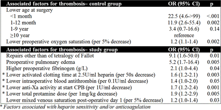
Article Information
vol. 132 no. Suppl 3 A18025
Published By:
American Heart Association, Inc.
Online ISSN:
History:
- Originally published November 6, 2015.
Copyright & Usage:
© 2015 by American Heart Association, Inc.
Author Information
- Cedric Manlhiot1;
- Leonardo R Brandao1;
- Helen M Holtby1;
- V. Ben Sivarajan1;
- Jennifer L Russell1;
- Luc Mertens1;
- Margaret L Rand1;
- Collen E Gruenwald1;
- Steven M Schwartz1;
- Seema Mital1;
- Lia Stenyk1;
- Martha Rolland1;
- Sunita O’Shea1;
- Glen S Van Arsdell1;
- Christopher A Caldarone1;
- Anthony K Chan2;
- Brian W McCrindle1
- 1Labatt Family Heart Cntr, The Hosp for Sick Children, Toronto, Canada
- 2Pediatric Hematology-Oncology, McMaster Univ, Hamilton, Canada
Abstract 18044: Predictors of Bicuspid Aortic Valve Associated Aortopathy in Pediatric Patients
Michael Grattan, Andrea Prince, Anders Franco-Cereceda, Michele Petrovic, Salah A Mohamed, Bart Loeys, Harry Dietz, Seema Mital, Cedric Manlhiot, Gregor Andelfinger, Luc Mertens
Circulation. 2015;132:A18044
Abstract
Bicuspid aortic valve (BAV) is the most common congenital heart disease and is commonly associated with significant ascending aorta dilation. Valve morphology and function have been proposed as potential risk factors; however evaluating their role is difficult, as BAV morphology and valve function are inherently related. Our objective was to determine whether BAV morphology and function are independently associated with ascending aorta dilation in pediatric patients.
We performed a multicenter, retrospective, cross-sectional study of pediatric patients with BAV that were followed after 2003. The last patient echocardiogram prior to valve intervention was analyzed for BAV morphology, presence and severity of aortic stenosis (AS) and insufficiency (AI), and aortic root and ascending aorta dimensions. The relationship of potential risk factors to ascending aorta dimensions at different ages was determined using linear regression.
Data was obtained from 1725 patients (68% male). The most common BAV morphology was right-left (R-L) coronary cusp fusion (65%) followed by right-non (R-N) fusion (34%) and left-non fusion (1%). Fourteen percent of patients had at least moderate AS and 7% had at least moderate AI. R-L fusion was associated with coarctation (55%), while R-N fusion was less associated with coarctation (19%) and more associated with valve dysfunction (24% and 12% with at least moderate AS and AI). Twenty-seven percent of patients had significant ascending aorta dilation (Z score >3). AS and AI were associated with more significant ascending aorta dilation and a faster Z-score progression over time. R-N fusion was associated with ascending aorta dilation; however, when valves with AS and AI were excluded, there were no differences related to morphology. In patients with no AS or AI, 15% had significant ascending aorta dilation.
In this large pediatric cohort of patients with BAV, we show that valve morphology is not independently associated with ascending aorta dilation. Valve dysfunction (both AS and AI) is associated with more significant dilation; however, even in valves with normal function, there is significant ascending aorta dilation. We suggest that there is an inherent arterial abnormality, possibly modified by aortic flow patterns.
Article Information
vol. 132 no. Suppl 3 A18044
Published By:
American Heart Association, Inc.
Online ISSN:
History:
- Originally published November 6, 2015.
Copyright & Usage:
© 2015 by American Heart Association, Inc.
Author Information
- Michael Grattan1;
- Andrea Prince2;
- Anders Franco-Cereceda3;
- Michele Petrovic4;
- Salah A Mohamed5;
- Bart Loeys6;
- Harry Dietz7;
- Seema Mital1;
- Cedric Manlhiot1;
- Gregor Andelfinger8;
- Luc Mertens1
- 1Paediatrics, Div of Cardiology, The Hosp for Sick Children, Toronto, Canada
- 2Pediatrics, Cntr hospitalier universitaire Sainte-Justine, Montreal, Canada
- 3Dept Molecular Medicine and Surgery, Karolinska Institutet, Stockholm, Sweden
- 4Cardiology, The Hosp for Sick Children, Toronto, Canada
- 5Dept of Cardio and Thoracic Vascular Surgery, Universitaetsklinikum Schleswig-Holstein, Campus Luebeck, Luebeck, Germany
- 6Cntr of Med Genetics, Univ of Antwerp/ Antwerp Univ Hosp, Antwerp, Belgium
- 7Medicine, Pediatrics, and Molecular Biology and Genetics, Johns Hopkins Univ Sch of Med./HHMI, Baltimore, MD
- 8Pediatrics, Sainte-Justine Rsch Cntr, Montreal, Canada
Abstract 17110: Risk Factors for Hepatic Fibrosis in Children and Adolescents After Fontan Operation
David J Goldberg, Lea F Surrey, Henry C Lin, Andrew C Glatz, Michael L O’Byrne, Jonathan J Rome, Katie Dodds, Elizabeth Rand, Pierre Russo, Jack Rychik
Circulation. 2015;132:A17110
Abstract
Introduction: Congestive hepatopathy is increasingly recognized as a complication of Fontan physiology. Prospective data regarding the incidence of hepatopathy and risk factors for its development are lacking.
Methods: Liver biopsies were obtained as part of a routine program of comprehensive end-organ evaluation offered to all patients >10 years after Fontan operation (FO) through the Single Ventricle Survivorship Program. Evaluation included cardiac catheterization and contemporaneous percutaneous liver biopsy. Quantitative determination of hepatic fibrosis was performed using Sirius red staining for collagen with automated calculation of percent positive staining per slide. Patient specific characteristics, echocardiographic findings, and hemodynamic measures were assessed and evaluated as potential risk factors for collagen deposition using Pearson correlation. Differences in collagen deposition (%CD) between subgroups were assessed using the student t-test.
Results: The cohort consisted of 67 patients (36 male) at mean age of 17.3 ±4.5 years and mean time from Fontan of 14.9 ±4.5 years. Right ventricular morphology was present in 37 subjects; 30 with hypoplastic left heart syndrome. Mean %CD by Sirius red staining was 23.8 ±9.5% (range 8.7-49.4%) as compared to 2.6 ±0.3% in controls. There was significant correlation between time from Fontan and degree of Sirius red staining (r=0.33, p=0.006). Serum liver enzymes and platelet count did not correlate with %CD. The median inferior vena cava (IVC) pressure was 13 mmHg (range 6-24 mmHg) and did not correlate with %CD. There was no difference in %CD between those with a measured IVC pressure ≥13 mmHg versus those with a pressure <13 mmHg. There was no difference in %CD based on ventricular morphology or severity of atrioventricular valve insufficiency.
Conclusion: In this cohort of predominantly asymptomatic children and adolescents electively evaluated after FO, all exhibited evidence for hepatic fibrosis as measured by collagen deposition in the liver. Time from FO was the only factor in this cohort that was significantly associated with collagen. These findings demonstrate that liver fibrosis is an inherent feature of Fontan physiology and that the degree of fibrosis increases over time.
Article Information
vol. 132 no. Suppl 3 A17110
Published By:
American Heart Association, Inc.
Online ISSN:
History:
- Originally published November 6, 2015.
Copyright & Usage:
© 2015 by American Heart Association, Inc.
Author Information
- David J Goldberg1;
- Lea F Surrey2;
- Henry C Lin3;
- Andrew C Glatz1;
- Michael L O’Byrne1;
- Jonathan J Rome1;
- Katie Dodds1;
- Elizabeth Rand3;
- Pierre Russo2;
- Jack Rychik1
- 1Cardiology, The Children’s Hosp of Philadelphia, Philadelphia, PA
- 2Pathology, The Children’s Hosp of Philadelphia, Philadelphia, PA
- 3Gastroenterology, The Children’s Hosp of Philadelphia, Philadelphia, PA
Abstract 17235: Trends in Endocarditis Hospitalizations at US Children’s Hospitals From 2003-2014: Impact of the 2007 American Heart Association Antibiotic Prophylaxis Guidelines
Katherine E Bates, Matthew Hall, Samir S Shah, Kevin D Hill, Sara K Pasquali
Circulation. 2015;132:A17235
Abstract
Introduction: Over the past decade, national organizations in several countries have released more restrictive guidelines for infective endocarditis (IE) prophylaxis, including the American Heart Association (AHA) 2007 guidelines. Multiple initial studies demonstrated no change in IE rates over time following release of these guidelines, however a more recent analysis over a longer time period in the UK suggested an increase in IE. This prior study primarily included adults. Current IE trends in the pediatric population are unknown.
Methods: Children (<18 years) hospitalized with IE at 29 US centers participating in the Pediatric Health Information Systems Database from 2003-2014 were eligible for inclusion. Our primary analysis focused on IE most directly related to the change in the AHA guidelines, and included community-acquired IE cases (antibiotics covering oral streptococcal species started within 7 days of admission) in those >5 years of age (most likely to be receiving dental care). Interrupted time series analysis was used to evaluate IE rates over time indexed to total hospital admissions.
Results: A total of 841 IE cases were identified. Median age was 13 years (interquartile range 9-15 years). In the pre-guideline period, the IE rate increased slightly over time (+0.013 IE cases/1000 hospitalizations per 6-month period, see Figure). In the post-guideline period there was a similar trend in IE rates (+0.012 IE cases/1000 hospitalizations per 6-month period) with no significant difference in slope in the pre vs. post period (p=0.9). Additional analyses in those with congenital heart disease, and in those hospitalized with any type of IE (not limited to oral streptococci) at any age, revealed similar results.
Conclusions: In contrast to a recent UK study, we found no evidence of a change in IE hospitalization rates associated with revised IE prophylaxis guidelines over an 11 year period across 29 US children’s hospitals.
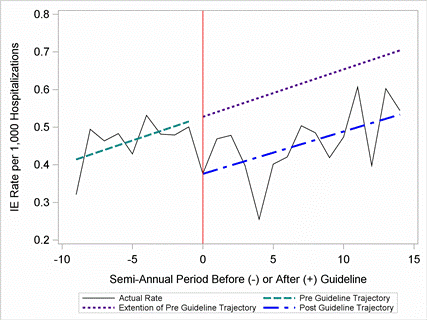
Article Information
vol. 132 no. Suppl 3 A17235
Published By:
American Heart Association, Inc.
Online ISSN:
History:
- Originally published November 6, 2015.
Copyright & Usage:
© 2015 by American Heart Association, Inc.
Author Information
- 1Congenital Heart Cntr, Univ of Michigan, Ann Arbor, MI
- 2Biostatistics, Children’s Hosp Association, Overland Park, KS
- 3Pediatrics, Cincinnati Children’s Hosp Med Cntr, Cincinnati, OH
- 4Pediatrics, Duke Univ Med Cntr, Durham, NC
Abstract 13582: Fetal Cardiac Intervention for Hypoplastic Left Heart Syndrome With Intact or Restrictive Atrial Septum: A Report From the International Fetal Cardiac Intervention Registry
David Jantzen, Anita J Moon-Grady, Aimee Armstrong, Christoph Berg, Joanna Dangel, Carlen Fifer, Michele Frommelt, Ulrich Gembruch, Ulrike Herberg, Edgar Jaeggi, Eftichia Kontopoulos, Owen Miller, Shaine A Morris, Renate Oberhoffer, Carlos Pedra, Simone Pedra, Ruben Quintero, Sarah Gelehrter
Circulation. 2015;132:A13582
Abstract
Background: Fetal cardiac intervention (FCI) for fetuses with hypoplastic left heart syndrome with restrictive or intact atrial septum (HLHS/IAS) has been reported in single-institution series as a modality that may improve outcomes in a high-risk subset of HLHS patients who historically have very high neonatal mortality.
Methods: The International Fetal Cardiac Intervention Registry was established in 2010. For this descriptive analysis, the database was queried for fetuses with HLHS/IAS. Maternal-fetal dyads from 2001 through March 2015 were included.
Results: Data from 15 institutions were submitted by data harvest. Of 87 cases of HLHS/IAS entered, 46 cases from 8 sites underwent FCI: 27 with atrial septal perforation/ balloon dilation and 19 with atrial septal stent placement. There were 34 procedural successes with a similar success rate between stents and balloons dilations (12/19=63% stent, 22/27=81% balloon dilation, p=0.19). Fetal complications were common (64% of cases), most frequently a pericardial effusion requiring treatment (51%), but no maternal complications. Procedure-related fetal demise occurred in 6 (13%). Seventeen had a clinical success (atrial septum not requiring urgent intervention at delivery), which was more likely in fetuses with a successful stent (75% stent vs 36% balloon dilation, p=0.03). Delivery by scheduled Cesarean section to facilitate immediate post-natal intervention was less common in those who underwent successful FCI compared with those with unsuccessful FCI or no FCI (3/34=8% FCI vs 18/46=39%, p=0.002). There was a trend toward improved survival to discharge for liveborn infants with HLHS/IAS in those who underwent successful FCI compared with those with unsuccessful FCI or no FCI (16/34=47% FCI vs 13/46=28%, p=0.10). Survival to hospital discharge was greater in those with clinical success compared to a procedural success but no clinical success (71% vs 24%, p=0.01).
Conclusion: The early multicenter experience of FCI for HLHS/IAS has good procedural success, with atrial septal stent placement more likely to result in clinical success at delivery. There was a trend toward better survival if FCI was successful, and significantly improved survival if FCI resulted in clinical success at delivery.
Article Information
vol. 132 no. Suppl 3 A13582
Published By:
American Heart Association, Inc.
Online ISSN:
History:
- Originally published November 6, 2015.
Copyright & Usage:
© 2015 by American Heart Association, Inc.
Author Information
- David Jantzen1;
- Anita J Moon-Grady2;
- Aimee Armstrong1;
- Christoph Berg3;
- Joanna Dangel4;
- Carlen Fifer1;
- Michele Frommelt5;
- Ulrich Gembruch6;
- Ulrike Herberg7;
- Edgar Jaeggi8;
- Eftichia Kontopoulos9;
- Owen Miller10;
- Shaine A Morris11;
- Renate Oberhoffer12;
- Carlos Pedra13;
- Simone Pedra13;
- Ruben Quintero14;
- Sarah Gelehrter1
- 1Pediatrics, Univ of Michigan, Ann Arbor, MI
- 2Pediatrics, Univ of California-San Francisco, San Francisco, CA
- 3Obstetrics and Gynecology, Univ of Bonn, Bonn, Germany
- 4Perinatal Cardiology, Med Univ of Warsaw, Warsaw, Poland
- 5Pediatrics, Children’s Hosp of Wisconsin, Milwaukee, WI
- 6Obstetrics and Gynecology, Univ Clinics of Bonn, Bonn, Germany
- 7Pediatric Cardiology, Univ of Bonn, Bonn, Germany
- 8Pediatrics, Hosp for Sick Children, Toronto, Canada
- 9Obstetrics and Gynecology, Jackson Memorial Hosp, Miami, FL
- 10Pediatrics, Evelina London Children’s Hosp, London, United Kingdom
- 11Pediatrics, Baylor College of Medicine, Houston, TX
- 12Pediatrics, Technical Univ of Munich, Munich, Germany
- 13Pediatric Cardiology, Hosp do Coração, Sao Paolo, Brazil
- 14Obstetrics and Gynecology, Univ of Miami Miller Sch of Medicine, Miami, FL
Abstract 18091: Growth and Puberty in Fontan Survivors (GAPS study): A Multicenter Cross-sectional Study
Shaji C Menon, Angela P Presson, Brian McCrindle, David J Goldberg, Ritu Sachdeva, Bryan Goldstein, Thomas Seery, Karen C Uzark, Anjali Chelliah, Ryan Butts, Heather Henderson, Tiffanie Johnson, Richard V Williams
Circulation. 2015;132:A18091
Abstract
Introduction: Chronic diseases may result in growth impairment and delayed puberty that contribute to psychosocial maladjustment. There are no data on the prevalence of short stature or delayed puberty in children and adolescents after Fontan operation, a cohort characterized by chronic low cardiac output.
Methods: This was a cross-sectional study of 299 Fontan patients (8-18 years) from 11 Pediatric Heart Network centers. We collected demographic data, anthropometric measurements and Tanner stage using a validated self-assessment questionnaire. Anthropometric measurements and pubertal stage were compared to United States normative data. Short stature was defined as height <5% and abnormal BMI as <5% or >95%. Delayed puberty was defined as failure to reach a stage of development at an age greater than the median age in the subsequent Tanner stage. Comparisons were made between study population and contemporary normal population data.
Results: Of the 299 subjects [42% female, median age at enrollment 13.9 years (IQR: 11.3, 16.1)], 98 (33%) had hypoplastic left ventricle and 24 (8%) had heterotaxy syndrome. Median age at Fontan was 3 years (IQR: 2, 4). PLE was present in 16 subjects (5%). Fontan survivors had a higher prevalence of short stature relative to normative data (20% vs. 5%, p<0.0001) and an increased prevalence of abnormal BMI (18% vs. 10%, p<0.0001). Abnormal BMI were split between low BMI (43%) and high BMI (57%). Both males (58%) and females (58%) had delay in ≥1 Tanner stage parameter with at least 2 yr differences between Fontan patients and population norms for most parameters (Figure).
Conclusion: Compared to the normal population, Fontan survivors have a 4-fold increase in the prevalence of short stature and nearly 2 fold abnormality in in BMI. Delayed puberty was common in both genders. As these factors may have a negative psychosocial impact, routine screening and management of short stature and delayed puberty should be a priority in Fontan survivors.
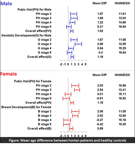
Article Information
vol. 132 no. Suppl 3 A18091
Published By:
American Heart Association, Inc.
Online ISSN:
History:
- Originally published November 6, 2015.
Copyright & Usage:
© 2015 by American Heart Association, Inc.
Author Information
- Shaji C Menon1;
- Angela P Presson1;
- Brian McCrindle2;
- David J Goldberg3;
- Ritu Sachdeva4;
- Bryan Goldstein5;
- Thomas Seery6;
- Karen C Uzark7;
- Anjali Chelliah8;
- Ryan Butts9;
- Heather Henderson10;
- Tiffanie Johnson11;
- Richard V Williams1
- 1Pediatrics, Univ of Utah, Salt Lake City, UT
- 2Pediatrics, The Hosp for Sick Children, Toronto, Canada
- 3Pediatrics, Children’s Hosp of Philadelphia, Philadelphia, PA
- 4Pediatrics, Emory Univ Sch of Medicine, Atlanta, GA
- 5Pediatrics, Cincinnati Children’s Hosp, Cincinnati, OH
- 6Pediatrics, Texas Children’s Hosp, Houston, TX
- 7Pediatrics, Univ of Michigan, Ann Arbor, MI
- 8Pediatrics, Columbia Univ Med Cntr, New York, NY
- 9Pediatrics, Children’s Hosp, Charleston, SC
- 10Pediatrics, Duke Univ Sch of Medicine, Durham, NC
- 11Pediatrics, Riley Hosp for Children, Indianapolis, IN
Abstract 17619: Interstage Growth Trajectory is Associated With Interstage Survival in the Absence of Post-operative Complications Following the Norwood Procedure
Charles F Evans, Danielle S Abraham, Sunjay Kaushal, Geoffrey L Rosenthal
Circulation. 2015;132:A17619
Abstract
Introduction: Low birth and procedure weight have been identified as risk factors for mortality after the Norwood operation. There are no strong data to show that growth during the interstage period is associated with interstage survival.
Hypothesis: We hypothesize that positive growth trajectory following the Norwood operation is associated with improved interstage transplant-free survival.
Methods: We conducted a retrospective cohort study using the National Pediatric Cardiology Quality Improvement Database. The exposure, growth trajectory, was the slope of the least-squares regression line of body mass index-for-age Z-scores over time. The outcome was interstage transplant-free survival. We constructed a multivariable logistic regression model to examine the association between interstage growth trajectory and interstage transplant-free survival.
Results: The study population included 1501 infants who were discharged alive from the Norwood operation between June 2008 and January 2015. The most common diagnosis was hypoplastic left heart syndrome with aortic and mitral atresia (34%). The most common operation was the Norwood procedure with a right ventricle to pulmonary artery shunt (55%). Interstage survival was 89%. Growth trajectory was positive in 945/1392 (68%) for whom sufficient data were available. The occurrence of at least one post-operative complication was identified as an effect measure modifier (p=0.02). When adjusted for sex, birth weight, presence of a secondary cardiac diagnosis, type of operation and post-operative arrhythmia, positive growth trajectory was significantly associated with interstage survival in the absence (OR=3.7, 95% CI 1.7, 8.0) but not in the presence (OR=1.1, 95% CI 0.7, 1.7) of post-operative complications.
Conclusions: Positive interstage growth trajectory is significantly associated with transplant-free interstage survival in the absence of post-operative complications.
Article Information
vol. 132 no. Suppl 3 A17619
Published By:
American Heart Association, Inc.
Online ISSN:
History:
- Originally published November 6, 2015.
Copyright & Usage:
© 2015 by American Heart Association, Inc.
Author Information
- 1Surgery, Div of Cardiac Surgery, Univ of Maryland Sch of Medicine, Baltimore, MD
- 2Epidemiology and Public Health, Univ of Maryland Sch of Medicine, Baltimore, MD
- 3Pediatrics, Div of Pediatric Cardiology, Univ of Maryland Sch of Medicine, Baltimore, MD
Abstract 14163: The Single Ventricle Reconstruction (SVR) Trial at 6 Years: Transplant-Free Survival, Catheter Interventions, and Morbidity
Jane W Newburger, Lynn A Sleeper, J. W Gaynor, Danielle Hollenbeck-Pringle, Peter Frommelt, Jennifer Li, William T Mahle, Ismee A Williams, Andrew M Atz, Kristin M Burns, Shan Chen, James F Cnota, Carolyn Dunbar-Masterson, Nancy S Ghanayem, Caren S Goldberg, Jeffrey P Jacobs, Alan B Lewis, Seema Mital, Christian Pizarro, Aaron W Eckhauser, Paul Stark, Richard G Ohye
Circulation. 2015;132:A14163
Abstract
Background: In the SVR trial, 1-year (y) transplant (tx)-free survival was better for the Norwood procedure with right ventricle-to-pulmonary artery shunt (RVPAS) vs modified Blalock-Taussig shunt (MBTS). At 6 y, we compared tx-free survival, unplanned interventions, morbidities, New York Heart Association (NYHA) Class, and RV ejection fraction (RVEF) by assigned shunt.
Methods and Results: The SVR trial treated 549 pts. Vital status and medical history were ascertained annually. Tx-free survival in the RVPAS (63.5%) vs MBTS (58.7%) groups did not differ (Figure; log-rank P=.13). Similarly, neither mortality nor tx alone differed by shunt type. By 6 y, RVPAS pts had a higher incidence of any catheter intervention (.38 vs .23/pt-yr, P<.001), balloon angioplasty (P=.014), stent (P=.009), and coiling (P<.001). The % of pts with morbidities by 6 y were similar in the groups, with overall rates: pacemaker 3%, thrombosis 16%, stroke 7%, seizures 13%, protein losing enteropathy 3%, plastic bronchitis 0.5%, and 6-y NYHA Class I 71%, II 21%, III 3%, and IV 5%. Among pre-specified subgroups, worse tx-free survival was associated with low birth weight (<2500 g); worse pre-Norwood tricuspid regurgitation (≥2.5 mm jet width); lower surgeon Norwood volume; preterm birth (<37 wks); and combined aortic atresia and pre-term birth (all P<.01). Subgroup x shunt interaction was significant only for surgeon volume levels (P<.05); in the highest volume group (n >15/y), the MBTS was beneficial (P<.04), and in 3 lower volume groups, the RVPAS was qualitatively better. In 6-y echoes read to date, RVEF was similar in the RVPAS vs MBTS groups (46±7, n=55 vs 46±6%, n=48; P=.9).
Conclusions: By 6 y, tx-free survival was an absolute 4.8% higher for pts assigned to the RVPAS vs MBTS group, but the difference no longer reached statistical significance, and they needed more catheter interventions. Rates of death, tx, and morbidities; distribution of NYHA Class; and RVEF were each similar in the shunt groups.
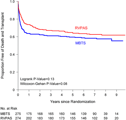
Article Information
vol. 132 no. Suppl 3 A14163
Published By:
American Heart Association, Inc.
Online ISSN:
History:
- Originally published November 6, 2015.
Copyright & Usage:
© 2015 by American Heart Association, Inc.
Author Information
- Jane W Newburger1;
- Lynn A Sleeper1;
- J. W Gaynor2;
- Danielle Hollenbeck-Pringle3;
- Peter Frommelt4;
- Jennifer Li5;
- William T Mahle6;
- Ismee A Williams7;
- Andrew M Atz8;
- Kristin M Burns9;
- Shan Chen10;
- James F Cnota11;
- Carolyn Dunbar-Masterson1;
- Nancy S Ghanayem4;
- Caren S Goldberg12;
- Jeffrey P Jacobs13;
- Alan B Lewis14;
- Seema Mital15;
- Christian Pizarro16;
- Aaron W Eckhauser17;
- Paul Stark18;
- Richard G Ohye19,
- for the Pediatric Heart Network Investigators
- 1Cardiology, Boston Children’s Hosp, Boston, MA
- 2Cardiovascular Surgery, Children’s Hosp of Philadelphia, Philadelphia, PA
- 3Statistician, New England Rsch Institutes, Inc., Watertown, MA
- 4Cardiology, Children’s Hosp of Wisconsin, Milwaukee, WI
- 5Pediatric Cardiology, Duke Univ Med Cntr, Durham, NC
- 6Sibley Heart Cntr Cardiology, The McGill Building, Emory Univ Sch of Medicine, Atlanta, GA
- 7Cardiology, Columbia Univ Med Cntr, New York, NY
- 8Pediatric Cardiology, Med Univ of South Carolina, Charleston, SC
- 9Div of Cardiovascular Services HLBI, NHLBI/NIH, Bethesda, MD
- 10Dept of Statistics, New England Rsch Institute, Inc., Watertown, MA
- 11Cardiology, Cincinnati Children’s Hosp, Cincinnati, OH
- 12Cardiology, C.S. Mott Children’s Hosp, Univ of Michigan Health System, Ann Arbor, MI
- 13Cardiovascular and Thoracic Surgery, Johns Hopkins All Children’s Heart Institute, St. Petersburg, FL
- 14Cardiology, Children’s Hosp of Los Angeles, Los Angeles, CA
- 15Pediatric Cardiology, Hosp for Sick Children, Toronto, Canada
- 16Nemours Cardiac Cntr, Alfred I. duPont Hosp for Children, Wilmington, DE
- 17Thoracic Surgery, Primary Children’s Hosp and the Univ of Utah Hosp, Salt Lake City, UT
- 18Cardiology, New England Rsch Institute, Watertown, MA
- 19Cardiovascular Surgery, C.S. Mott Children’s Hosp, Univ of Michigan Health System, Ann Arbor, MI
Abstract 11041: Use of Digoxin is Associated With Reduced Interstage Mortality: Results From the Pediatric Heart Network Single Ventricle Reconstruction Trial
Matthew Oster, Michael Kelleman, Courtney McCracken, Richard P Ohye, William T Mahle
Circulation. 2015;132:A11041
Abstract
Introduction: Despite medical and surgical advances over the past few decades, mortality for infants with single ventricle congenital heart disease remains as high as 8-12% during the interstage period, the time between discharge after the Norwood procedure and before the stage II palliation. The objective of our study was to determine the effect of digoxin use on interstage mortality in infants with single ventricle congenital heart disease.
Hypothesis: We hypothesized that digoxin would be associated with lower interstage mortality.
Methods: We conducted a retrospective cohort study using the Pediatric Heart Network Single Ventricle Reconstruction Trial public use dataset, which includes data on infants with single right ventricle congenital heart disease randomized to receive either a Blalock-Taussig shunt or right ventricle-to-pulmonary artery shunt during the Norwood procedure at 15 institutions in North America from 2005-2008. Parametric survival models were used to compare the risk of interstage mortality between those discharged to home on digoxin vs. those discharged to home not on digoxin, adjusting for center volume, ascending aorta diameter, shunt type, and socioeconomic status. Further comparisons were made to compare the number of other adverse events in the two groups.
Results: Of the 330 infants eligible for this study, 102 (31%) were discharged home on digoxin. Interstage mortality for those not on digoxin was 12.3%, compared to 2.9% among those on digoxin (Figure), with a number needed to treat of 11 patients to prevent one death. The adjusted hazard ratio was 3.5 (95%CI 1.1-11.7, p=0.04). There were no differences in complications between the two groups during the interstage period.
Conclusions: Digoxin use in infants with single ventricle congenital heart disease is associated with significantly reduced interstage mortality and should be considered for all such infants unless otherwise contraindicated.
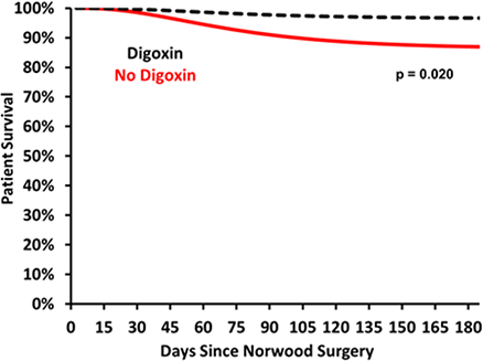
Article Information
vol. 132 no. Suppl 3 A11041
Published By:
American Heart Association, Inc.
Online ISSN:
History:
- Originally published November 6, 2015.
Copyright & Usage:
© 2015 by American Heart Association, Inc.
Author Information
- 1Sibley Heart Cntr Cardiology, Children’s Healthcare of Atlanta, Atlanta, GA
- 2Pediatrics, Emory Univ, Atlanta, GA
- 3Pediatrcs, Emory Univ, Atlanta, GA
- 4Surgery, Univ of Michigan, Atlanta, GA