TCTAP A-072 Early Results of Percutaneous Closure of the Atrial Septal Defects with Occlutech Figulla Devices: A Single Center Experience
Hye Won Kwon, Hyo Bin Lim, Seong-Ho Kim, Sang Yun Lee, So Ick Jang, Su-Jin Park and Mi Kyoung Song
vol. 67 no. 16 Supplement S31-S32
DOI:
https://doi.org/10.1016/j.jacc.2016.03.094
Published By:
Journal of the American College of Cardiology
Print ISSN:
Online ISSN:
History:
- Published online March 13, 2018.
Copyright & Usage:
© 2016
Author Information
- Hye Won Kwon1,
- Hyo Bin Lim1,
- Seong-Ho Kim1,
- Sang Yun Lee1,
- So Ick Jang1,
- Su-Jin Park1 and
- Mi Kyoung Song1
- 1Sejong General Hospital, Korea (Republic of)
Background
Percutaneous closure of secundum type atrial septal defect (ASD) has gained widespread use in recent years. We evaluated the safety and efficacy of the c devices for ASD closure in a single center.
Methods
We retrospectively reviewed medical records of all patients who underwent percutaneous ASD closure in Sejong general hospital from July 2014 to January2016. We evaluated the feasibility and short-term results of transcatheter closure of ASD using the FSO device.
Results
The FSO device was implanted in 63 patients (mean ± SD [range] age: 32.6 ± 22.1 [2-74] years; weight: 48.9 ± 22.4 [11.9 – 119.3] kg). All patients had right atrial and ventricular volume overload with a mean Qp/Qs ratio of 2.4 ± 1.0 (range 1.5 – 6.7). Mean ASD diameter was 22.0 ± 6.7 (range 10 – 38) mm and the size of the implanted FSO was 23.1 ± 7.3 (range 10.5 – 39) mm. Mean procedure time was 64.7 ± 26.4 (range20-187) minutes and the mean fluoroscopy time was 16.8 ± 15.4 (4.7 – 113) minutes. The patients were followed-up for mean 4.8 ± 4.2 (range 1 – 17) months. There were 3 cases of device embolization during the procedure or immediate after the procedure. Four patient experiences frequent premature atrial contractions and 1 patient had transient junctional tachycardia after the procedure. Although one patient died of subdural hemorrhage after the procedure, there were no other serious complications associated with the device.
Conclusion
The FSO device appears to be easy to use and effective for percutaneous closure of ASD. Long-term follow-up is required to identify future risk for developing arrhythmias or device-related complications.
TCTAP A-070 Long-Term Safety and Efficacy of Closure of Atrial Septal Defects with Cocoon Septal Occluder
Rajasekhar Durgaprasad, Vamsidhar Akkulagari, Vanajakshamma Velam, Latheef Kasala, Obul Reddy Gajjala, Boochi Babu Mannuva and Vasudevachetty Pakala
vol. 67 no. 16 Supplement S31
DOI:
https://doi.org/10.1016/j.jacc.2016.03.092
Published By:
Journal of the American College of Cardiology
Print ISSN:
Online ISSN:
History:
- Published online March 13, 2018.
Copyright & Usage:
© 2016
Author Information
- Rajasekhar Durgaprasad1,
- Vamsidhar Akkulagari1,
- Vanajakshamma Velam1,
- Latheef Kasala1,
- Obul Reddy Gajjala1,
- Boochi Babu Mannuva1 and
- Vasudevachetty Pakala1
- 1Sri Venkateswara Institute of Medical Sciences, India
Background
Transcatheter closure of ostium secundum Atrial septal defect (ASD II) isusually done worldwide with Amplatzer septal occluder (ASO) (St. Jude Medical, St. Paul Minnesota, USA). Though complications are rare, it is associated with life threatening complications like tissue erosions and severe nickel allergy. The Cocoon septal occluder (CSO) (Vascular Innovations Co., Nonthaburi, Thailand) is an improved new generation double disk device with distinct features making it an attractive alternative to ASO. The aim of this study is to evaluate long-term safety and efficacy of CSO in ASD II including large and complex defects.
Methods
This is a single center, retrospective, observational study of 239 consecutive ASD II patients between November 2008 and January 2015, who were attempted with CSO at Sri Venkateswara Institute of Medical Sciences, Tirupati, India. The patients were followed at 1-month and thereafter every 3 months up to October 2015. They were followed for adverse events–cardiac mortality, arrhythmias, residual leak, device embolization and tissue erosion. The procedure of closing ASDs with the CSO is similar to that of using the ASO. CSO is a new generation double disk device that is self-expanding, self-centering and repositionable. It has nanoplatinum coating making it superior bio compatible, softer and preventing leakage of nickel.
Results
A total of 236 patients who were successfully treated with CSO were followed for median duration of 38 months (range: 9-78 months). Mean age was 28.3 ± 16.8 years, pediatric group had 80 patients (33.5%) and females were 67.4%. Mean ASD size was 20.5 ± 6.5 mm (range: 7-37 mm), 24.3% had defects >30 mm. Intra procedural Transesophageal echocardiography, balloon sizing and Greek maneuver were done in 161 (67.4%), 61 (25.5%) and 8 (3.3%) patients respectively. Mean device size was 25.3 ± 7.2 mm (range: 10-42 mm). Device was embolized in 3 (1.3%) patients with large defects and at least 2 deficient rims. One (0.4%) patient had air embolism, 2 (0.8%) had complete heart block, 2 (0.8%) had transient atrial fibrillation, 2 (0.8%) had pericardial effusion and 3 (1.3%) patients had trivial residual leak which disappeared at 1-month follow-up. There was no cardiac mortality and tissue erosion.
Conclusion
Our study shows that Cocoon septal occluder (CSO) is safe in simple and complex ostium secundum Atrial septal defects (ASD II) at mid and long-term follow-up.
TCTAP A-068 Complications of Cardiac Catheterization in Structural Heart Disease in a Tertiary Referral Center
Gi-Beom Kim, Eun-Jung Bae and Bo Sang Kwon
vol. 67 no. 16 Supplement S30
DOI:
https://doi.org/10.1016/j.jacc.2016.03.090
Published By:
Journal of the American College of Cardiology
Print ISSN:
Online ISSN:
History:
- Published online March 13, 2018.
Copyright & Usage:
© 2016
Author Information
- 1Seoul National University Children’s Hospital, Korea (Republic of)
- 2Seoul National University Hospital, Korea (Republic of)
Background
Cardiac catheterization is used to diagnose structural heart disease (SHD) and to perform transcatheter treatment. This study aims to evaluate complications of cardiac catheterization and the associated risk factors in a tertiary Korean center over recent 10 years.
Methods
Total 2,071cardiac catheterizations performed at the Seoul National University Children’s Hospital from January 2004 to December 2013 were included in this retrospective study.
Results
The overall complication, severe complication, and mortality rates were 16.2%, 1.15%, and 0.19%, respectively. The factors that significantly associated risk of overall and severe complications were use of anticoagulant before procedure (odds ratio[OR] 1.83, p=0.012 and OR 6.45, p<0.001, respectively), prothrombin time (OR2.30, p<0.001 and OR 5.99, p<0.001, respectively), use of general anesthesia during procedure (OR 1.84, p=0.014 and OR 5.31, p=0.015, respectively), and total procedure time (OR 1.01, p<0.001 and OR 1.02, p<0.001, respectively). Low body weight (OR 0.99, p=0.003), severe SHD (OR1.37, p=0.012), repetitive procedures (OR 1.7, p=0.009), and total fluoroscopy time (OR 1.01, p=0.005) were significantly associated with the overall complication risk. High activated partial thromboplastin time (OR 1.04, p=0.001), intensive care unit admission state (OR 14.03, p<0.001), and concomitant electrophysiological study during procedure (OR 3.41, p=0.016) were significantly associated with severe complication.
Conclusion
The use of cardiac catheterization in SHD is increasing and becoming more complex nowadays; this could cause complications despite the efforts to reduce complications. Patient selection for therapeutic catheterization should be done carefully and technique and management during the peri-procedural period need to be improved to reduce complications.
TCTAP A-069 Guidewire Assisted Device Placement in Large Atrial Septal Defect with Aortic Rim Deficiency – An Easy and Simple Method
Kenji Suda
vol. 67 no. 16 Supplement S30-S31
DOI:
https://doi.org/10.1016/j.jacc.2016.03.091
Published By:
Journal of the American College of Cardiology
Print ISSN:
Online ISSN:
History:
- Published online March 13, 2018.
Copyright & Usage:
© 2016
Author Information
- Kenji Suda1
- 1Kurume University School of Medicine, Japan
Background
It is sometimes difficult to place a device in large atrial septal defect (ASD), especially aortic rim deficiency and several useful equipment or techniques are developed and reported to securely place a device in such ASD. However, usually these equipment or techniques require additional cost of equipment or femoral vein access. Therefore the aim of this study was to develop more simple method to place a device in such ASD.
Methods
Subjects were 3 patients; Case 1, 4.9 y.o. 19 kg 20 mm device; Case 2, 7.8 y.o., 22 kg, 18 mm device; Case 3, 70 y.o. device 22 mm. All ASDs have wide-range aortic rim deficiency and were attempted to place an Amplatzer septal occluders. After placed a >= 9 Fr long sheath into the left atrium, we intentionally deployed both side discs, then, placed a stiff guide wire, that used for sizing, into the left upper pulmonary vein through the same long sheath. Keeping a guidewire in position, we pulled back both discs that allowed the discs aligned inter-atrial septum preventing from prolapse of the discs. Pulled back the right atrial disc and waist into long sheath with left atrial disc attached on the left side of inter-atrial septum and then re-deployed body and right atrial disc to properly place device. After confirmed secure position of the device, we pulled back the guidewire and finished procedure.
Results
We could have easily placed devices in all 3 patients without additional equipment or additional femoral vein access. In 2 of 3 patients, we failed to place device even with specially made long sheath such as Hausdorf sheath.
Conclusion
Our guide wire assisted device placement does not require additional equipment or placing additional sheath and is an easy and simple method to place a device in large atrial septal defect with aortic rim deficiency.
TCTAP A-073 Procedural, Early and Long-Term Outcomes After Transcatheter Closure of Atrial Septal Defects: Comparison Between Adult Patients with Large and Very Large Defect Groups
Se Yong Jung, Jeong Yoon Kim, Nam Kyun Kim, Jo Won Jung, Lucy Youngmin Eun and Jae Young Choi
vol. 67 no. 16 Supplement S32
DOI:
https://doi.org/10.1016/j.jacc.2016.03.095
Published By:
Journal of the American College of Cardiology
Print ISSN:
Online ISSN:
History:
- Published online March 13, 2018.
Copyright & Usage:
© 2016
Author Information
- 1Yonsei University Severance Cardiovascular Hospital, Korea (Republic of)
- 2Gangnam Severance Hospital, Korea (Republic of)
Background
Transcatheter closure of atrial septal defect (ASD) has become a preferred treatment modality for most patients with secundum ASD. Many studies have demonstrated transcatheter closure of moderate to large sized ASD is safe and effective. However, there are lacking of evidence regarding the feasibility and safety of transcatheter ASD closure for very large defects. This study aimed to compare procedural, early and long-term outcome of device closure of adult patients with large and very large ASDs.
Methods
We performed a retrospective study of adult patients with large ASD (group 1, defined as defect diameter of 25 mm ≤ ASD < 35mm) and very large ASD (defines as defect diameter ≥35 mm) treated by transcatheter closure using Amplatzer septal occluder during 12-year period (May 2003-February 2015) at a single tertiary center. A total of 269 adult patients had a large ASD with defect diameter ≥ 25mm before closure. The studied population was divided into two groups according to the pre-procedural maximal ASD diameter; group 1 (n=216) was composed of patents with defect diameter of 25 mm ≤ ASD < 35mm, and group2 (n=53) consisted of patients with defect diameter ≥35mm. We compared procedural parameters, early complications and long-term follow-up results between two groups.
Results
The need of modified implantation techniques (MIT) was higher in group2 (23.6% vs 37.7%, p = 0.034). Procedural success rate was considerably high in both groups (99.1% in group 1 vs 100% in group 2, p=0.620).Major complications were occurred in 4 (1.5 %) patients (1.4% vs 1.9%, p=0.804).Minor complication rate was not different between two groups. During long term follow-up (47.2 ± 32.0 months, range 6.0-135.5), there was one major complication (0.4%) of stroke in a patient with atrial arrhythmia. Most common long-term minor event was new-onset or aggravated migraine headache (3.9%) followed by arrhythmias (1.9%) without statistical difference between two groups.
Conclusion
Although modified implantation technique (MIT) is more frequently required in very large ASD groups, the procedural, early and long-term outcomes after transcatheter closure of ASD are similar in both groups. These results suggest transcatheter closure of ASD may be a good treatment option in selected group of patients with very large defect.
TCTAP A-071 Cardiac Intervention with Off-Label Use of Medical Devices in Congenital Heart Disease
Young Hwa Kong, Jin Young Song, June Huh and I-Seok Kang
vol. 67 no. 16 Supplement S31
DOI:
https://doi.org/10.1016/j.jacc.2016.03.093
Published By:
Journal of the American College of Cardiology
Print ISSN:
Online ISSN:
History:
- Published online March 13, 2018.
Copyright & Usage:
© 2016
Author Information
- 1Samsung Medical Center, Korea (Republic of)
Background
Catheter-based cardiac intervention in congenital heart disease has been established as an alternative treatment method with good outcome. However, the relatively small population and variety of specific lesions make the investment in this field difficult compared to that of adults with acquired heart disease. The off-label use of medications and medical devices is common in pediatric disease. The growth of catheter intervention in congenital heart disease has increased the off-label utilization of approved devices in different ways from their intended applications. Although it is possible to amend the labeling of devices to add other approved uses, it might be too inefficacious in terms of cost and time. To our knowledge, there are few investigations regarding the off-label use of medical devices implanted in congenital heart disease. The most commonly used implantable medical devices in congenital heart disease are stent and occlusion devices, such as the Amplatzer duct occluder, coil and Amplatzer vascular plug. The off-label use of implantable devices could be more important because implanted devices should keep those function for a long time. Therefore, we investigated the present condition of off-label use of implanted transcatheter devices and their outcomes in congenital heart disease.
Methods
Our study is a retrospective review of all cardiac catheterization in CHD at Samsung medical center. Definition of “off-label” was not included as indications in the FDA-approved labeling. 144(8.32%) of 1730 patients with CHD underwent cardiac interventions with off-label devices.
Results
Median age was 51 months (15days-86years). VSD closure (12 of 12 patients, 100%) and stenting (16 of37 patients, 43%) were common. And Amplatzer Vascular Plugs and coils were most commonly used off-label. Sixteen underwent stent implantations used off-label devices: Four underwent PDA stenting; in seven, stenting of right ventricular outflow tract; in two, stenting in LPA/RPA; in one, stenting in coarctation of aorta; and one underwent stenting of atrial septal defect. Nine (6.3%) of 144were failed the interventions used off-label devices. Seven (4.9%) underwent operation due to complications of interventions.
Conclusion
Our study indicates that interventions used the off-label devices were safe as far as it goes. But still we need to take a more cautious interventional approach of the off-label devices.
TCTAP C-241 Transactheter Closure of Atrial Septal Aneurysm (ASA) Associated with Multiple Atrial Septal Defect (ASD)
Jihun Ahn and Do Hoi Kim
vol. 67 no. 16 Supplement S395-S396
DOI:
https://doi.org/10.1016/j.jacc.2016.03.459
Published By:
Journal of the American College of Cardiology
Print ISSN:
Online ISSN:
History:
- Published online March 13, 2018.
Copyright & Usage:
© 2016
Author Information
- 1SoonChunHyang University Gumi Hospital, Korea (Republic of)
[Clinical Information]
Patient initials or identifier number
64 year-old male
Relevant clinical history and physical exam
The patient was 64 year-old male and presented with transient loss of conciousness.Transthoracic echocardiography and trans esophagealechocardiography revealed the right atriam (RA) and right ventricle (RV) was moderately dilated, the ventricular septum moved paradoxically, a atrial septal aneurysm (ASA) associated with multiple atrial septal defect (ASD) with marked mobility intothe right atrium.
Relevant test results prior to catheterization
Transthoracicechocardiography demonstrated a ratio of pulmonary blood flow to systemic bloodflow of 1.5.
The defect size on TEE was 1.76 cm. Usually ASD defect size is underestimated on TEE. As a result of this measurement, we chose a 2.2cm sized Amplatzer device.
Relevant catheterization findings
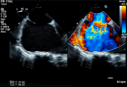
After the Amplatzer device has been fully opened, we performed the TEE and confirmed that both atrial disks are flattened or nearly flattened and that the left atrial disk is entirely in the left atrium and the right atrial disk entirely in the right atrium without remnant shunt. After the stability of the device has been confirmed, the delivery cable was released by counterclock wise rotation. Amplatzer spetal occluder device is a useful treatment option of ASA with multiple ASD.
[Interventional Management]
Procedural step
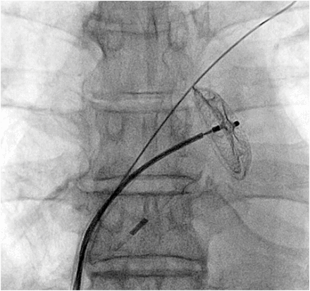
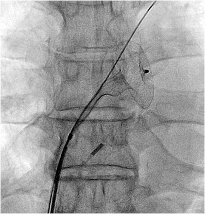
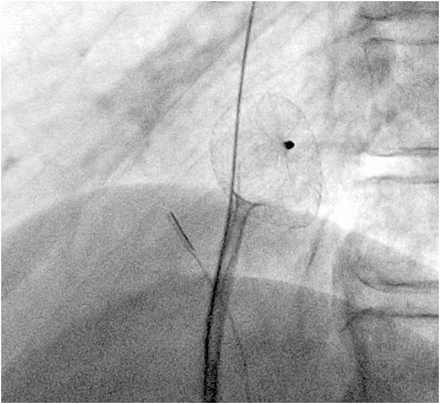
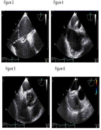
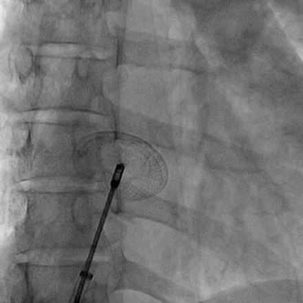
After general anesthesia, an 8Fr sheath was inserted through right femoral vein. Intraprocedural TEE wasused for guidance in the catheterization laboratory. Initially multi purpose catheter was advanced into left atrium through ASD. And then the multi purpose catheter was replaced with a 0.035inch J-tipped exchange length guidewire, thetip of which is preferably located in a left upper lobe pulmonary vein forstability. The defect size on TEE was 1.76 cm. Usually ASD defect size is underestimated on TEE. As a result of this measurement, we chose a 2.2cm sized Amplatzer device. Selected Amplatzer device was loaded into the delivery tube. And then, delivery sheath, a long dilator, was inserted into left atrium over a 0.035 inch guidewire. After which the prepared device was loaded into delivery sheath. Most important procedural point is to puncture the central area of the shunt (figure 3, 4). Because of multiple shunt and aneurysmal change make it difficult to cover the whole defect area if the guiding sheath was positioned obliquely. After positioned the sheath at left atrium through the central area of the defect site, the Amplatzer device was advanced until it reached the tip of the sheath and then deploy the Amplatzer device (figure 5, 6).
Case Summary
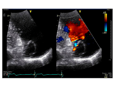
As the atrial septum returns to its natural position, the device typically springs superiorly and leftward radiologically and significant interatrial shunting through the device on TEE was eliminated on color Doppler. We think central puncture of the ASA with multiple ASD and using enough big size Amplatzer spetal occluder device is a useful treatment option of ASA with multiple ASD.
TCTAP C-244 Emergency Creation of Atrial Communication in Bilateral Occluded Femoral Veins
Manish Shrestha, Worakan Promphan and Thanarat Layangool
vol. 67 no. 16 Supplement S398-S399
DOI:
https://doi.org/10.1016/j.jacc.2016.03.462
Published By:
Journal of the American College of Cardiology
Print ISSN:
Online ISSN:
History:
- Published online March 13, 2018.
Copyright & Usage:
© 2016
Author Information
- 1Queen Sirikit National Institute of Child Health, Thailand
[Clinical Information]
Patient initials or identifier number
SL
Relevant clinical history and physical exam
Five years old boy with complex congenital heart disease presented with severe cyanosis and shortness of breath – functional class III. He was tachypneic and had grade III clubbing but no pedal edema. His oxygen saturation in room air was 68%, precordium was active and systolic ejection murmur grade III was heard at left upper sternal border. Bilateral lung field revealed conducted breath sound with no definite wheezing. Liver was palpable 2 cm from right costal margin at mid-clavicular line.
Relevant test results prior to catheterization
Chest X-ray revealed perihilar infiltration with pulmonary congestion andthere was no cardiomegaly. Transthoracic echocardiography revealed congenitally corrected transposition of great arteries, left atrioventricular valve atresia, restrictive atrial septal defect (ASD), and large ventricular septal defectwith severe subvalvar pulmonary stenosis.
Hence decompression of left atrium (LA) is utmost necessary for hissurvival at that time.
Relevant catheterization findings
Inaccessible bilateral femoral veins. Venogram revealed total occlusion of both femoral veins with multiple collaterals connected to azygos system.
Transhepatic venous approach was considered.
| Site | Pressure (mm Hg) | |
| Pre-procedure | Post-procedure | |
| RA | 7 | 9 |
| PV | 21 | – |
| LA | 21 | 16 |
| Ao | – | 77/44 |
Table 1
Hemodynamic data of the patient (pre and post procedure)
[Interventional Management]
Procedural step
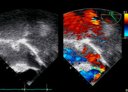
Transhepatic approach was performed by experienced radio-interventionistunder ultrasound guidance at right anterior axillary position. After theguide wire was securely placed into RA, a 6 Fr. introducing sheath was placedat IVC. Multiple attempts with different wires and catheters were made to enterinto LA. Finally ASD was successfully crossed by 5 Fr. “Judkins Right (JR)”catheter and parked atleft upper pulmonary vein (LUPV) via JR catheter. Static balloon dilatation ofASD was performed by “Admiral” Balloon10 x 20 mm with maximum inflation pressure of 6 ATM. The ASD waist disappearedafter the last inflation, however the mean pressure of LA was stillconsiderably high (21mmHg) with pull back pressure gradient of 15 mmHg andtransesophageal echocardiogram (TEE) still showed restrictive atrialcommunication. Therefore, the IAS was re-attempted to cross in different angle. Under TEE guidance, once the JR catheter was impinging onto thin part of IAS, the “Conquest Pro CTO”coronaryguide wire mounted with FineCross Microcatheter successfully punctured the IAS.All the system was then placed into the LUPV. The “Rosen”guide wire was exchanged, and then second attempt of static balloondilatation was performed. The mean LA pressure decreased to 16 mmHg with pullback pressure gradient of 7 mmHg. TEE showed two jets across IAS withsignificant decrease of LA dimension.
After successful balloon dilatation, transhepatic tract was occluded byusing gelfoam.
Case Summary
Our patient considerably improved after the procedure and discharged back home. We conclude:
Creating an atrial communication in this type of lesion is lifesaving. Transhepatic access is an effective alternative approach in those patients whose femoral venous access is in accessible.
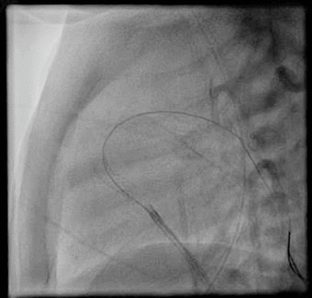
TCTAP C-243 Percutaneous Retrieval of Migrated Atrial Septal Defect Occluder After Implantation of Device
Sang Jin Ha, Won-Kyung Lee, Yeo-Jeong Song, Woo Dae Bang, Sang-Yong Yoo and Sangsig Cheong
vol. 67 no. 16 Supplement S397-S398
DOI:
https://doi.org/10.1016/j.jacc.2016.03.461
Published By:
Journal of the American College of Cardiology
Print ISSN:
Online ISSN:
History:
- Published online March 13, 2018.
Copyright & Usage:
© 2016
Author Information
- 1GangNeung Asan Hospital, Korea (Republic of)
[Clinical Information]
Patient initials or identifier number
PSJ
Relevant clinical history and physical exam
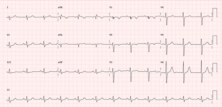
A 45-year-old female presented with mild dyspnea andpapitation. Physical examinations were non-specific except for the fixedsplitting of second heart sound on auscultation. Electrocardiography showednormal sinus rhythm with non-specific intraventricular conduction delay.
Relevant test results prior to catheterization
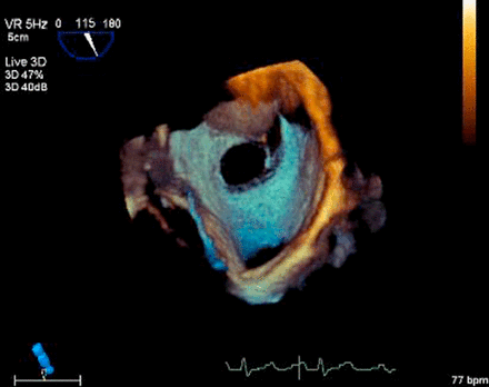
Transthoracic(TTE) and transesophagealechocardiography (TEE) revealed dilated right ventricle and atrium, abnormalleft to right shunt flow through maximally 20 mm-sized Atrial septal defect ofsecundum type, and the calculated ratio of pulmonary (Qp) to systemic bloodflow (Qs) was 1.8. A defect showed the 8 mm sized anterio-superior rim, 11mmsized anterio-inferior rim, and 15mm sized posterior-inferior rim.
Relevant catheterization findings
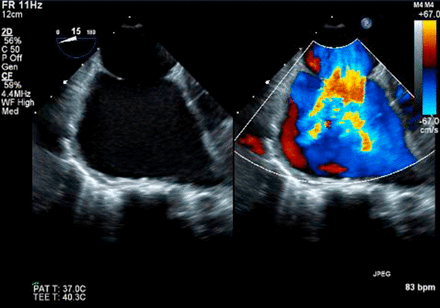
Under the TTE and fluoroscopic guidance, percutaneousdevice closure of ASD was performed using Occlutech®Figulla® FlexII. Balloon-sizing diameter of each ASD was measured as 21 mm using stop-flowtechnique, and deployed after push test
[Interventional Management]
Procedural step
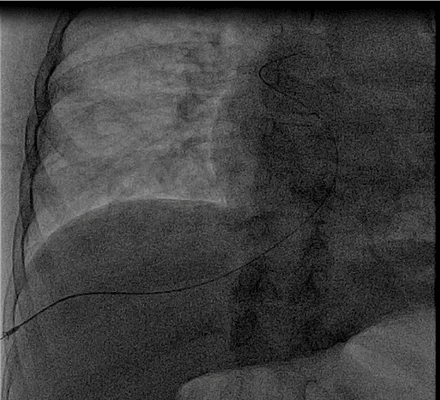
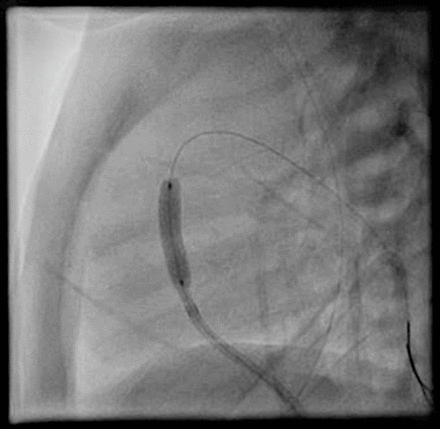
However, ASD device migrated into Left ventricle in aminute. The migrated Figulla was trapped in Left ventricular outflow tract. Afterconfirmation of migration, percutaneous retrieval of the embolized Figulla was performedusing snare. The embolized Figulla was successfully retrieved by snaring thescrew on the right atrial disc of Figulla. And then under TEE and fluoroscopicguidance, percutaneous device closure was successfully and Final TEE revealedgood apposition of two ASO devices with minimal residual shunt flow.
Case Summary
Embolization of the occlusion device after percutaneous closure of atrial septal defect (ASD) is a potential disastrous complication. Here, we report a successful percutaneous retrieval case of the embolized Figulla to the left ventricular outflow tract after the successful deployment of ASD closure device using snare. Before detachment of device, TEE confirmation was important for procedural success.
TCTAP C-242 Stent Migration After Right Ventricular Outflow Tract Stenting in the Severe Cyanotic Tetralogy of Fallot Patient
Tamaki Hayashi, Saleem Akhtar and Mazeni Alwi
vol. 67 no. 16 Supplement S396-S397
DOI:
https://doi.org/10.1016/j.jacc.2016.03.460
Published By:
Journal of the American College of Cardiology
Print ISSN:
Online ISSN:
History:
- Published online March 13, 2018.
Copyright & Usage:
© 2016
Author Information
- 1Institut Jantung Negara, Malaysia
- 2Aga Khan University Hospital, Pakistan
[Clinical Information]
Patient initials or identifier number
338939
Relevant clinical history and physical exam
A one-month-old baby was referred to our hospital with severe cyanosisand mild tachypnea. The body weight was 3.3kg and SpO2 was 69%. Theechocardiography revealed tetralogy of Fallot with Patent ductus arteriosus (PDA) and patent foramen ovale. The infundibular stenosis was severe and hypoxia wassignificant. The patient was brought in the intervention alcatheterizationlaboratory as an emergency case for PDA or right ventricular outflow tract(RVOT) stenting.
[Interventional Management]
Procedural step
Right ventricle angiography showed pulmonary stenosis. Valve annulus was3.7mm (Z score -3.09) and it was difficult to localize the narrowest part inthe infundibulum by fluoroscopy. Echocardiography revealed the infundibularstenosis as 2.1mm. Valve was crossed with a coronary guide wire. PTCA balloon5mm X 12mm was placed over the coronary wire and balloon dilatation of thepulmonary valve was done. However, no significant improvement in the saturationwas observed. Then, test angiogram in the RVOT was done. Thepre-mountedcoronary stent 3.5mm x 12mm was deployed. Initially the stent appeared stablebut once guide wire was removed, the stent migrated proximally to the tricuspidvalve and caused heart block in the child. Saturation also did not improve.Temporary pacemaker was inserted. PDA stenting was done subsequently, andsaturation improved. Heart block resolved spontaneously and the temporarypacemaker was removed after 72 hours of procedure. On follow up, the stent isstable but at displaced position and not causing any valve regurgitation orstenosis.
Case Summary
The pulmonary valve sizewas acceptable in our case and the narrowest part was in the proximal rightventricular outlet tract. Therefore, we decided to preserve the valve function, and the stent was placed in the right ventricular outlet tract. This contributedthe instability of stent because the infundibulum is made from a contractilemuscle and ended up with migrating. PDA stenting was performed as analternative remedy.