Ari was our second child, but despite having the experience of our son under my belt, I was still sensitive to the perception that I was overreacting. I didn’t want to be viewed as the quintessential over-protective or hysterical mother.
Looking back, I see the absurdity clearly. Mother’s intuition is really about honed observation skills. Trust your “instinct”. Had I heeded the voice that I was overreacting, that it could wait until Monday when I could get in to see the pediatrician, there is zero doubt that Ari would be dead.
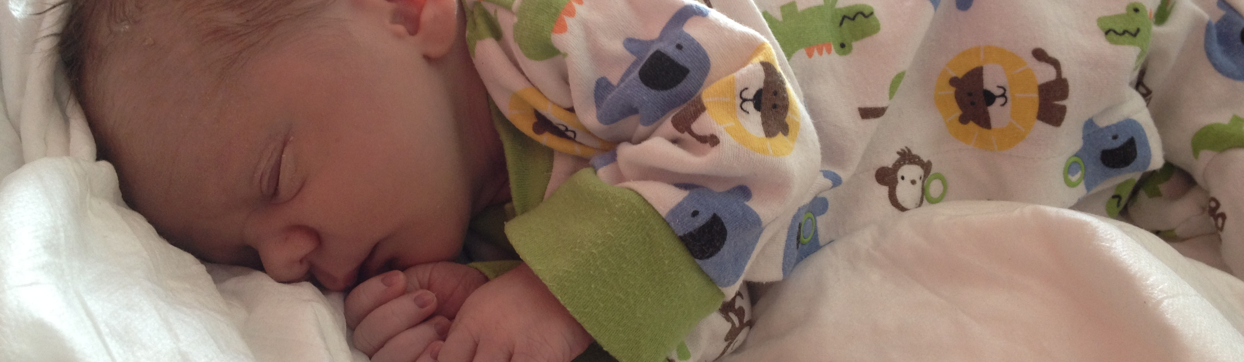
Signs of Possible CHDs in Babies:
- Feeding difficulties
- Weight problems
- Fatigue (sleeps a lot- even for a newborn)
- Shortness of breath, rapid breathing
- Pale gray or blue skin color, especially around lips and fingertips (it can be very subtle)
Additional Tests for Diagnosing CHDs
In addition to detecting possible congenital heart defects at birth, the pulse oximetry test can be used well after birth to identify underlying cardiac malformations.
Echocardiograms not only detect Congenital Heart Defects, but they allow doctors to diagnosis the majority of CHDs, even those that require immediate surgery.
Echocardiography uses sound waves to create 2D images of the heart. It has evolved significantly over the last 5 decades, and provides a comprehensive anatomic and functional understanding of a patient’s cardiac anatomy.
An echocardiogram is non-invasive, relatively inexpensive and carries little downside. It involves placing a few stickers on a child’s chest. A cardiologist will then take an ultrasound probe- like the one used at the 20-week prenatal ultrasound- to create a movie of the heart.
Other Tests
Transesophageal Echocardiogram (TEE)
A TEE is extension of the echocardiogram, the most significant difference being that a much smaller version of the probe is inserted through the mouth and into the esophagus. Because the esophagus is so close to the upper chambers of the heart, it is possible to generate very clear images of the heart using the same Doppler technology.
However, because it is a more invasive imaging technique, sedation is required.
Cardiac Catheterization
Cardiac catheterization is a procedure whereby a tube (or catheter) is inserted typically through a vein in the groin, and is then threaded through the artery to the heart. Using contrast dye, images can be created of the heart. Catheterization can also be used to perform repairs to the heart, even repairs that, until recently, required open-heart surgery.
Sedation is typically required for children, and significant radiation exposure has increasingly become a point of concern.
Magnetic Resonance Imaging (MRI)
Every person’s heart is as distinctive as her face, and sometimes as a result of certain characteristics of her heart’s topography or the type of defect, it is necessary to use more advanced imaging equipment to draw an accurate picture of her heart.
MRI and CT are the most advanced type of imaging available to doctors, and they are both used rather sparingly and only when considered absolutely necessary for treatment.
MRI uses magnetic waves to evaluate the heart, vessels and surrounding structures. It does not require any exposure to radiation.
However, MRI typically requires sedation, is very expensive, and can take around an hour to perform. That said, new methods for imaging are being developed that reduce the amount of time required to utilize the equipment (thus reducing cost) and even eliminate the need for sedation.
That said, the quality of the 2D images created with MRI is so high that they can then be used to create 3D models, which can then be printed and even utilized to practice surgery in anticipation of surgery.
Computerized Topography (CT)
CT is the other advanced imaging tool that is used sparingly to diagnose the most complex hearts. Although patients are exposed to some radiation, new equipment significantly reduces exposure (a fraction of radiation levels in the catheterization lab), making it an increasingly attractive advanced imaging tool. And like MR imaging, CT images are detailed enough to convert from 2D images to 3D-printed models.